RCOM RADIOLOGICAL CASE OF THE MONTH
Applied Radiology — Vol. 35 , Issue 12 , pp. 36 -38
DOI: 10.37549/AR1471
Published: December 1, 2006
Categories
CASE SUMMARY
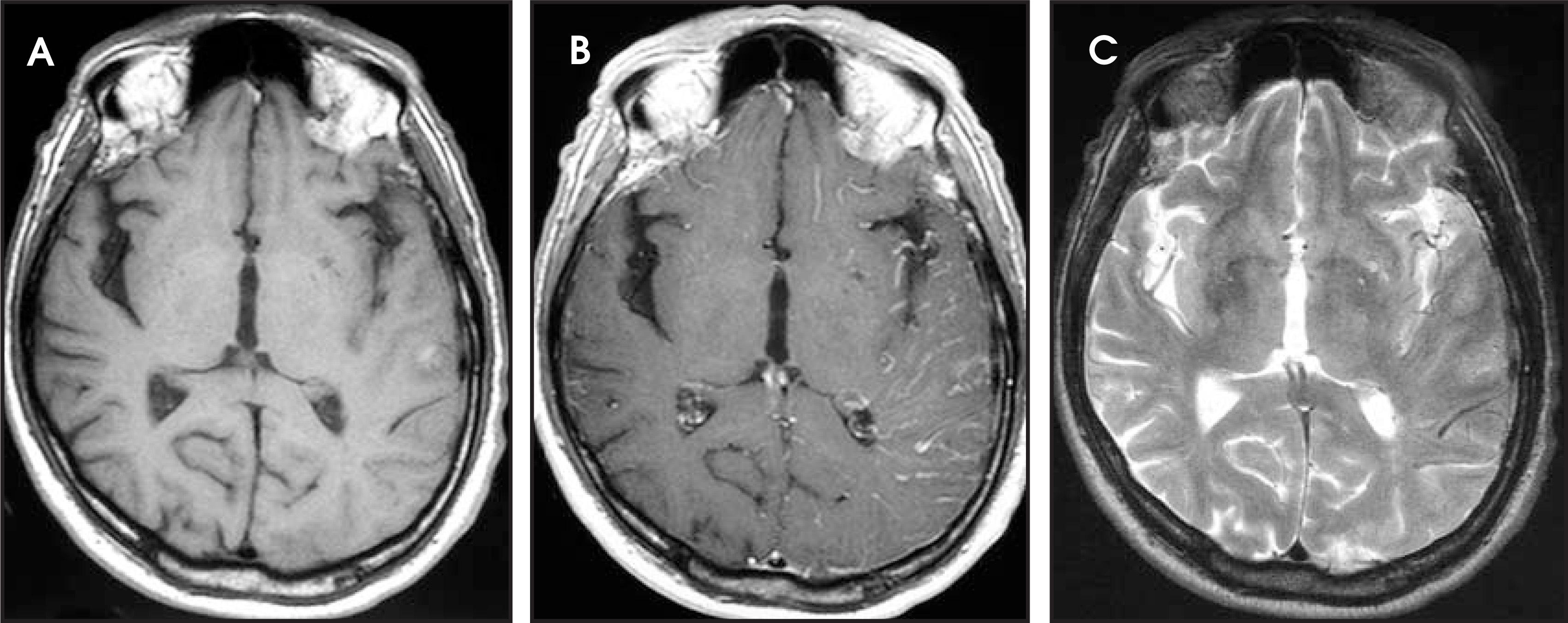
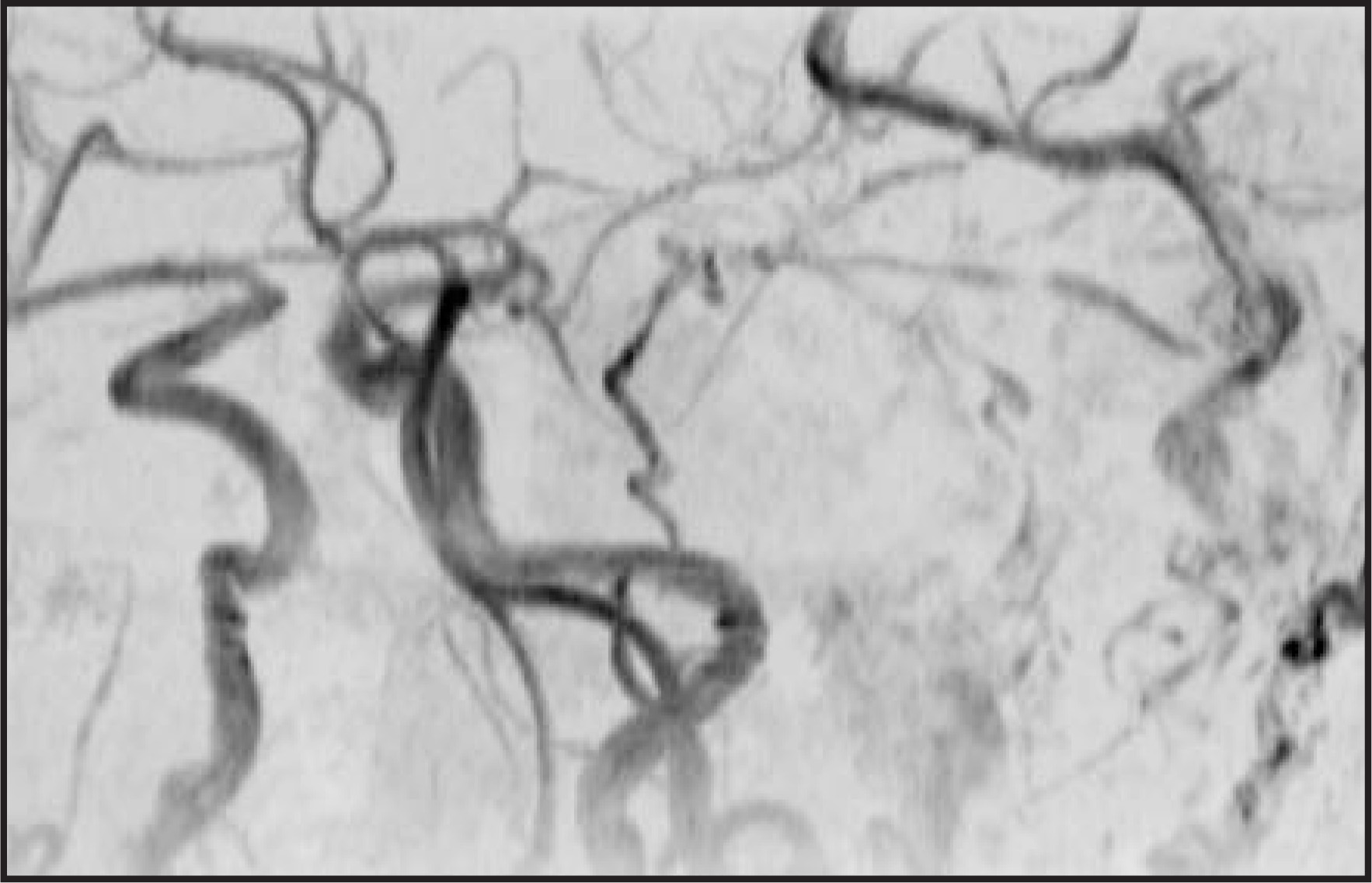
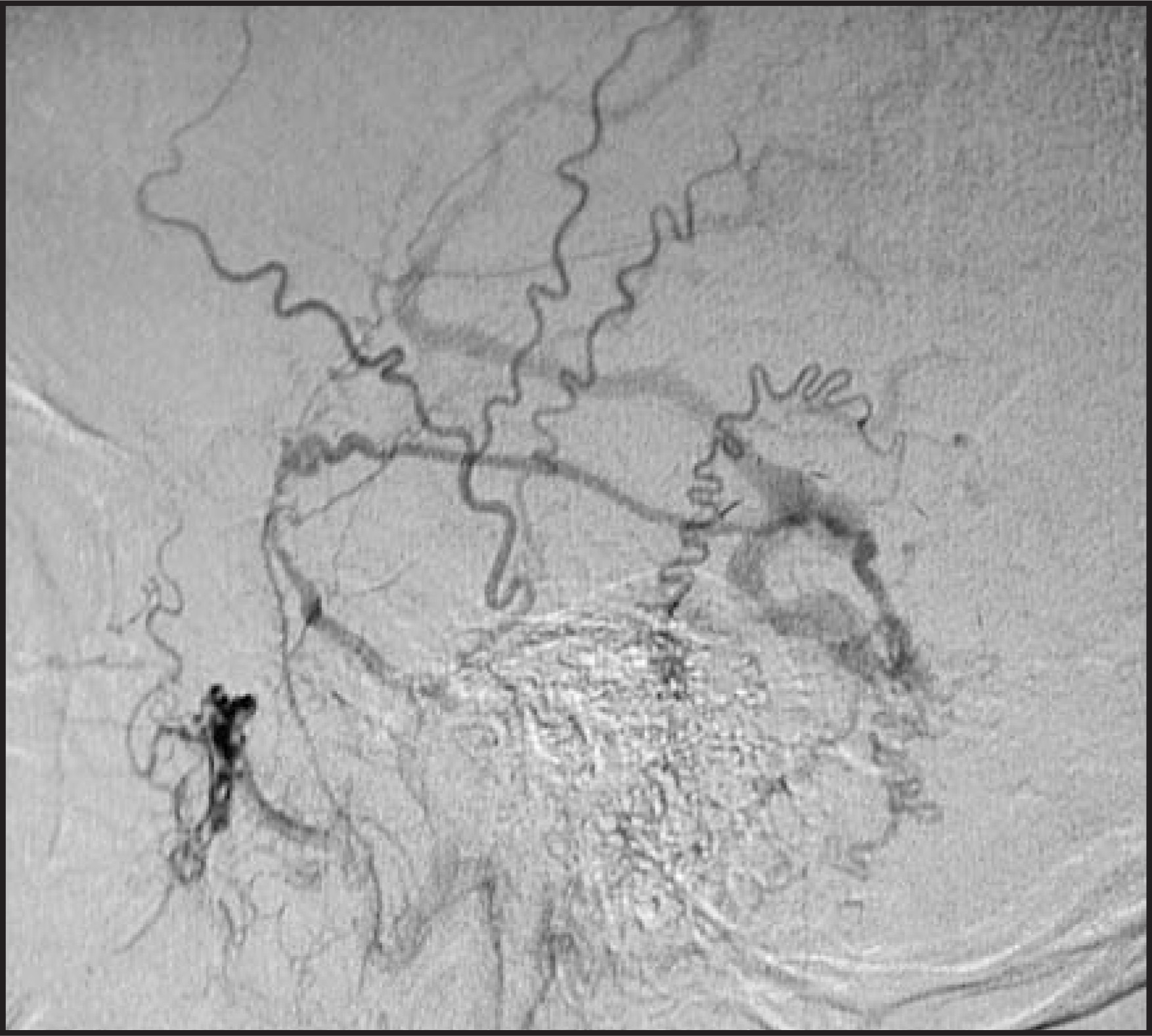
A 75-year-old man who was transferred from an outside hospital presented with slurred speech and a headache. No prior trauma, surgery, infection, drug use, or vascular disease was reported. The patient was afebrile and had a blood pressure of 141/89 mm Hg. No focal neurological deficits were noted on examination. The noncontrast computed tomographic (CT) image from an outside hospital had shown a left temporoparietal region of hemorrhage (not shown). The carotid ultrasound was negative. Magnetic resonance imaging (MRI) was performed (Figure 1), followed by magnetic resonance angiography (MRA) (Figure 2). For further evaluation, a cerebral angiogram was obtained (Figure 3). The patient was discharged without complaints after treatment.
IMAGING FINDINGS
The circular area of increased signal intensity in the left posterior temporoparietal region involving the cortical gray matter on the T1weighted (T1W) image with peripheral hypointensity is consistent with sub-acute hemorrhage and edema (Figure 1A). The postgadolinium T1W MRI demonstrated tortuous and engorged pial vessels in the left temporoparietal region (Figure 1B). The MRA revealed a dural arteriovenous fistula (AVF) between the left middle meningeal artery and a cortical vein with retrograde drainage into Labbé’s vein (Figure 2). The occipital artery, the posterior branch of the middle meningeal artery, and the transmastoid branch of the occipital artery flowed in a retrograde fashion into ectatic cortical veins with drainage into Labbé’s vein and then Trolard’s vein (Figure 3).
DIAGNOSIS
Type IV dural arteriovenous fistula
DISCUSSION
Dural AVFs comprise 10% to 15% of intracranial arteriovenous lesions.1 The pathophysiology of a dural AVF includes high-flow shunting that results in angiopathy on both the venous and arterial sides, with segmental dilatation of blood vessels that may lead to hemorrhage and/or venous congestion.2 The venous congestion may result from ectasia or thrombosis of the dural AVF.2 The term fistula implies an acquired nature, such as from infection, trauma, surgery, or vascular disease. Ironically, as in this example, often no etiology is elicited from the patient.3 The presentation of a dural AVF usually correlates with the site of venous congestion, hemorrhage, or infarction.4 In this case study, the transient slurred speech may have resulted from the venous congestion of the left frontal temporoparietal region, as shown by the pseudophlebitic pattern that is discussed subsequently.
Dural AVFs are classified into 5 categories.5 The Type I dural AVF drains into the sinus with normal antegrade flow. The Type II dural AVF drains into the sinus with insufficient antegrade venous drainage and subsequent reflux. The Type III dural AVF drains directly into a nonectatic cortical vein. The Type IV dural AVF drains into an ectatic cortical vein. The Type V dural AVF drains into the spinal perimedullary veins. Type IV dural AVFs were present in 29 of 205 patients (14%) with dural AVFs studied in a retrospective series by Cognard et al.5
The Type III and IV dural AVFs are associated with the highest risk for hemorrhage and tumorlike symptoms because of their cortical venous drainage.5 In the series of 205 patients by Cognard et al,5 97% of the patients with Type IV dural AVFs had aggressive neurological symptoms and 66% had hemorrhage. Therefore, complete occlusion with endovascular treatment, surgery, radiosurgery, or a combination thereof is required in Type III and Type IV dural AVF, as was performed in this patient.
The term pseudophlebitic pattern (PPP) describes tortuous engorged pial vessels that correlate with venous congestion in patients with dural AVFs. The PPP is thought to represent a response to venous congestion that results in venous rerouting.4 In a study of dural AVFs by Willinsky et al,4 73% of the patients with a PPP had hemorrhage, a neurological deficit, or a seizure. Panasci and Nelson6 described cortical venous congestion as a “corkscrew” appearance that is typically over the cerebral convexities and is usually seen on proton-density MRI as abnormal signal voids in the brain parenchyma. Gadolinium-enhanced MRI may be used, as in this case, to make the cortical pial vessels more conspicuous.6 MR angiography does not increase the sensitivity for cortical venous drainage but does reveal feeding arteries and venous shunting (Figure 2).6 In the case presented here, a feeding artery and dilated draining veins were shown on MRA, allowing for the diagnosis of a dural AVF. In a study by Wetzel et al,7 MRA accurately detected the presence of dural AVFs in all patients studied. However, it may miss smaller dural AVFs or those without dilated venous drainage.3 Therefore, angiography is considered the gold standard.
Differential diagnostic considerations for a spontaneous intracranial hemorrhage would include cerebral amyloid angiopathy, anticoagulation, hypertension, a neoplasm, a cortical vein thrombosis, and an arteriovenous malformation.8 Since there was no history of anticoagulation or fluid/fluid levels identified, a coagulopathic bleed was excluded. The patient was elderly; however, no foci of prior hemorrhage were noted, which argued against cerebral amyloid angiopathy. Hypertension is usually located in the basal ganglia or cerebellum. A cortical vein thrombosis is possible, yet, on MRI, no “cigar-shaped” blooming effect of a thrombosed vessel or hyperdense cortical vein were identified.8 An arteriovenous malformation may be cryptic when small, although no blooming effect of hemosiderin deposits nor a “bag of worms” appearance from flow voids within the region of hemorrhage were noted. The lack of either significant mass effect or vasogenic edema helped exclude a neoplasm. Finally, the PPP is indicative of venous ectasia that is typically seen with dural AVFs. Since the PPP was identified, a dural AVF was the most likely differential consideration, and an MRA was then performed.
CONCLUSION
The symptoms of a dural AVF often correlate to the anatomic location of venous drainage or hemorrhage as seen on CT or MRI. In addition, the PPP is a diagnostic and prognostic aid that indicates venous hypertension in a dural AVF, which is associated with an increase in patient morbidity. The PPP is seen as either “corkscrew” flow voids on T1W images or as enhancing engorged pial vessels on postgadolinium images that are identified over the cerebral convexity. MR angiography is capable of revealing the feeding arteries and draining veins of the dural AVF. Angiography, however, is reserved for cases in which intervention is warranted or clinical suspicion is elevated, since small AVF may be missed on MRA.
References
- Houser O, Campbell J, Campbell R, Sundt T. Arteriovenous malformation affecting the transverse dural venous sinus—an acquired lesion. Mayo Clin Proc. 1979;54:651-661.
- Taveras J, Pile-Spellman J. Neuroradiology. 1996:1074-1083.
- Malek A, Halbach V, Dowd C, Higashida R. Diagnosis and treatment of dural arteriovenous fistulas. Neuroimaging Clin N Am. 1998;8:445-468.
- Willinsky R, Goyal M, terBrugge K, Montanera W. Tortuous, engorged pial veins in intracranial dural arteriovenous fistulas: Correlations with presentation, location, and MR findings in 122 patients. AJNR Am J Neuroradiol. 1999;20:1031-1036.
- Cognard C, Gobin Y, Pierot L. Cerebral dural arteriovenous fistulas: Clinical and angiographic correlation with a revised classification of venous drainage. Radiology. 1995;194:671-680.
- Panasci D, Nelson P. MR imaging and MR angiography in the diagnosis of dural arteriovenous fistulas. Magn Reson Imaging Clin N Am. 1995;3:493-508.
- Wetzel S, Bilecen D, Lyrer P. Cerebral dural arteriovenous fistulas: Detection by dynamic MR projection angiography. AJR Am J Roentgenol. 2000;174:1293-1295.
- Osborn A, Blaser S, Salzman K. Diagnostic Imaging: Brain. 2004.
Citation
. RCOM RADIOLOGICAL CASE OF THE MONTH. Applied Radiology. 2006;35(12):36-38. doi:10.37549/AR1471.