RCOM RADIOLOGICAL CASE OF THE MONTH
Applied Radiology — Vol. 35 , Issue 12 , pp. 40 -42
DOI: 10.37549/AR1474
Published: December 1, 2006
Categories
CASE SUMMARY
A 53-year-old white male presented to the emergency department with recurrent, atypical chest pain. He had a history of long-standing hypertension and aortic coarctation that had been surgically repaired 20 years earlier, but subsequently he had been lost to follow-up. His history of prior coarctation repair with interposition grafting prompted contrast-enhanced magnetic resonance (MR) angiography of the aorta (Figures 1 and 2).
IMAGING FINDINGS
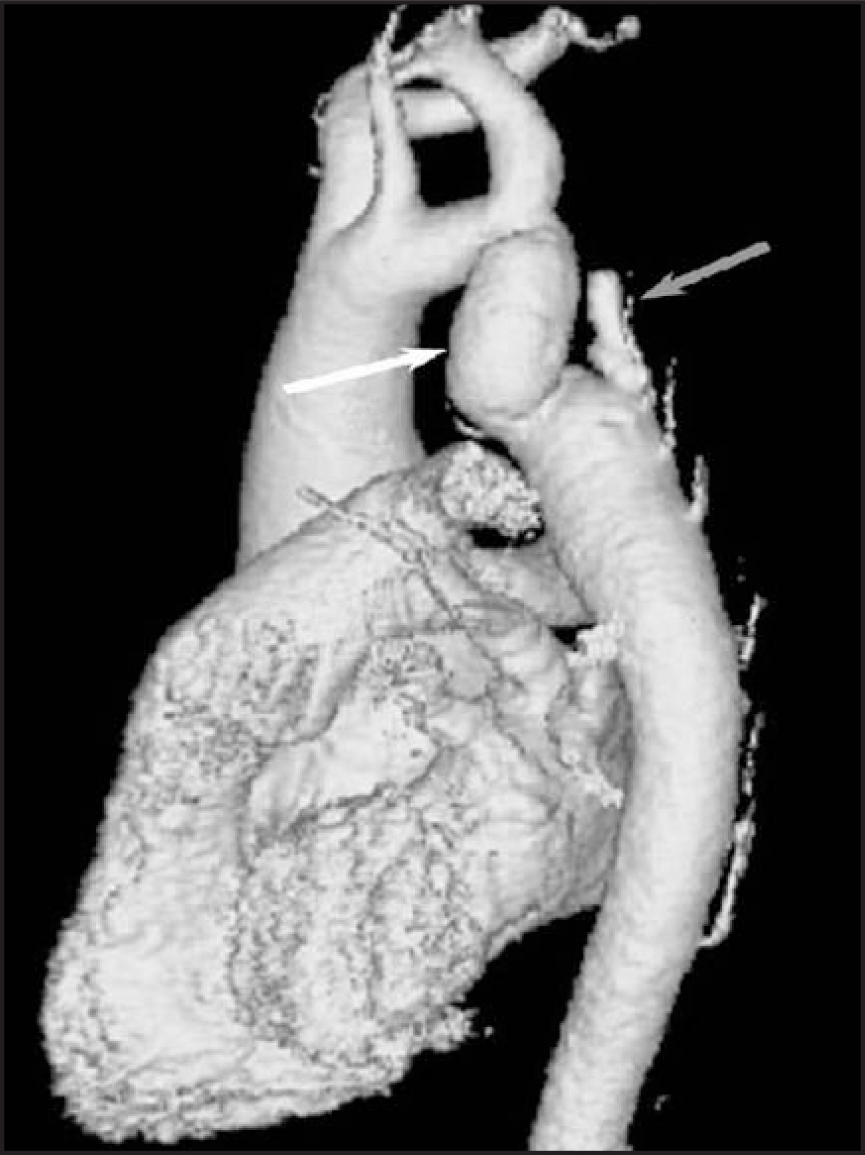
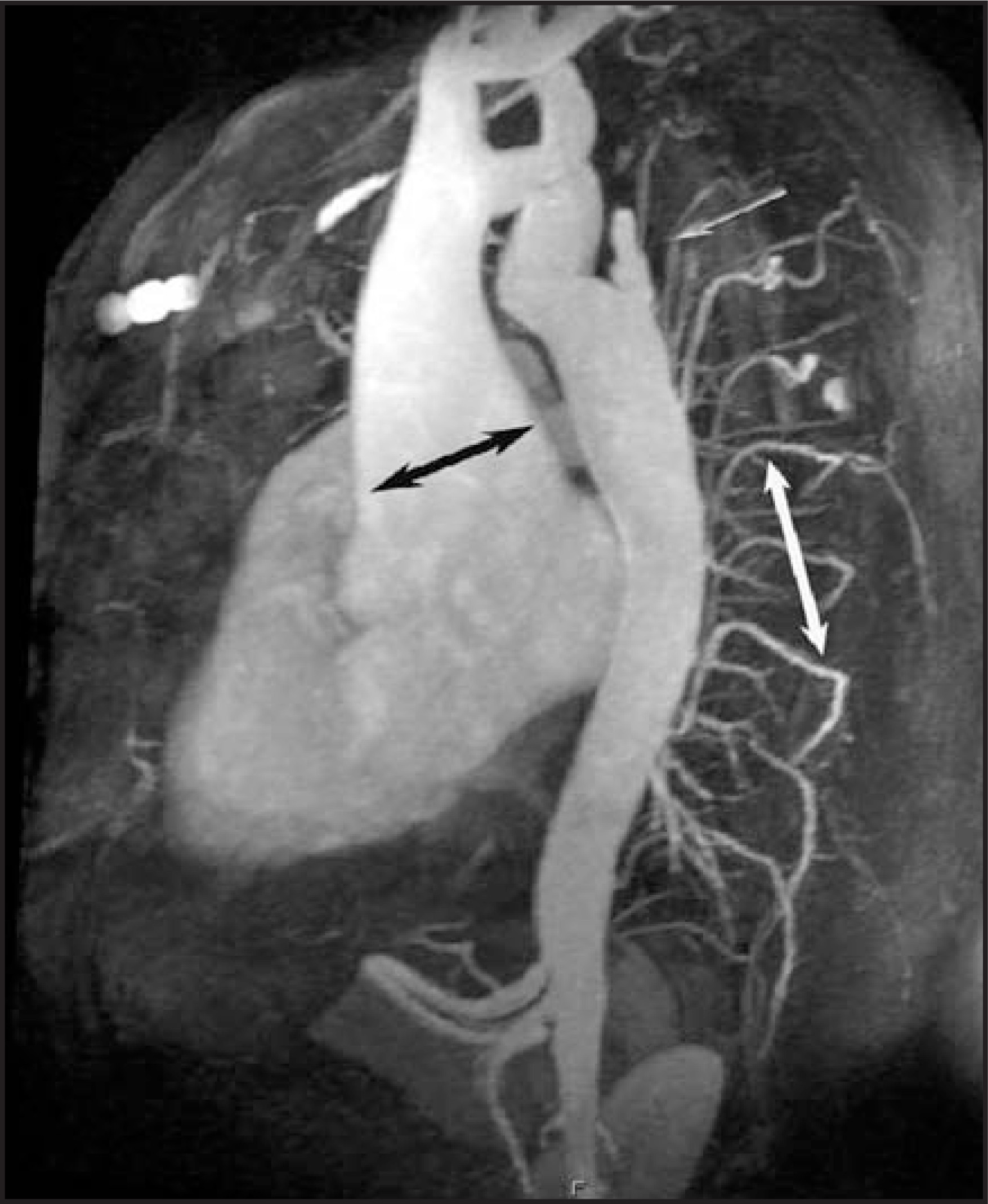
Three-dimensional surface-rendered reconstruction of the MR angiogram revealed the interposition graft with a moderate degree of stenosis at both the proximal and distal anastomoses as well as a pseudoaneurysm (at the distal suture line) that measured 27 mm in length and 7 mm at the neck (Figure 1). Velocity-encoded phase-contrast MR imaging measured the gradient across the graft at 15 mm Hg (not shown), which was confirmed at cardiac catheterization. Prominent intercostal vasculature was noted; also noted were the pseudoaneurysm and an ascending aortic aneurysm that measured 5.8 cm (Figure 2).
DIAGNOSIS
Ascending aortic aneurysm, descending aortic pseudoaneurysm
DISCUSSION
An aortic coarctation is a common cardiovascular lesion that accounts for 5% to 7% of all congenital heart disease.1 Coarctation is more common in males than females and is known to occur in conjunction with a variety of conditions, including Turner’s syndrome, Shone complex, ventricular septal defect, bicuspid aortic valve, and aneurysms of the circle of Willis.2 If left untreated, complications are common and can include aortic dissection, infective endocarditis, severe aortic insufficiency, hypertension, coronary artery disease, and intracranial hemorrhage.3 In patients with uncorrected coarctation, as many as 90% die by the age of 60 years.4
Following surgical repair of aortic coarctation, close follow-up of patients is recommended, as surgery is, in many ways, not “curative.” Late complications, as a consequence of the surgery itself or the systemic arteriopathy, are not uncommon. Irrespective of the success of the repair, hypertension frequently develops and is a major contributor to long-term cardiovascular morbidity, although early surgical intervention may reduce the risk of developing late hypertension and other cardiovascular sequelae.5,6 In a large surgical series, the most common causes of death in patients with successful coarctation repair were coronary artery disease (37%), congestive heart failure (9%), and complications of reoperation (7%).2 The most frequent indications for reoperation include recurrent coarctation, ascending aortic aneurysm, valvular heart disease, and pseudoaneurysm formation.3
Aortic imaging techniques are critical in ruling out pathology in patients with a history of coarctation repair. MR imaging has evolved into the modality of choice for screening these patients, as it provides safe, high-quality images of the aorta.7 In the case reported here, several abnormalities associated with this disease process have been identified, including an ascending aortic aneurysm, narrowing of the graft at both the proximal and distal anastomosis, and the unusual-appearing pseudoaneurysm at the distal suture site. Although not identified here, true aneurysm formation at the site of repair is not uncommon in patients who have undergone prior synthetic patch aortoplasty, while it is infrequent in those repaired with end-to-end anastamosis or interposition grafting.8 Some have advocated routine aortic screening with MR imaging every 12 to 24 months after coarctation repair, particularly for those with prior patch aortoplasty.7
Following balloon angioplasty of native coarctation, the incidence of aneurysm formation and recurrent coarctation is higher than with surgical repair.9,10 Long-term experience and follow-up of patients with aortic stent implantation in native coarctation is limited,11,12 but short-term outcomes have been encouraging.
CONCLUSION
Aortic coarctation is a potentially life-threatening congenital lesion that often requires surgical or percutaneous intervention. However, repair is frequently not “curative” in the traditional sense, as long-term complications from hypertension, aneurysm formation, associated valvular heart disease, and recoarctation are common. Close follow-up by a cardiovascular specialist is warranted in all cases.
References
- Jenkins N, Ward C. Coarctation of the aorta: Natural history and outcome after surgical treatment. QJM. 1999;92:365-371.
- Attenhofer Jost C, Schaff H, Connolly H. Spectrum of reoperations after repair of aortic coarctation: Importance of an individualized approach because of coexistent cardiovascular disease. Mayo Clin Proc. 2002;77:646-653.
- Brickner M, Hillis L, Lange R. Congenital heart disease in adults. First of two parts. N Engl J Med. 2000;342:256-263.
- Campbell M. Natural history of coarctation of the aorta. Br Heart J. 1970;32:633-640.
- Seirafi P, Warner K, Geggel R, Payne D, Cleveland R. Repair of coarctation of the aorta during infancy minimizes the risk of late hypertension. Ann Thorac Surg. 1998;66:1378-1382.
- Brouwer R, Erasmus M, Ebels T, Eijgelaar A. Influence of age on survival, late hypertension, and recoarctation in elective aortic coarctation repair. Including long-term results after elective aortic coarctation repair with a follow-up from 25 to 44 years. J Thorac Cardiovasc Surg. 1994;108:525-531.
- Swan L, Wilson N, Houston A, Doig W, Pollock J, Hillis W. The long-term management of the patient with an aortic coarctation repair. Eur Heart J. 1998;19:382-386.
- Knyshov G, Sitar L, Glagola M, Atamanyuk M. Aortic aneurysms at the site of the repair of coarctation of the aorta: A review of 48 patients. Ann Thorac Surg. 1996;61:935-939.
- Shaddy R, Boucek M, Sturtevant J. Comparison of angioplasty and surgery for unoperated coarctation of the aorta. Circulation Circulation.
- Rao P, Galal O, Smith P, Wilson A. Five- to nine-year follow-up results of balloon angioplasty of native aortic coarctation in infants and children. J Am Coll Cardiol J Am Coll Cardiol.
- Ebeid M, Prieto L, Latson L. Use of balloon-expandable stents for coarctation of the aorta: Initial results and intermediate-term follow-up. J Am Coll Cardiol. 1997;30:1847-1852.
- Magee A, Brzezinska-Rajszys G, Qureshi S. Stent implantation for aortic coarctation and recoarctation. Heart. 1999;82:600-606.
Citation
. RCOM RADIOLOGICAL CASE OF THE MONTH. Applied Radiology. 2006;35(12):40-42. doi:10.37549/AR1474.