RCOM RADIOLOGICAL CASE OF THE MONTH
Applied Radiology — Vol. 36 , Issue 1 , pp. 39 -41
DOI: 10.37549/AR1476
Published: January 1, 2007
Categories
CASE SUMMARY
An 81-year-old white man presented to the Emergency Department at Mount Sinai Medical Center in mild distress with complaints of abdominal pain, nausea, coffee-ground emesis, and abdominal bloating for 2 days. The patient also reported that he had not had a bowel movement since the symptoms appeared. The patient denied any history of diarrhea, fever, chills, diaphoresis, hematochezia, or hemoptysis. His medical history was significant for colon cancer, non–insulin-dependent diabetes mellitus, benign prostatic hyperplasia, hypertension, and a previous small bowel obstruction. His surgical history was significant for a right colectomy with gastrostomy secondary to colon cancer at age 66, a total hip replacement, and a left inguinal hernia repair. He had no known drug allergies. Upon admission, the patient’s medications were ramipril, clopidogrel bisulfate, pantoprazole sodium, glipizide, pindolol, and pancrelipase. An initial physical examination revealed a confused gentleman in mild-to-moderate distress, who was afebrile with stable vital signs except for a heart rate of 130 bpm. He was found to have dry lips and oral mucosa. His abdominal examination was remarkable for findings of abdominal distention, diffuse tenderness, and hypoactive bowel sounds in all quadrants. He had a notable abdominal wall deformity that resembled a ventral hernia. The stomach was palpable and tympany was noted in the left upper quadrant. No rebound tenderness or guarding was appreciated. A nasogastric tube was inserted, and 4 liters of coffee-ground material were evacuated. Laboratory studies ordered included a complete blood cell count, metabolic profile, liver function test, amylase, lipase, and coagulation profile. Abnormal laboratory results included a white blood cell count of 19.9 × 10⁹ cells/L, amylase of 135 U/L, and lipase of 435 U/L. Abdominal radiographs were also ordered (not shown).
IMAGING FINDINGS
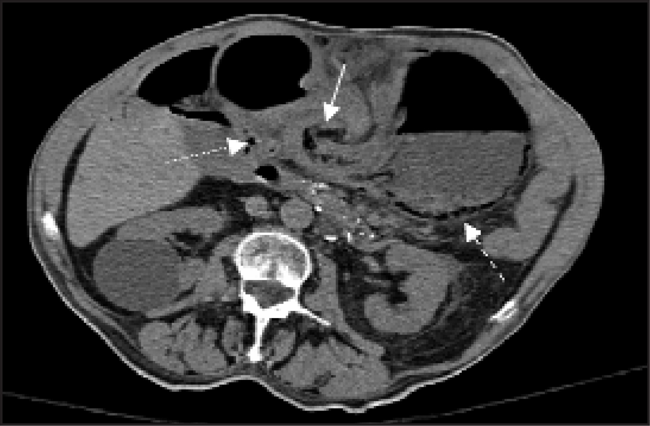
An X-ray revealed a distended stomach and a markedly dilated loop of bowel both with an air-fluid level (not shown). A computed tomography (CT) scan was then performed, which revealed marked gastric distention with a focal area of torsion (solid arrow in Figure 1) that is seen to involve the gastric body that separated the gastric cavity into 2 portions. Pneumatosis of the gastric wall (dashed arrow in Figure 1) and air were visualized in the portal system (dotted arrow in Figure 1). All findings were consistent with an obstruction secondary to partial mesenteroaxial gastric volvulus.
DIAGNOSIS
Mesenteroaxial gastric volvulus
DISCUSSION
First described by Berti1 in 1866, a gastric volvulus is a rare condition that is caused by a rotation of the stomach >180° along a trans-verse or longitudinal axis. The age of peak incidence for gastric volvuli occurs in the 5th decade of life.2,3 A primary volvulus is idiopathic in nature with no anatomical abnormalities present.4 Secondary volvuli are more common than primary and are typically attributed to a diaphragmatic abnormaity, a hiatal hernia, a lung resection, or abdominal adhesions.4,5 The direction of the axis of rotation is used for classification of the 2 major types—mesenteroaxial and organoaxial—the latter being more common.2 Mesenteroaxial gastric volvuli are characterized by an axis of rotation around a line that runs from the center of the greater curvature to the porta hepatis (transverse axis), with the site of obstruction being located in the pyloric antrum.5,6 Generally, mesenteroaxial gastric volvuli are chronic or intermittent, idiopathic in nature, and less likely to strangulate.5 In contrast, organoaxial gastric volvuli are more common and are characterized by an axis of rotation around a line that extends from the pylorus to the esophogastric junction (longitudinal axis).5 This type is more likely to produce the clinical symptoms referred to as Borchardt’s triad, which includes severe epigastric pain and distention, unproductive vomiting, and difficulty or inability to pass a nasogastric tube.4,5,7 Additionally, organoaxial gastric volvuli often produce ischemia caused by their torsional pattern around the pylorus and cardia of the stomach, which can result in gangrene.4,5 Organoaxial and mesenteroaxial gastric volvuli have been reported to occur concurrently in 2% of cases.2,4 The most common sites of intestinal volvulus are the sigmoid colon and the cecum.4,8 Gastric volvulus, however, is a rare condition but, when present, frequently leads to operative repair because of an acute abdomen. As in this case, it is imperative that an early diagnosis be made and that a successful operative reduction be performed.5 In one article, the barium study was shown to have the highest diagnostic yield for diagnosing gastric volvulus, and CT was proven not to be helpful.9 In the case we present, the diagnosis was made with plain films and CT, with a clear swirl sign present on CT that is diagnostic of gastric volvulus. Consistent with the literature, this patient did not present with Borchardt’s triad, which has been shown to be more commonly present in the organoaxial type.10 The patient, however, presented with signs of an upper gastrointestincal bleed. Bleeding is usually the result of congestive gastritis, ulceration, and ischemia, and is often a late finding.11 Contrary to the majority of cases of mesenteroaxial volvulus cited in the literature, this particular case was acute and produced necrosis, findings that are more consistent with an organoaxial volvulus.5,10 Treatment options for acute gastric volvuli depend on the type and extent of the damage. The first successful operative report of a gastric volvulus was in 1896 by Berg.12 The literature reports that the reduction of an acute volvulus can be achieved if passage of a nasogastric tube is possible and the stomach contents emptied, which is more possible in mesoenteroaxial volvuli.5 Acute cases require urgent surgical intervention to try to avoid consequent necrosis and subsequent resection. The literature has reported cases of laparoscopic9,13 and endoscopic14-16 reduction; however, subsequent open exploration has followed. The open surgical procedure of choice is an anterior gastroplexy. In cases in which ischemia or necrosis is encountered, gastric resection is indicated.4 A high index of suspicion will lead to early operative intervention and prevent the need for resection. Finally, it must be noted that subsequent to the reduction of the volvulus, the underlying defect, most notably a diaphragmatic hernia, must be repaired.2,4,5,11,12,17
CONCLUSION
Physicians should be aware of the presentation of this rather rare cause of an acute abdomen. The clinical presentation of the classic triad of severe epigastric pain and distention, unproductive vomiting, and difficulty or inability to pass a nasogastric tube should raise the suspicion, but this presentation is not always present in a gastric volvulus. In this case, clear radiographic evidence was diagnostic. Prompt recognition of the condition and urgent intervention may prevent widespread necrosis and avoid subsequent surgical resection.
References
- Berti A. Singolar attortigliamento dele’esofago col duodeno seguita da rapida morte. Gass Med Ital. 1866;9:139-141.
- Wastell C, Ellis H. Volvulus of the stomach. Br J Surg. 1971;58:557-562.
- Shriki J, Nguyen K, Rozo J. Rare chronis gastric volvulus associated with left atrial and mediastinal compression. Tex Heart Inst J. 2002;29:324-328.
- Wasselle J, Norman J. Acute gastric volvulus: Pathogenesis, diagnosis, and treatment. Am J Gastroenterol. 1993;88:1780-1784.
- Carter R, Brewer L, Hinshaw D. Acute gastric volvulus. Am J Surg. 1980;140:99-106.
- Katkhouda N, Mavor E, Achanta K. Laparoscopic repair of chronic intrathoracic gastric volvulus. Surgery. 2000;128:784-790.
- Borchardt M. Zun pathologie and therapie des magnevolvus Arch Klin Chir. 1904;74:243-248.
- Gean A, Deluca S. Acute gastric volvulus. Am Fam Phys. 1986;34:99-100.
- Teague W, Ackroyd R, Watson D, Devitt P. Changing patterns in the management of gastric volvulus over 14 years. Br J Surg. 2000;87:358-361.
- Taylor M. Gastrointestinal Emergencies. 1992:167-170.
- Haubrich W, Schaffner F, Berk J. Bockus Gastroenterology. 1995:830-833.
- Berg J. Zwei falle von axendrehung des magens, operation, heilung. Nord Med Arkiv. 1897;30:1-6.
- Koger K, Stone J. Laparoscopic reduction of acute gastric volvulus. Am Surg. 1993;59:325-328.
- Kodali V, Mass L. Endoscopic reduction of acute gastric volvulus. J Clin Gastroenterol. 1995;21:331-332.
- Tsang T, Walker R, Yu D. Endoscopic reduction of gastric volvulus: The alpha-loop maneuver. Gastrointest Endosc. 1995;42:244-248.
- Akamatsu T, Nakamura N, Kiyosawa K. Gastric volvulus in living, related liver transplantation donors and usefulness of endoscopic correction. Gastrointest Endosc. 2002;55:55-57.
- Siu W, Leong H, Li M. Lapsaroscopic gastropexy for chronic gastric volvulus. Surgical Endoscopy. 1998;12:1356-1357.
Citation
. RCOM RADIOLOGICAL CASE OF THE MONTH. Applied Radiology. 2007;36(1):39-41. doi:10.37549/AR1476.