RCOM RADIOLOGICAL CASE OF THE MONTH
Applied Radiology — Vol. 36 , Issue 1 , pp. 36 -48
DOI: 10.37549/AR1478
Published: January 1, 2007
Categories
CASE SUMMARY
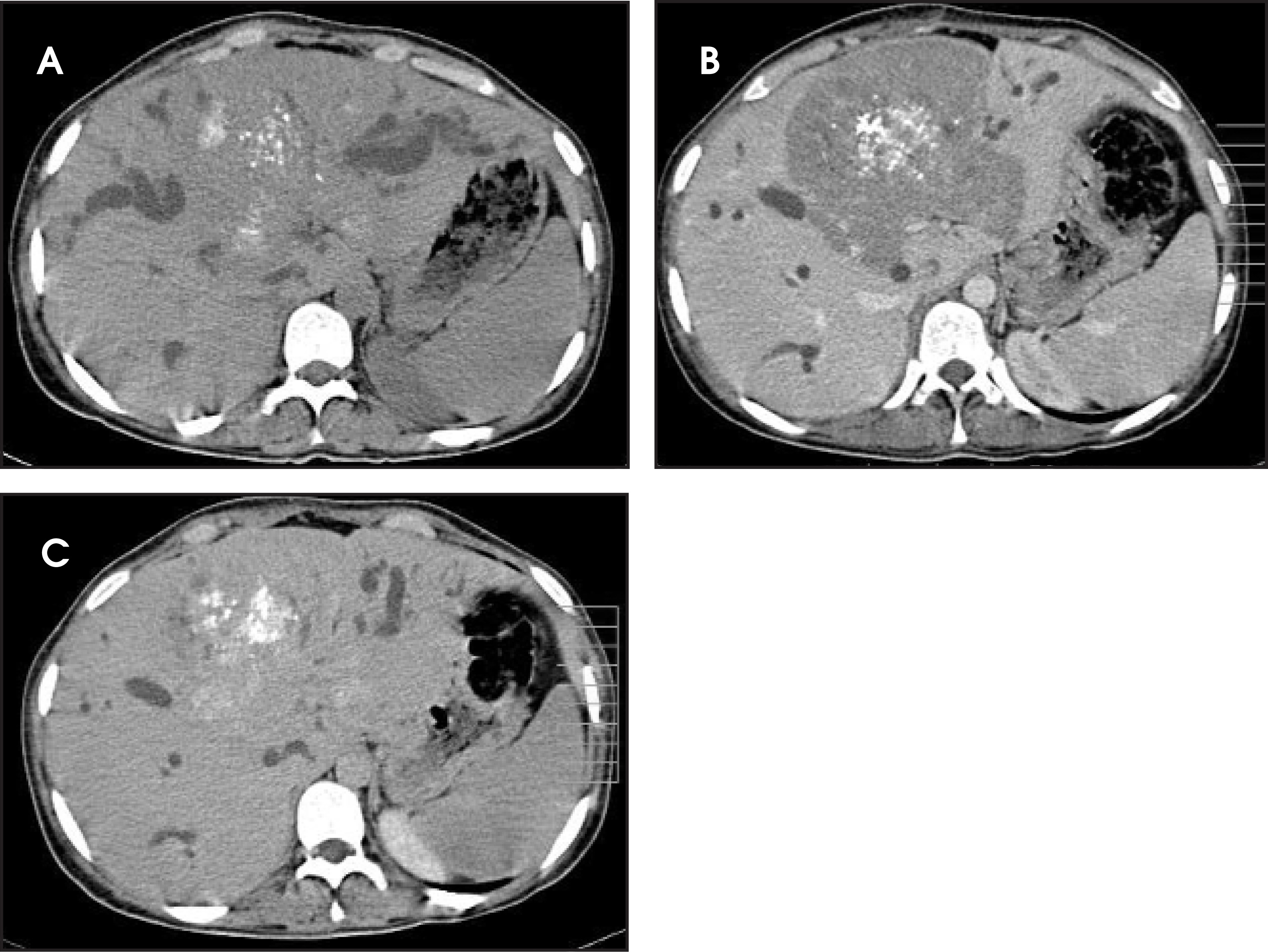
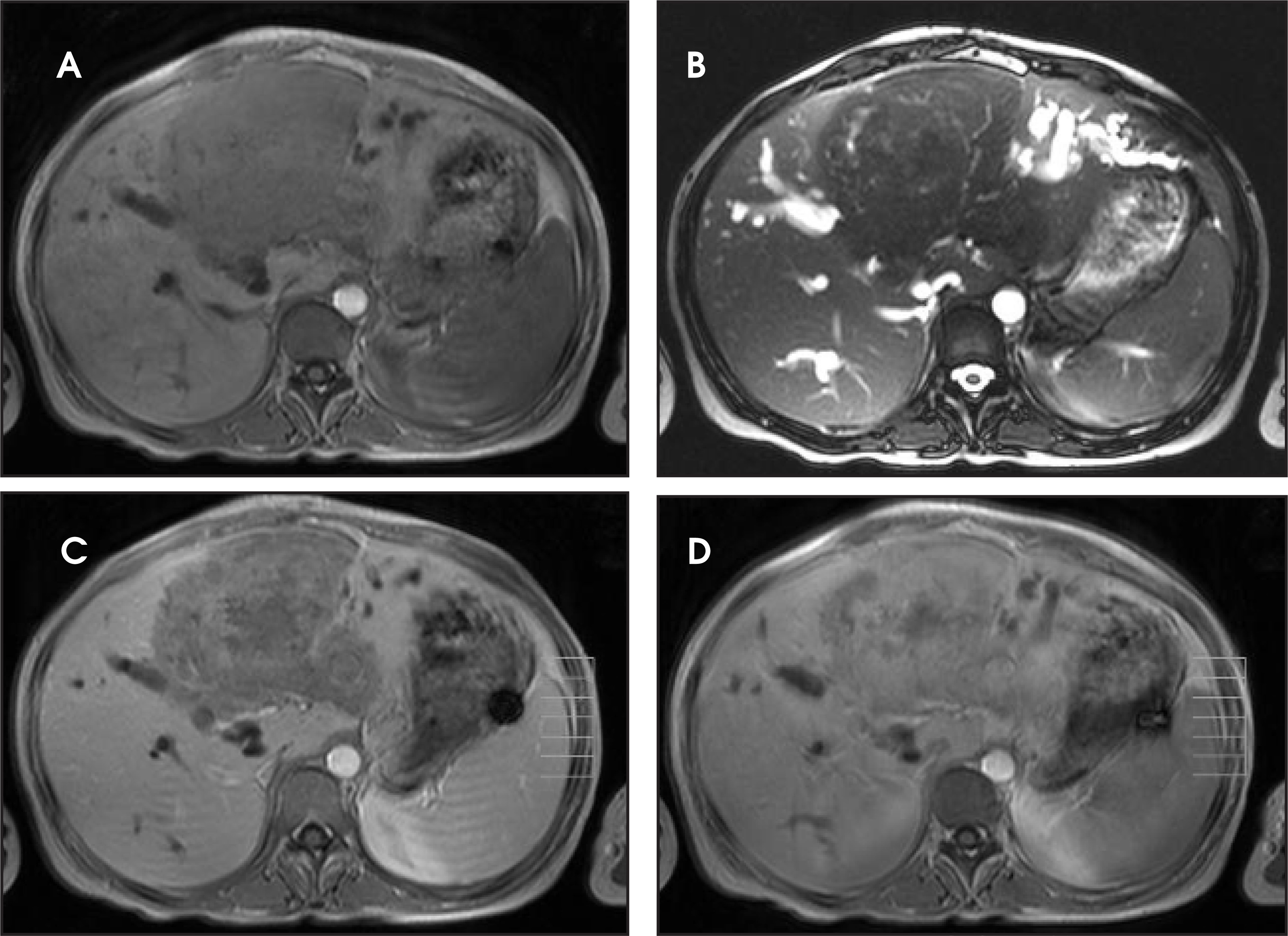
A 15-year-old girl was admitted to the hospital with right upper-quadrant pain and abdominal swelling of 3 months’ duration. A physical examination revealed a mass filling the epigastrium and the right subcostal region. Abnormal laboratory findings included erythrocyte sedimentation rate (ESR) of 60 mm/h, gamma glutamyl transferase of 67 IU/L, alkaline phosphatase of 310 IU/L, total bilirubin of 2.1 mg/dL, direct bilirubin of 1.1 mg/dL, and indirect hemagglutination of 1/160. An abdominal computed tomography (CT) scan (Figure 1) and a magnetic resonance imaging (MRI) examination (Figure 2) were performed.
IMAGING FINDINGS
A precontrast abdominal CT scan revealed heterogenous isodensity, vascular displacement, and ductal dilatation. Calcifications were seen mostly on the central region of the lesion (Figure 1A). Postcontrast CT scans revealed prominent contrast enhancement on delayed images (Figure 1, B and C). MRI revealed a hypointense 16 × 15 × 12-cm mass on T1-weighted images (T1WI) and T2-weighted images (T2WI) (Figure 2, A and B). With gadolinium administration, the lesion exhibited heterogeneous enhancement in the portal and late phases (Figure 2, C and D). The mass extended into the portal hilum and had a central cystic necrotic area that was hypointense on T1WI and hyperintense on T2WI. Both T1WI and T2WI showed heterogenous hypointensity of the affected area when compared with the normal liver parenchyma. The margins of the lesion were well-delineated on T1WI, whereas the borders were somewhat indistinct on both CT and T2WI. The vascular structures, either within or outside the affected area, were displaced.
DIAGNOSIS
Hepatic infestation of Echinococcus alveolaris (EA)
DISCUSSION
Infestation of Echinococcus alveolaris is a rare parasitic condition caused by Echinococcus multilocularis. The infestation mimics a slow-growing tumor and infiltrates the liver and the surrounding structures, especially the portal hilus, the hepatic veins, the inferior vena cava, and the biliary system. The displacement of the hepatic veins, the portal vein, and the biliary tree is common and is caused by mass effect. Central necrosis frequently develops as the “tumor mass” increases in size. Microcalcifications are common; and the mass may also extend into the extrahepatic area.1
CT findings of liver infestation by EA include heterogeneous hypodense areas without contrast enhancement, isodense areas with or without contrast enhancement, calcifications, pseudocystic necrotic areas, and dilatation of the intrahepatic bile ducts.1,2 MRI is important for diagnosing alveolar echinococcosis because of its ability to reveal its fibrous and infiltrative nature as well as the extent of the lesion. Although not used routinely, MRI allows better visualization of mass contours, central necrosis, vascular relationships, and extrahepatic extension of alveolar echinococcosis than does CT. Fibrosis and parasitic tissue exhibit low signal intensity on T1WI, and the margins of the mass are well visualized with this sequence. T2-weighted images are best for detecting small cystic peripheral extensions and for defining central necrotic zones.3 The irreversible acellular keloid scarlike fibrosis observed in EA infestation is the ultimate result of cytotoxic and fibrogenetic events that are related to the immune response of the host and initially occur in the granulomatous area surrounding the young parasite larvae.4
In this case, the extensive inflammatory reaction and focal calcifications that were seen on the histologic examination (not shown) were consistent with the hypointensity on both T1WI and T2WI and the contrast enhancement on postcontrast T1WI. The number of cysts and the degree of necrosis were minimal, and, thus, high signal intensity on T2WI was not seen. The lesion showed heterogeneous enhancement following gadolinium administration, an interesting and unexpected finding that, to our knowledge, has not been described before. The contrast enhancement may have been related to the extensive inflammatory cells (plasma cells, neutrophils, and leukocytes) that were seen at microscopic examination.
The CT patterns of EA lesions have been reported to be similar to those seen in primary hepatic neoplasms and metastasis.5 CT findings of a hypodense lesion with calcifications and a lack of contrast enhancement are helpful in differentiating EA infestation from liver neoplasms. MRI may give additional clues to the diagnosis of infestation of EA. A hypointense hepatic lesion on both T1WI and T2WI without contrast enhancement is characteristic of an EA infestation. However, a mass with calcifications and contrast enhancement in late-venous phases on both CT and MRI may represent a hepatic neoplasm, such as cholangiocarcinoma.
CONCLUSION
We suggest that EA infestation should not be disregarded in the differential diagnosis of focal hepatic lesions showing contrast enhancement and should be considered in the differential diagnosis of enhancing focal hepatic lesions.
References
- Balci N, Tunaci A, Semelka R. Hepatic alveolar echinococcosis: MRI findings. Magn Reson Imaging. 2000;18:537-541.
- Claudon M, Bessieres M, Regent D. Alveolar echinococcosis of the liver: MR findings. J Comput Assist Tomogr. 1990;14:608-614.
- Harman M, Arslan H, Kotan C. MRI findings of hepatic alveolar echinococcosis. Clin Imaging. 2003;27:411-416.
- Katranci N, Elmas N, Yilmaz F, Mentes A. Correlative CT, MRI and histological findings of hepatic Echinococcus alveolaris: A case report. Comput Med Imaging Graph. 1999;23:155-159.
- Didier D, Weiler S, Rohmer P. Hepatic alveolar echinococcosis: Correlative US and CT study. Radiology. 1985;154:179-186.
Citation
. RCOM RADIOLOGICAL CASE OF THE MONTH. Applied Radiology. 2007;36(1):36-48. doi:10.37549/AR1478.