RCOM RADIOLOGICAL CASE OF THE MONTH
Applied Radiology — Vol. 36 , Issue 3 , pp. 36 -38
DOI: 10.37549/AR1491
Published: March 1, 2007
Categories
CASE SUMMARY
A 44-year-old white man with a long-standing history of diabetes, chronic pancreatitis, and polysubstance abuse presented to our emergency room with complaints of fever and jaundice of several days’ duration. The patient was well known to our institution, having been treated multiple times for benign biliary stenosis attributed to repeated bouts of pancreatitis. Because this patient refused to undergo a curative bypass operation, his common bile duct stent had been replaced periodically during the previous 7 years whenever the stent became nonfunctional.
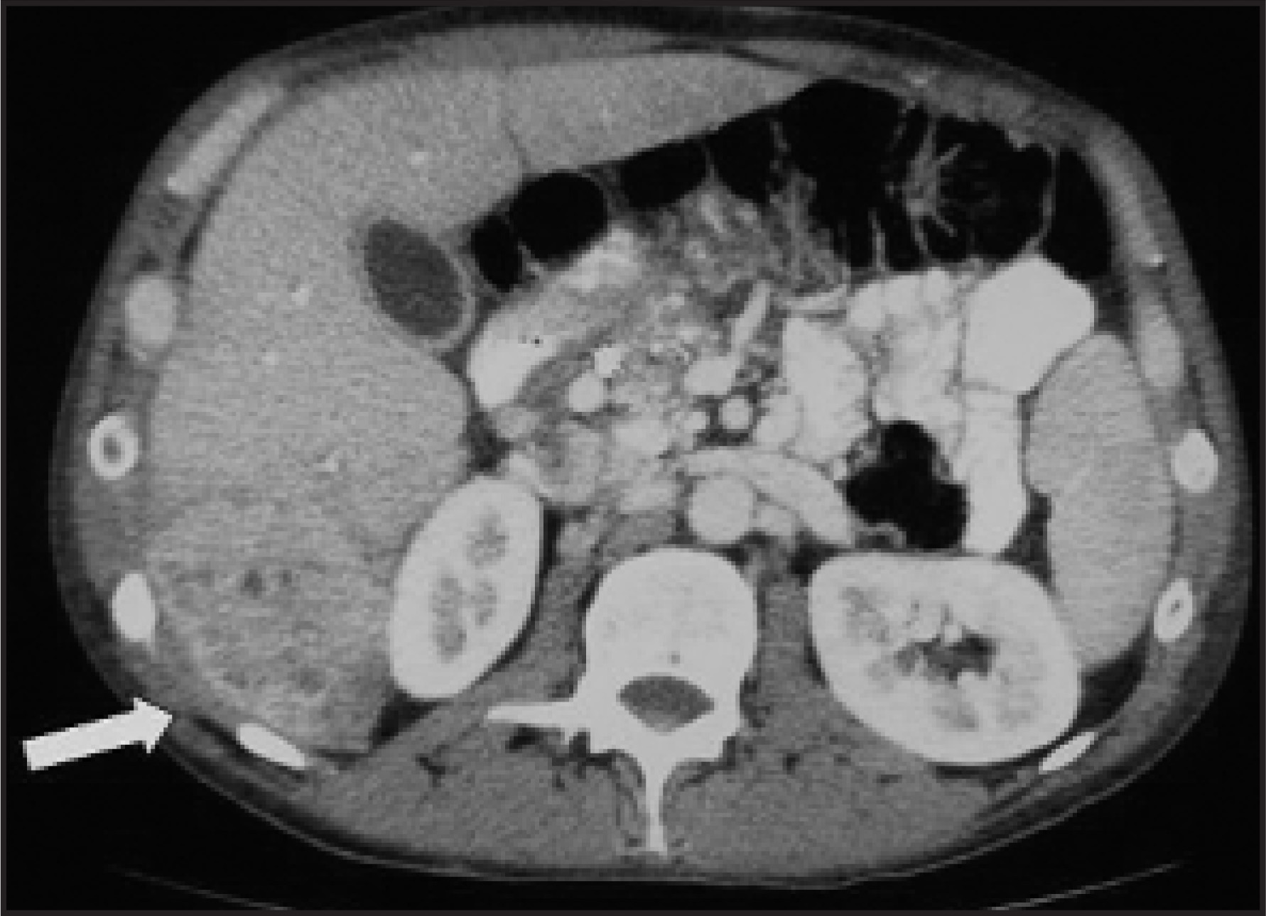
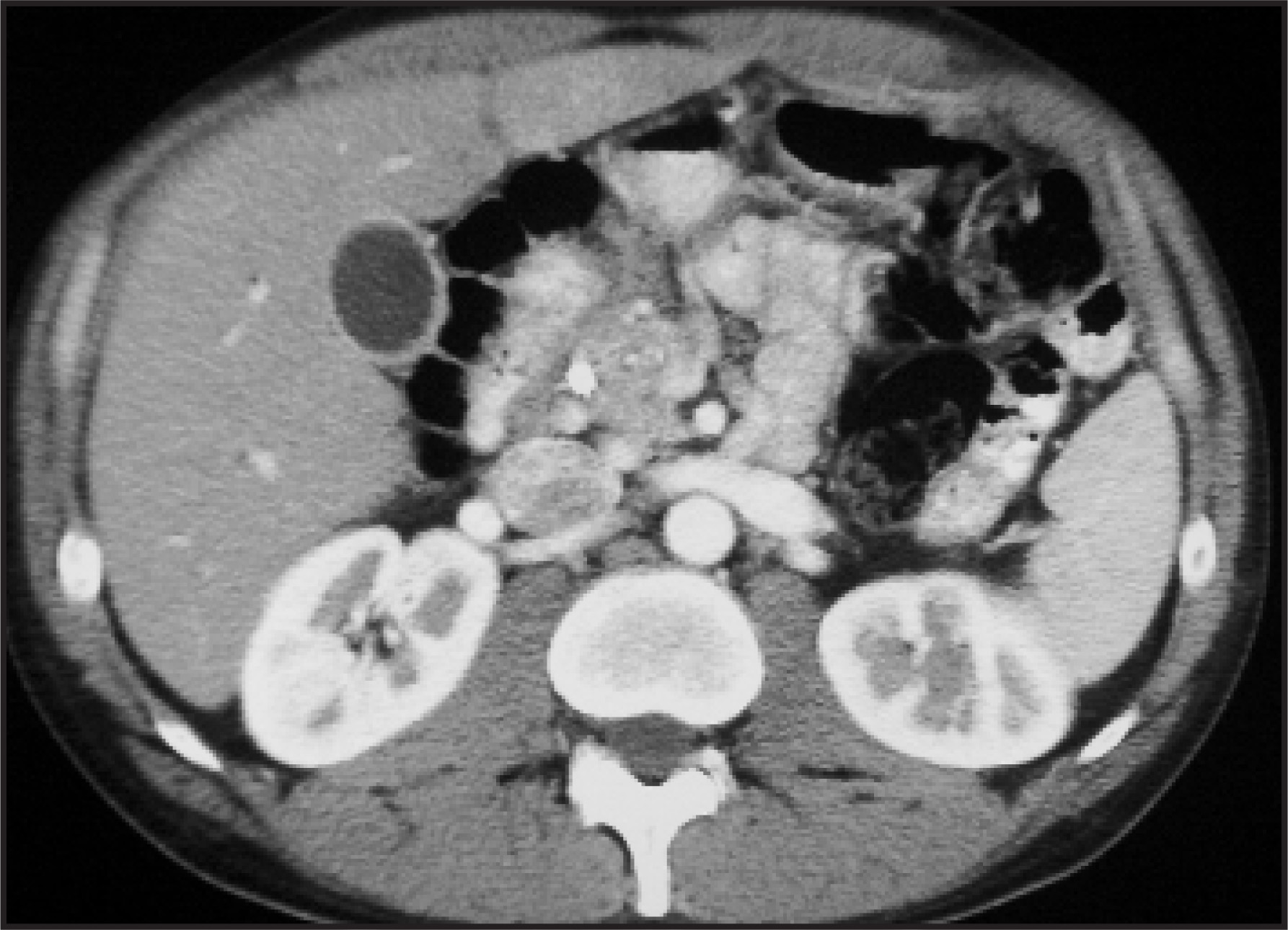
At this hospital admission, the patient was placed on an antibiotic regimen, and the biliary stent was replaced. Following the stent replacement, a computed tomographic (CT) scan of the abdomen was performed, without and with contrast (Figures 1 and 2).
IMAGING FINDINGS
Significant CT findings revealed the development of a heterogenously enhancing lesion in the posterior segment of the right hepatic lobe that measured up to 6.4 × 5.7 cm (Figure 1) when compared with a CT scan that was taken 10 months earlier (Figure 2). These findings were interpreted as suggestive of metastatic disease or a primary hepatocellular carcinoma. Multiple enlarged abdominal lymph nodes that were suggestive of neoplasms were also described. The CT scan was additionally remarkable for continued pancreatic duct dilatation with pancreatic calcifications, which were consistent with the patient’s known chronic pancreatitis. The re-placement of the common bile duct stent with interval development of intrahepatic pneumobilia was also noted.
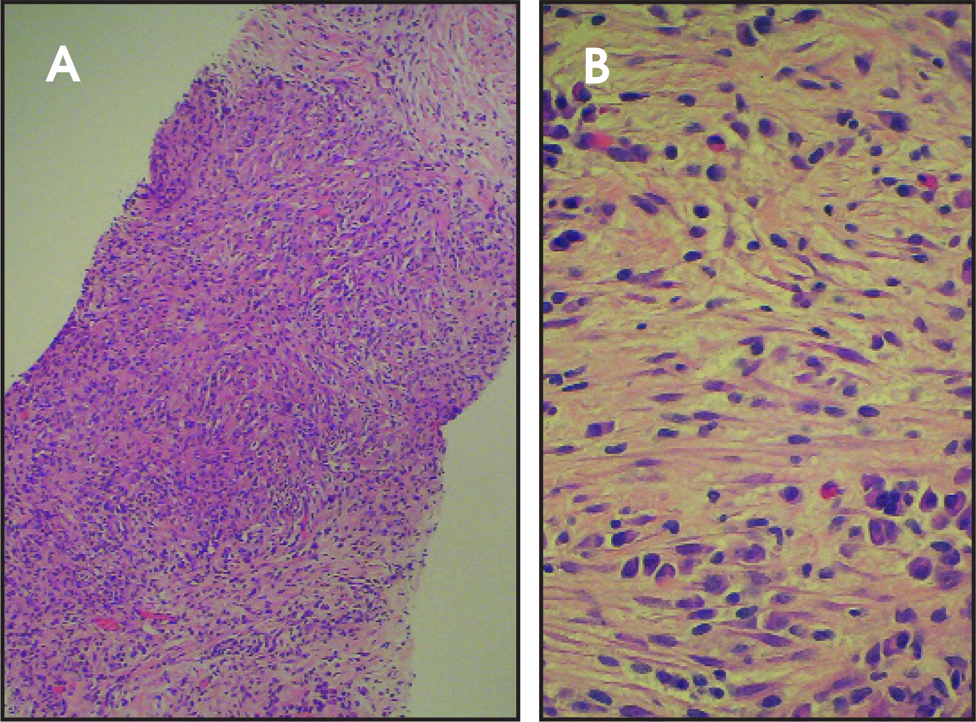
A percutaneous CT-guided core biopsy was performed on the liver mass. The histopathologic analysis revealed a spindle cell proliferation containing a heavy chronic inflammatory cell infiltrate composed mainly of plasma cells; lesser numbers of eosinophils, lymphocytes, and histiocytes were also present. A few large bile ducts and several foci of residual benign hepatic parenchyma were also found. There was no evidence of malignancy (Figure 3). A portion of the sample was sent for microbiologic cultures; the results were reported as negative for acid-fast bacilli, fungi, and bacteria.
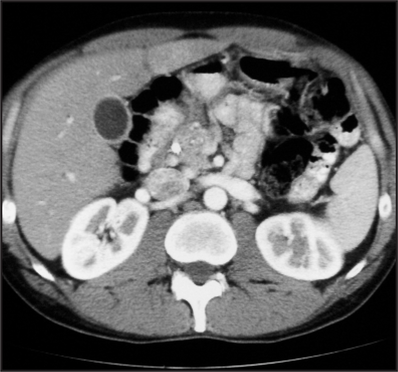
The patient completed a 14-day course of antibiotics without additional treatment. A repeat CT scan performed 2 months later showed that there had been significant spontaneous regression in the size of the lesion to 2 cm. The enlarged lymph nodes had also decreased in size (Figure 4).
DIAGNOSIS
Inflammatory pseudotumor (IPT) of the liver
DISCUSSION
An infrequent benign tumorlike lesion, IPT is most common in the lung, but has been described in other body sites as well.1 Liver involvement was first described in 1953 by Pack and Baker.2 Since that time, additional reports of IPT of the liver have been reported.1,3-5 The lesion is most common in younger age groups and has a male predominance. Presenting symptoms usually include fever, abdominal pain, and jaundice.1 The CT features are nonspecific and usually of a hypoattenuating mass with some peripheral hepatic enhancement. Since the CT images commonly mimic malignancy, tissue sampling is required to establish the diagnosis.6 Histologically, the lesion consists of fibrous stroma and a mixture of chronic inflammatory cells with a predominance of polyclonal plasma cells.
The etiology of IPT of the liver is not clear. Some reports suggest that IPT of the liver represents a spectrum of lesions ranging from inflammatory to low-grade neoplasm.1 Others speculate that IPT of the liver represents an inflammatory response to infection of the biliary tract or other extrahepatic sites.6 For example, IPT of the liver has been reported in patients with biliary stenosis,1 gallstones,1 pyogenic cholangitis,6 intra-abdominal abscesses,7 and gastrointestinal malignancies.8 Each of these conditions may predispose a patient to hepatic infection, either directly through irritation and stasis of the biliary tract, or hematogenously through the portal vein.6 The relationship between infection and IPT of the liver has not been conclusively linked because, as in our case, usually no infectious agents are recovered from the tissue. However, the successful isolation of pathogenic microorganisms from lesional tissue has been infrequently reported.7
Additionally, there are published case reports of patients with both IPT of the liver and diabetes mellitus,9 but a direct causal relationship between these diseases has not been established. Our patient had diabetes, but in this case, we believe that the development of IPT was unrelated to his diabetes. Rather, his chronic biliary stenosis is most likely the predisposing factor.
Concurrently, IPT of the liver can also involve other organs, such as the spleen and lymph nodes.10 Although we did not take a tissue sample from our patient’s enlarged lymph nodes, the synchronous decrease in size of our patient’s liver mass and lymphadenopathy provides evidence that our patient’s lymph nodes were also involved with IPT.
Since the natural history of IPT is not well understood, many cases undergo surgical resection. However, the literature suggests that the clinical course of IPT is typically resolution, and, therefore, a trial of conservative medical treatment may be considered initially in compliant patients.11 Surgery should be reserved for patients who fail nonsurgical management. In the patient reported here, the CT findings of a decrease in lesion size over several months support the case for nonoperative management.
CONCLUSION
Radiologists need to include IPT in the differential diagnosis of liver masses. Since routine CT findings are not specific, a percutaneous-core tissue biopsy is needed to establish the diagnosis. Since IPT may resolve without surgery, this approach will allow the clinician the option of a trial of conservative medical management. Periodic follow-up CT scans can be used to monitor the lesion size.
ACKNOWLEDGMENT
The authors thank Andrea Gwosdow, PhD, for her editorial assistance.
References
- Anthony P, MacSween R, Burt A, Portmann B. Pathology of the Liver. 2002:756-757.
- Pack G, Baker H. Total right hepatic lobectomy: Report of a case. Ann Surg. 1953;138:523-258.
- Horiuchi R, Uchida T, Kojima T, Shikata T. Inflammatory pseudo-tumor of the liver. Clinicopathologic study and review of the literature. Cancer. 1990;65:1583-1590.
- Standiford S, Sobel H, Dasmahapatra K. Inflammatory pseudo-tumor of the liver. J Surg Oncol. 1989;40:283-287.
- Lopez J, Eizaguirre B, Nevado M, Ruiz-Jaureguizuria J. Inflammatory pseudotumor of the liver. Report of a case and literature review. APMIS. 1990;98:1022-1026.
- Yoon K, Ha H, Lee J. Inflammatory pseudotumor of the liver in patients with recurrent pyogenic cholangitis: CT-histopathologic correlation. Radiology. 1999;211:373-379.
- White J, Chase C, Kelley J. Inflammatory pseudotumor of the liver associated with extrahepatic infection. South Med J. 1997;90:23-29.
- Lo O, Poon R, Lam C, Fan S. Inflammatory pseudotumor of the liver in association with a gastrointestinal stromal tumor: A case report. World J Gastroenterol. 2004;10:1841-1843.
- Younis N, Khaleeli A, Soran H, Monteith P. Inflammatory pseudotumour of the liver associated with diabetes mellitus. Int J Clin Pract. 2001;55:717-719.
- Miras-Parra F, Parra-Ruiz J, Gomez-Morales M. Inflammatory pseudotumor of lymph nodes with focal infiltration in liver and spleen. Dig Dis Sci. 2003;48:2003-2004.
- Koea J, Broadhurst G, Rodgers M, McCall J. Inflammatory pseudotumor of the liver: Demographics, diagnosis, and the case for nonoperative management. J Am Coll Surg. 2003;196:226-235.
Citation
. RCOM RADIOLOGICAL CASE OF THE MONTH. Applied Radiology. 2007;36(3):36-38. doi:10.37549/AR1491.