RCOM RADIOLOGICAL CASE OF THE MONTH
Applied Radiology — Vol. 36 , Issue 3 , pp. 40 -41
DOI: 10.37549/AR1495
Published: March 1, 2007
Categories
CASE SUMMARY
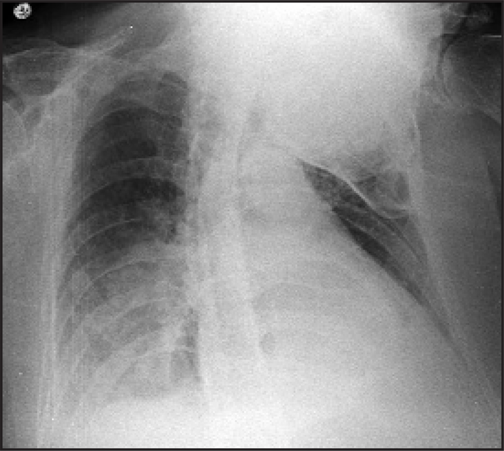
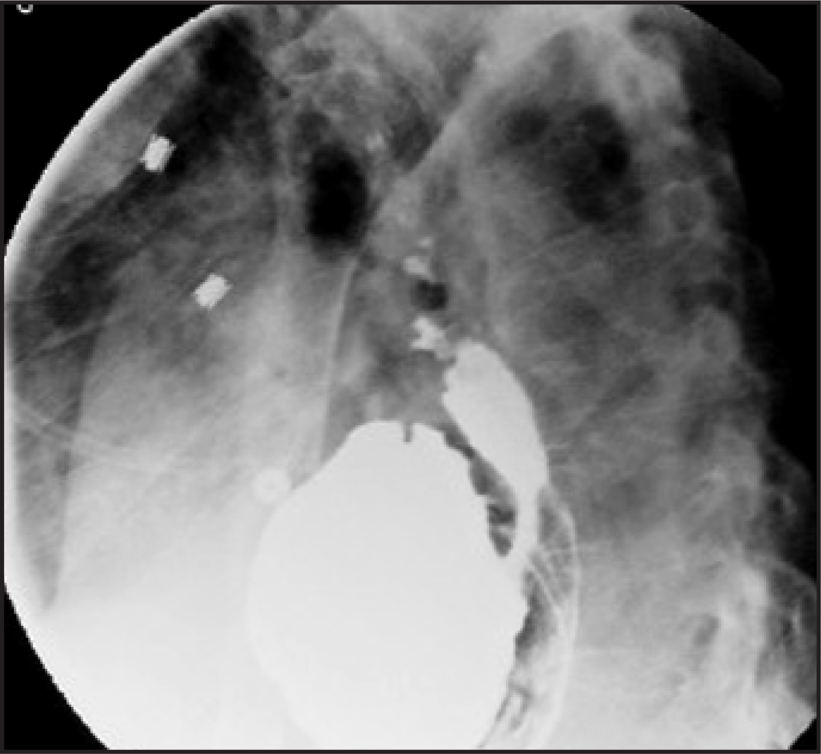
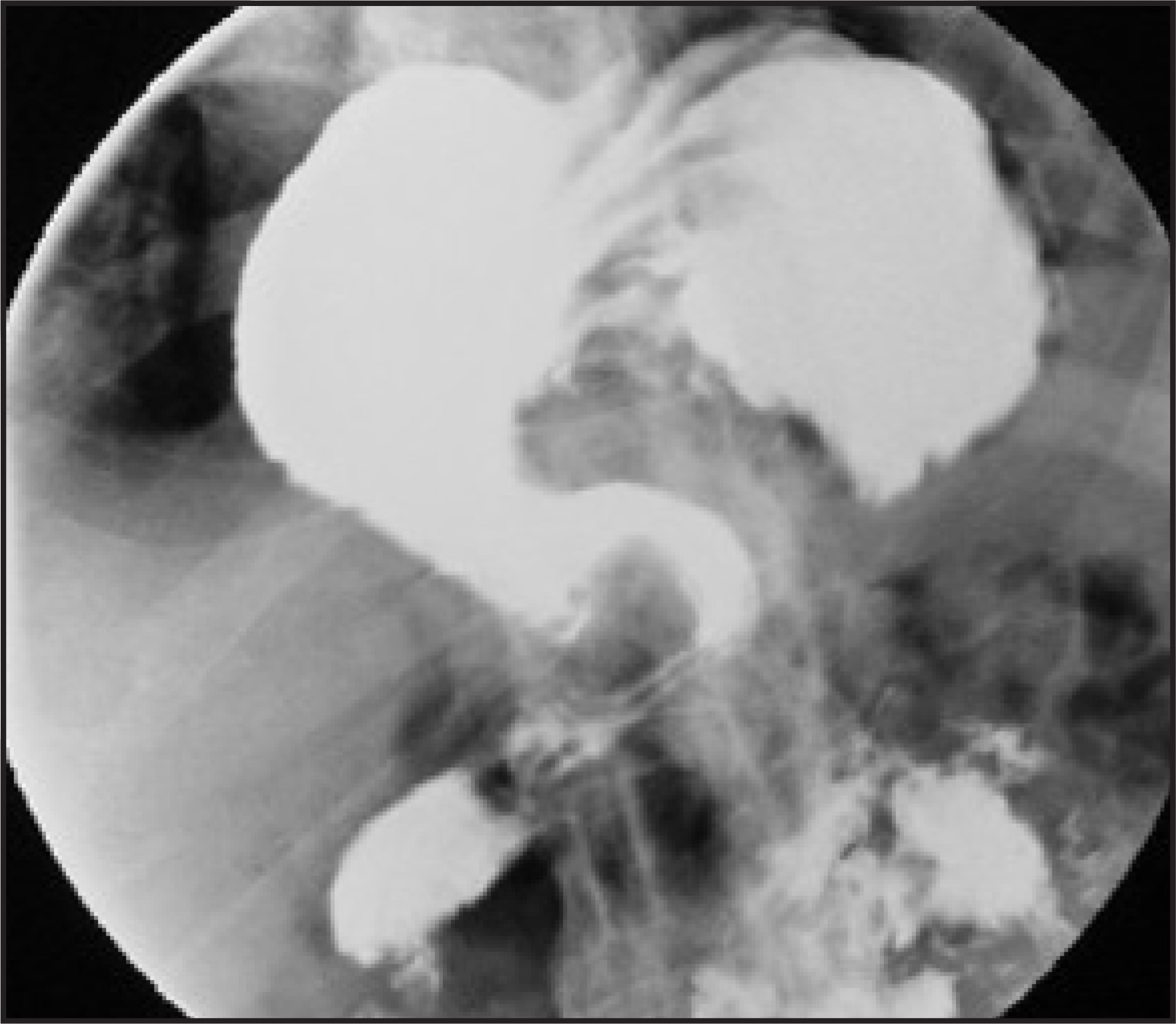
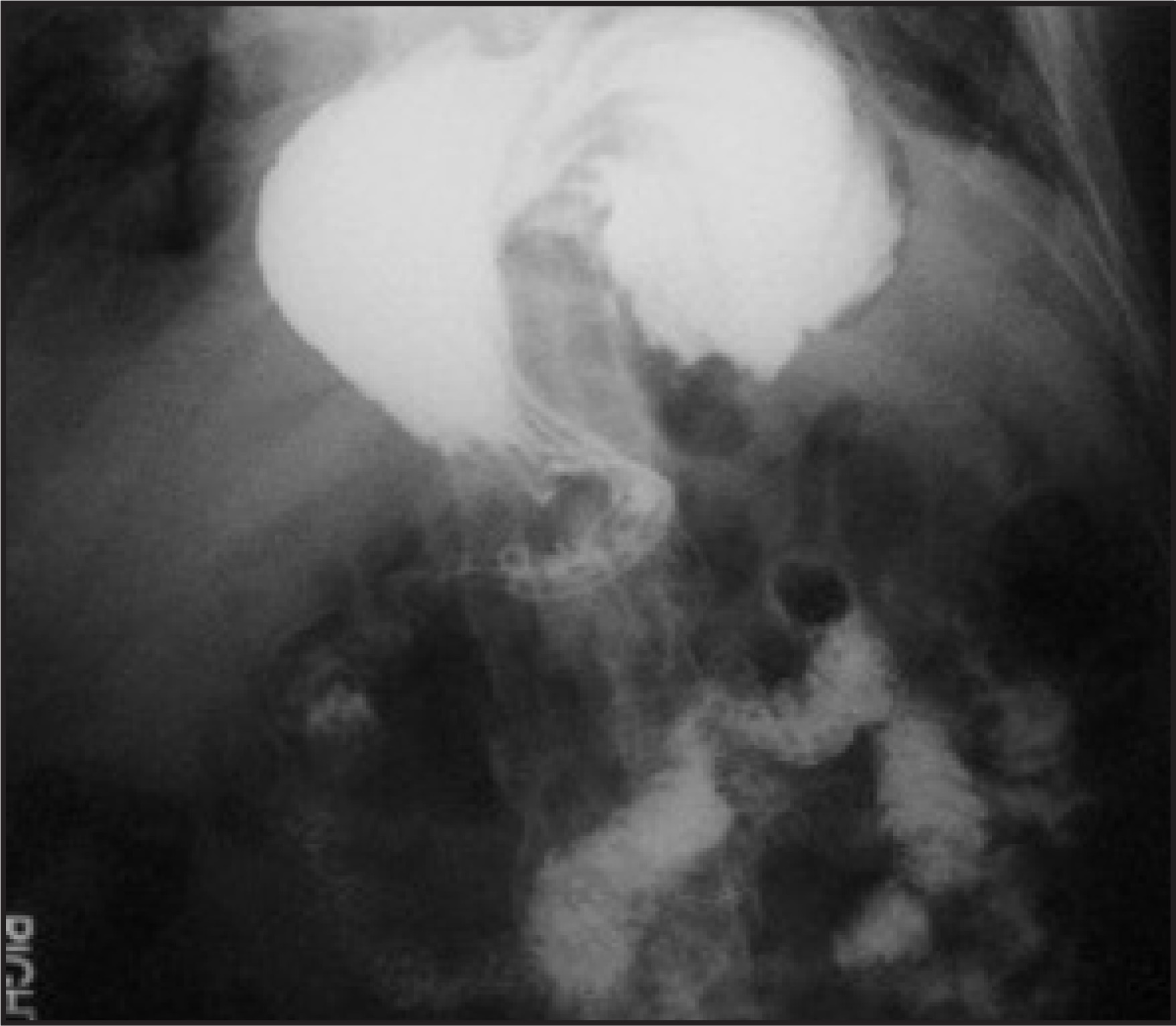
A 71-year-old man with a history of a hiatal hernia, Paget’s disease, dementia, and failure to thrive was admitted to the hospital for dehydration. On admission, a chest X-ray was ordered (Figure 1). After having several episodes of nausea and vomiting, an esophagogastroduodenoscopy was attempted, but there was difficulty in passing the endoscope. An upper gastrointestinal (GI) series was then ordered (Figures 2 through 4).
IMAGING FINDINGS
An admission chest X-ray revealed a hiatal hernia (Figure 1). A lateral view from the upper GI series showed a hiatal hernia, with components of a sliding and a paraesophageal hernia (Figure 2). The frontal view from the upper GI series shows a significant component of the greater curvature to be intrathoracic in location (Figure 3). The greater curvature of the stomach is superior to the lesser curvature of the stomach. The antrum is also superior to its expected inferior position. An additional frontal view shows no evidence of obstruction (Figure 4).
DIAGNOSIS
Organoaxial gastric volvulus
DISCUSSION
There are 2 major types of gastric volvulus, organ-oaxial and mesenteroaxial. In an organoaxial volvulus, the stomach rotates on its longitudinal axis (from cardia to pylorus).1 In mesenteroaxial volvulus, the stomach rotates around the gastrohepatic omentum in a left/right or right/left direction.2 If this occurs acutely, patients become symptomatic from vascular compromise or gastric outlet obstruction with the classic Borchardt’s triad (severe epigastric pain and distention, vomiting then retching without production of any vomitus, and difficulty or inability to pass a nasogastric tube). If vascular compromise occurs, there is a 30% mortality rate.3
In addition to the 2 major types of gastric volvulus, gastric volvulus can be classified as primary or secondary.3 In the primary form, there is no diaphragmatic defect. In the secondary, the more common form, a diaphragmatic defect is present.3 Organoaxial volvulus is more common in the elderly and is the result of ligamentous laxity.4 Patients often have a long-standing hiatal hernia, and severe symptoms are rare.5 Radiological signs of gastric volvulus include a double air-fluid level on upright films, inversion of the stomach with the greater curvature above the level of the lesser curvature, positioning of the cardia and pylorus at the same level, and downward pointing of the pylorus and duodenum.6,7
CONCLUSION
Gastric volvulus is a life-threatening emergency. In patients with a clinical history of abdominal or chest pain, retching without vomiting, and the finding of a hernia, the diagnosis should be excluded. When there has been complete volvulus, a nasogastric tube cannot be passed and rapid therapy should be sought.
References
- Wasselle J, Norman J. Acute gastric volvulus: Pathogenesis, diagnosis, and treatment. Am J Gastroenterol. 1993;88:1780-1784.
- Campbell J, Rappaport L, Skerker L. Acute mesentero-axial volvulus of the stomach. Radiology. 1972;103:153-156.
- Carter R, Brewer L, Hinshaw D. Acute gastric volvulus. A study of 25 cases. Am J Surg. 1980;140:99-106.
- Ajao O. Gastric volvulus: A case report and a review of literature. J Natl Med Assoc. 1980;72:520-522.
- Scott R, Selker R, Rimer-Muram H. The differential cardiac air fluid level: A sign of intrathoracic gastric volvulus. J Can Assoc Radiol. 1986;37:119-121.
- Eisenberg R. Gastrointestinal Radiology. A Pattern Approach. 1983:286-288.
- Menuck L. Plain film findings in gastric volvulus herniating into the chest. AJR Am J Roentgenol. 1976;126:1169-1174.
Citation
. RCOM RADIOLOGICAL CASE OF THE MONTH. Applied Radiology. 2007;36(3):40-41. doi:10.37549/AR1495.