RCOM RADIOLOGICAL CASE OF THE MONTH
Applied Radiology — Vol. 36 , Issue 4 , pp. 48H -48J
DOI: 10.37549/AR1497
Published: April 1, 2007
Categories
CASE SUMMARY
An 83-year-old woman was referred with an 18-month history of dysphagia for solids. In the previous 6 months, she had to change her style and speed of eating, replacing solid foods with purees and swallowing slowly to avoid regurgitation, because of progressive worsening of the symptoms. She also complained of heartburn and bloating.
IMAGING FINDINGS
Videofluoroscopy was performed with the administration of thin and thick liquid barium. The oral phase of deglutition was within normal limits while the pharyngeal phase showed significant abnormalities. The pharyngeal peristalsis was reduced with impaired bolus passage beyond the superior esophageal sphincter, which failed to relax during swallowing. This resulted in a large, smooth, rounded posterior indentation on the barium column at the level of C5–C6 determining a luminal compromise of about 80% (Figures 1 and 2).
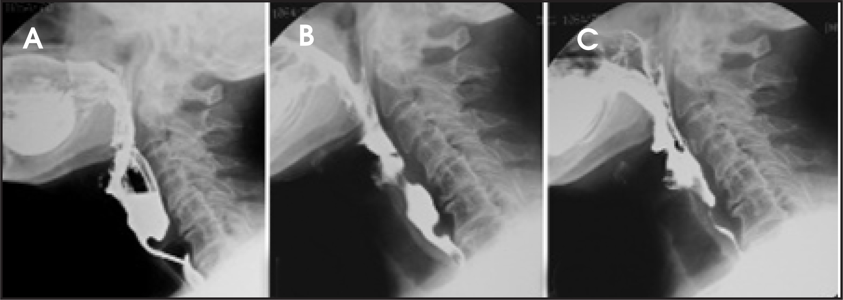
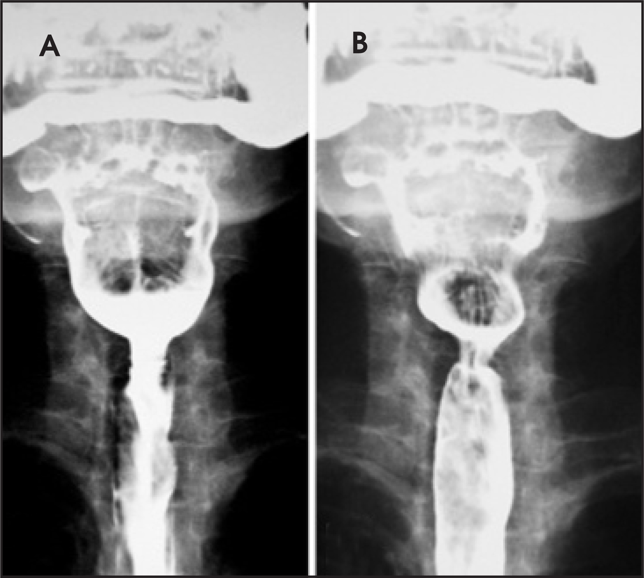
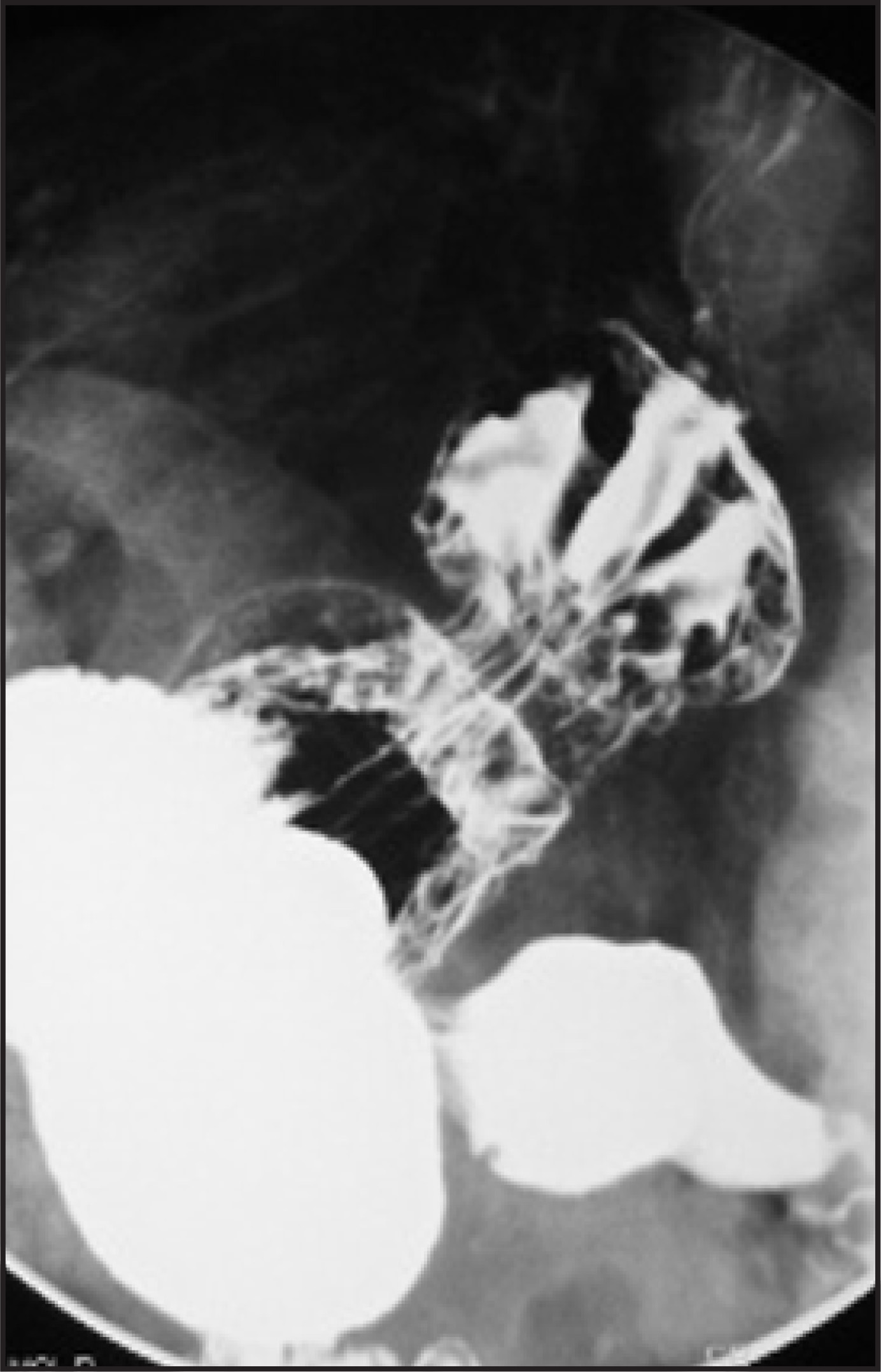
Bolus passage in the cervical esophagus was delayed, and multiple deglutitions were necessary to clear the contrast media from the hypopharynx. No inhalations were seen; instead nasal regurgitation occurred (Figure 1, B and C). Examination of the esophageal transit revealed an incarcerated gastric hiatal hernia (Figure 3) with gastroesophageal reflux.
DIAGNOSIS
Cricopharyngeal bar and incarcerated hiatal hernia
DISCUSSION
The cricopharyngeus (CP) muscle is the main component of the upper esophageal sphincter (UES), which consists of the CP muscle with the inferior pharyngeal constrictor and the proximal cervical esophagus.
The normal function of the sphincter is to maintain esophageal closure during inspiration and between swallows. This prevents air passage into the hypopharynx during inspiration and protects the airways from esophagogastric content reflux after deglutition.
In the resting state, the CP muscle is under tonic contraction and relaxes only momentarily during swallowing, belching, or vomiting. The muscle is not under voluntary control, but it can be made to function indirectly by initiating the deglutitory act. An exception is represented by sword swallowers who can keep it open at will for several seconds.1
Under normal conditions, the CP muscle can be recognized by an indentation on the posterior aspect of a barium swallow study at the level of the 5th or 6th vertebral body. Various degrees of posterior indentation ranging from mild to severe may be present. When it is very prominent, it has been called the cricopharyngeal bar, which may become a physical barrier affecting the normal deglutition, leading to dysphagia.
The pathogenesis of CP bar formation is not completely understood, and its clinical significance is not always clear, since a CP bar can be seen in 5% of asymptomatic individuals2 and a prominent posterior hypopharyngeal wall protrusion can be found in a high percentage of elderly cadavers.3 Some authors believe that a CP bar develops because of reduced muscle compliance,4 which limits normal distension of the UES during swallowing.
This muscle dysfunction could be induced by aging or by inflammatory-induced sclerosis secondary to chronic gastroesophageal reflux disease (GERD). This data is supported by anatomopathologic specimens from CP myotomy in patients with GERD who showed mild-to-severe muscle fibrosis.5
When a very prominent CP bar develops, it can result in impaired bolus passage in the esophagus with dysphagia and, if the obstruction is particularly severe, aspiration of pharyngeal contents may occur. Moreover, a noncompliant, fibrotic CP muscle may predispose a patient to the formation of Zenker’s diverticulum because of the increased intrapharyngeal pressure caused by the functional obstruction.6
The presence of a symptomatic CP bar associated with tracheal aspiration requires measures to prevent pneumonia and relieve dysphagia. Treatment should be tailored to the patient’s disease severity. The gold standard treatment is myotomy; other therapeutic options include balloon dilatation or botulinum toxin injection. Endoscopic dilatation can provide long-term relief of dysphagia and can be considered as an alternative to myotomy in patients who have a high perioperative risk.7 Botulinum toxin injection can temporarily decrease CP spasm, but the treatment must be repeated multiple times to achieve long-term results.8
Since the CP muscle hypertrophy may represent a compensatory response induced by chronic GERD, if a hiatal insufficiency or hernia is present, as in this case, treatment should also be addressed to solve this issue in order to avoid pharyngeal exposure to gastric secretions after the correction of the CP bar.
CONCLUSION
Cricopharyngeal bars have a characteristic appearance on barium swallow examinations and may represent a cause of pharyngeal dysphagia. The pathogenesis of CP bar formation is not fully understood but may result from muscle fibrosis induced by chronic GERD.
This case again shows that it is necessary to examine the entire swallowing chain when performing a videofluoroscopy study because esophagogastric alterations may be the cause of a pharyngeal disease.
References
- Jones B, Donner M, Jones B, Donner M. Normal and Abnormal Swallowing: Imaging in Diagnosis and Therapy. 1991:77-84.
- Seaman W. Cineroentgenographic observations of the cricopharyngeus. AJR Am J Roentgenol. 1966;96:922-931.
- Leaper M, Zhang M, Dawes P. An anatomical protrusion exists on the posterior hypopharyngeal wall in some elderly cadavers. Dysphagia. 2005;20:8-14.
- Dantas R, Cook I, Dodds W. Biomechanics of cricopharyngeal bars. Gastroenterology. 1990;99:1269-1274.
- Henderson R, Hanna W, Henderson R, Marryatt G. Myotomy for reflux-induced cricopharyngeal dysphagia. Five-year review. J Thorac Cardiovasc Surg. 1989;98:428-433.
- Veenker E, Andersen P, Cohen J. Cricopharyngeal spasm and Zenker’s diverticulum. Head Neck. 2003;25:681-694.
- Wang A, Kadkade R, Kahrilas P, Hirano I. Effectiveness of esophageal dilation for symptomatic cricopharyngeal bar. Gastrointest Endosc. 2005;61:148-152.
- Parameswaran M, Soliman A. Endoscopic botulinum toxin injection for cricopharyngeal dysphagia. Ann Otol Rhinol Laryngol. 2002;111:871-874.
Citation
. RCOM RADIOLOGICAL CASE OF THE MONTH. Applied Radiology. 2007;36(4):48H-48J. doi:10.37549/AR1497.