RCOM RADIOLOGICAL CASE OF THE MONTH
Applied Radiology — Vol. 36 , Issue 4 , pp. 48A -48D
DOI: 10.37549/AR1498
Published: April 1, 2007
Categories
CASE SUMMARY
A 27-year-old Hispanic man presented with an 8-year history of left foot pain, palpable nodules, and fistulae of the left foot. Physical examination revealed a swollen ankle and foot with several draining soft tissue subcutaneous nodules.
IMAGING FINDINGS
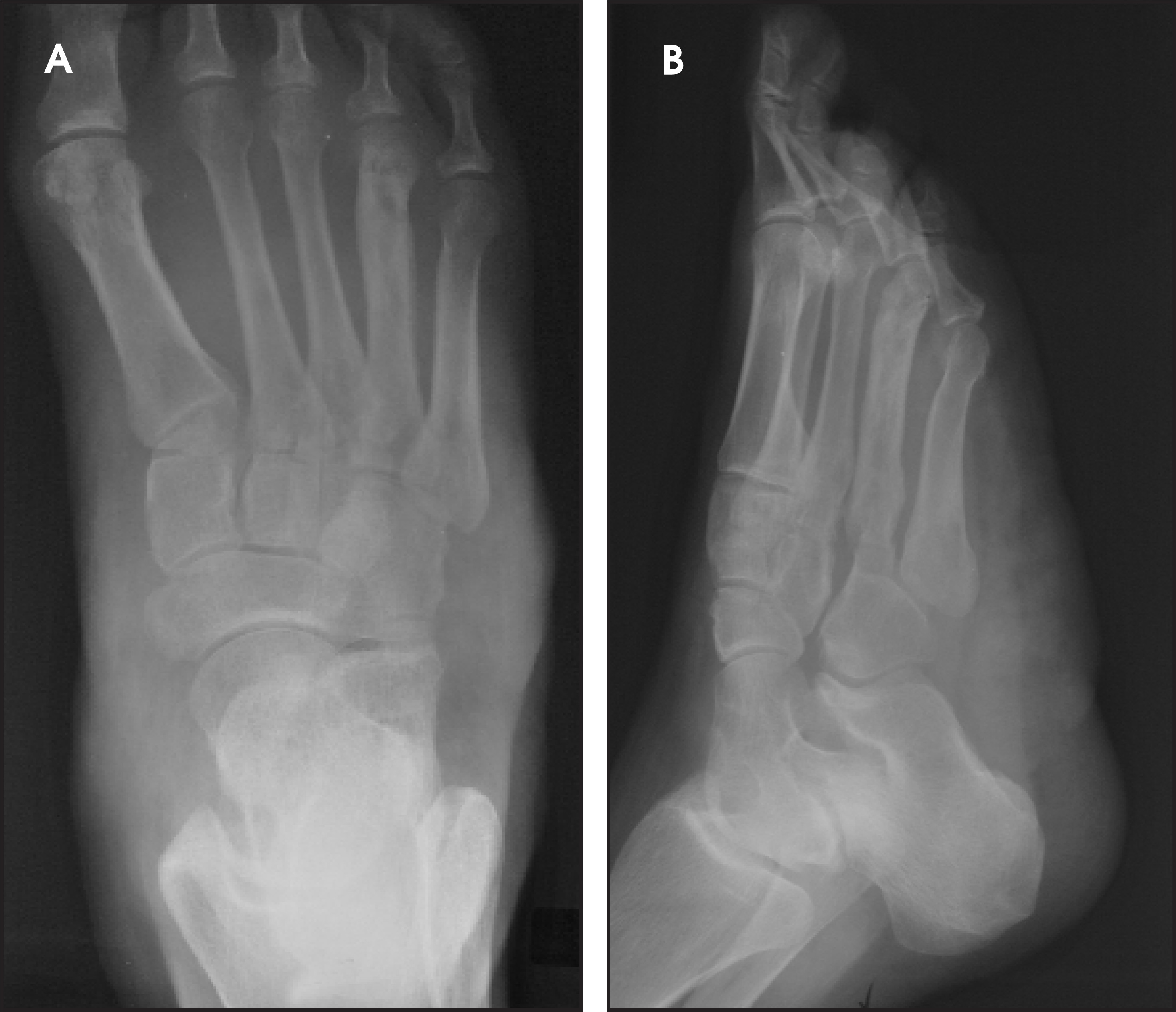
A plain radiograph shows changes of periosteal reaction and cortical thickening of the shaft of the 4th metatarsal with no cortical disruption (Figure 1A). The appearance is consistent with changes of chronic osteomyelitis. Soft tissue swelling and nodularity are present along the plantar aspect (Figure 1B).
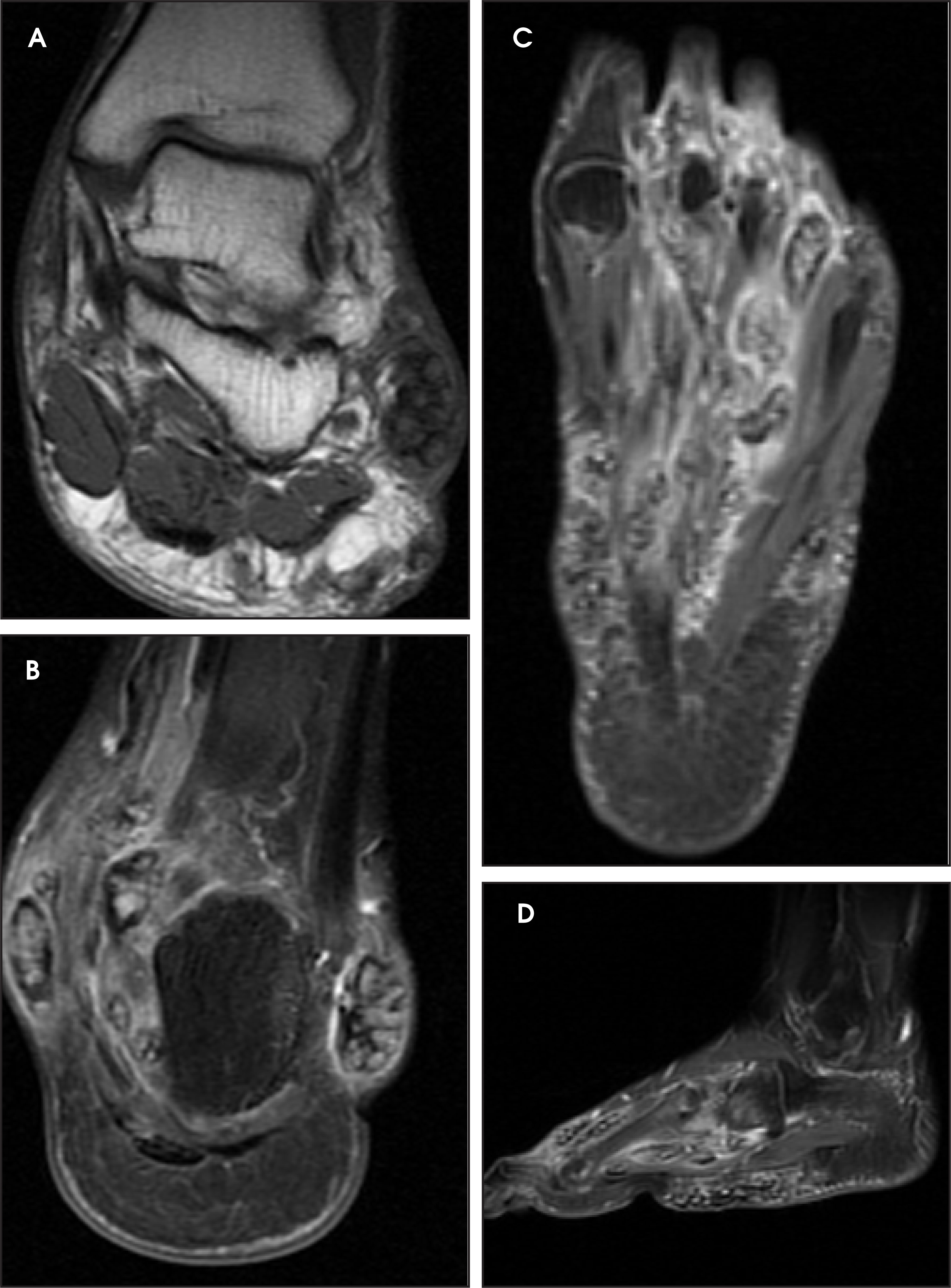
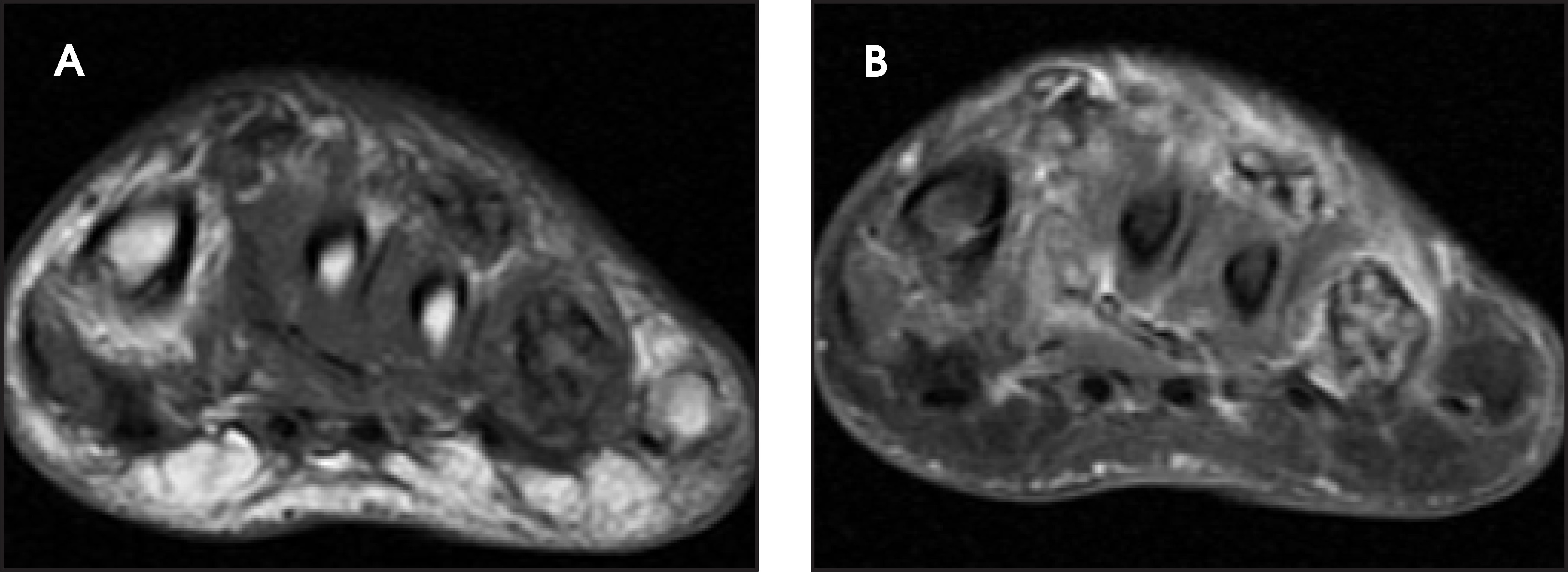
On magnetic resonance imaging (MRI), multiple loculated nodular soft tissue masses were seen surrounding the ankle and the plantar aspect. These masses were of intermediate signal intensity on coronal T1-weighted images (Figure 2) and enhanced with high signal intensity on coronal views of the ankle and foot (Figure 2, A–C) and on a fat-saturated T1-weighted postgadolinium sagittal view (Figure 2D). A focal area of abnormally dark bone marrow signal intensity is noted within the shaft of the 4th metatarsal bone on the axial T1-weighted fast spin-echo sequence (Figure 3A) and bright signal on the postgadolinium fat-saturated sequence (Figure 3B). A small, well-circumscribed multiloculated mass similar to that in the soft tissues is seen within the shaft of the metatarsal bone in this area of abnormal bone marrow signal (Figure 3).
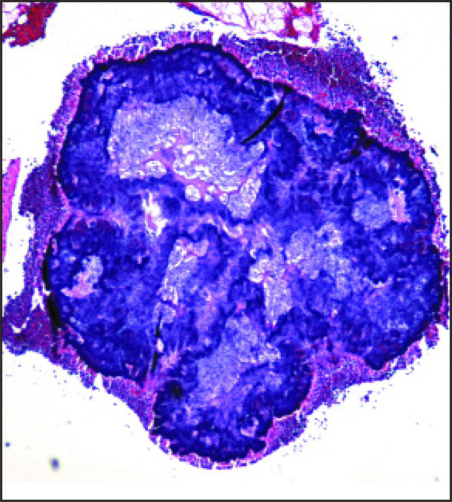
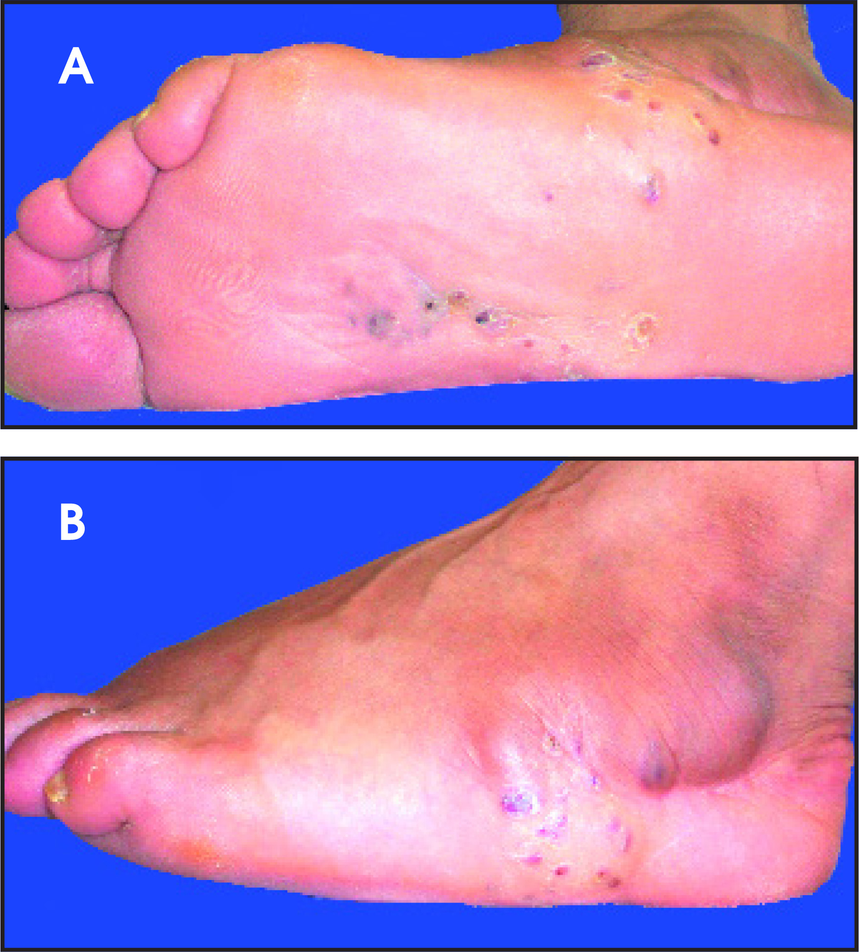
Histologic examination following biopsy of the draining nodule shows a dense mass of filamentous organisms surrounded by a rim of inflammatory cells (Figure 4). Photographs of the foot show multiple draining subcutaneous nodules, with a characteristic yellow powdery substance on the skin surface (Figure 5).
DIAGNOSIS
Maduromycosis, or Madura foot, with fungal osteomyelitis of the 4th metatarsal
DISCUSSION
Maduromycosis (mycetoma) is a chronic granulomatous fungal disease usually affecting the feet. This is common in the town of Madura in India from which the name is derived. Different species have been described in the United States, the common strain is Petriellidium boydii. Infection of the foot is secondary to trauma with contamination by the organism, which is normally present in the soil. The organism invades the muscles, tendons, bones, and joints, resulting in sinus tracts. Long-standing infection leads to swelling, deformity, and sinus tracts. Secondary bacterial infection or lymphatic dissemination may occur. Mycetomas can be caused by Actinomycetales, bacteria, and true fungi. Actinomycotic mycetomas can be caused by Nocardia brasiliensis, Streptomyces somaliensis, Streptomyces madurae, and Streptomyces pelleteri. Several fungi can cause mycetomas. Among them are Madurella mycetomi, Madurella grisea, Allescheria boydii, Cephalosporium sp., Phialophora jeanselmei, Pyrenochaeta romeroi, Leptosphaeria senegalensis, and Neotestudina rosatti. Fungal morphology and special stains indicated that the etiologic agent in this patient was Actynomices sp. Cultures were not obtained.
On imaging, multiple localized osseous defects, soft tissue and bony disruption, periostitis, and sclerosis are seen in long-standing cases. Intra-articular osseous fusion (referred to as a “melting snow” appearance) is noted. The differential diagnosis includes other causes of osteomyelitis, including fungal or parasitic etiologies, neuropathic osteoarthropathy, and neoplasm.
CONCLUSION
Maduromycosis is a rare cause of osteomyelitis. The clinical picture of multiple draining subcutaneous fistulae with a yellow “sulphur”-like substance and a characteristic MRI appearance of multiple loculated enhancing soft tissue masses are diagnostic.
References
- Binford C, Connor D. Pathology of Tropical and Extraordinary Diseases. 1976;2:557-561.
- Resnick D. Diagnosis of Bone and Joint Disorders. 2002;3:2563-2584.
Citation
. RCOM RADIOLOGICAL CASE OF THE MONTH. Applied Radiology. 2007;36(4):48A-48D. doi:10.37549/AR1498.