RCOM RADIOLOGICAL CASE OF THE MONTH
Applied Radiology — Vol. 36 , Issue 4 , pp. 38 -40
DOI: 10.37549/AR1500
Published: April 1, 2007
Categories
CASE SUMMARY
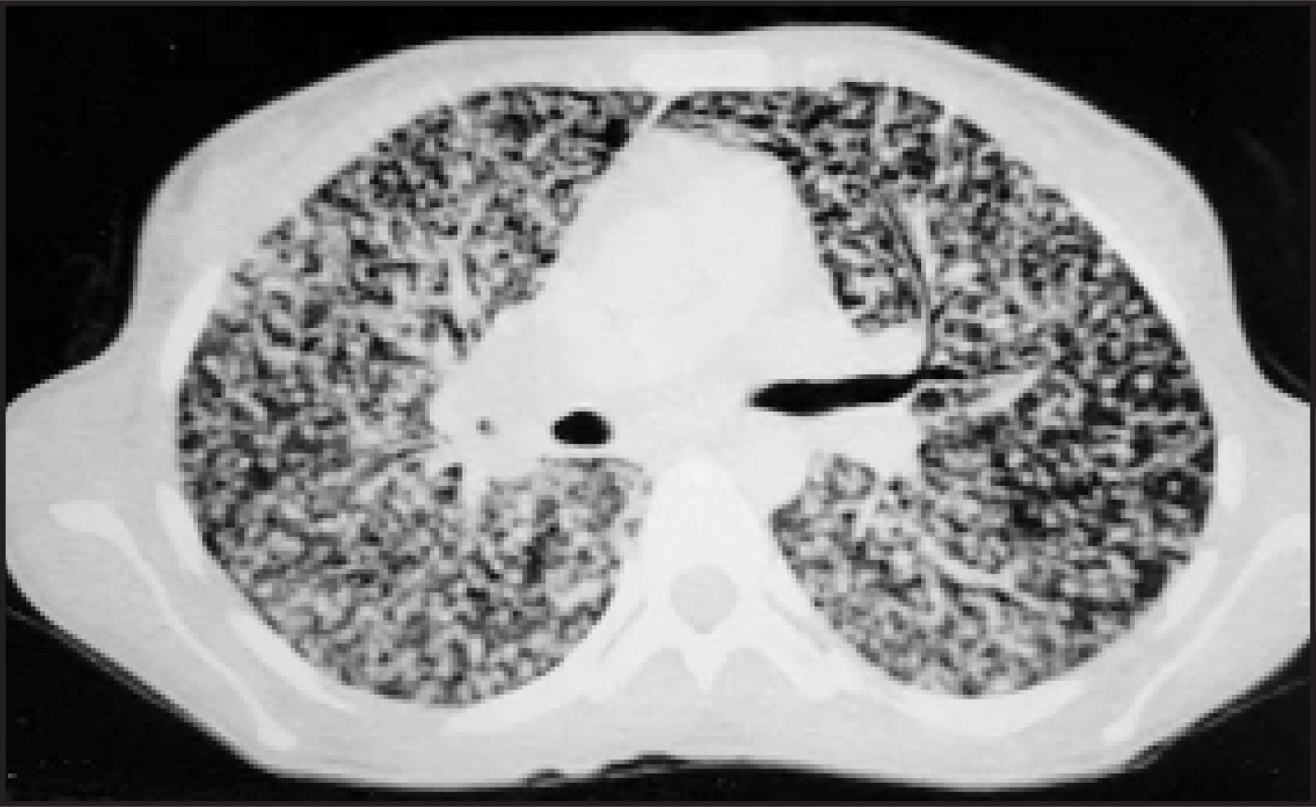
A 23-year-old housewife presented with a history of breathlessness, cough, night sweats, weakness, anorexia, weight loss for 1 month, and fever of 2 days’ duration. She had bacillus Calmette-Guérin (BCG) scars on her left upper extremity but no smoking history. The patient had a temperature of 38.5 to 39°C, pallor, dyspnea, and general malaise. There were cervical, submandibular, supraclavicular, and axillary lymphadenomegaly on physical examination, crackles in both lungs on pulmonary examination, and hepatosplenomegaly on abdominal examination. Erythrocyte sedimentation rate was 57 mm/h, hemoglobin 7.3 g/dL, white blood cell count 7190/mm3, serum albumin: 22 g/L (normal 35 to 55), alanine aminotransferase 50 U/L, aspartate aminotransferase 86 U/L, lactate dehydrogenase 805 U/L (normal 230 to 460), alkaline phosphatase 304 U/L (normal 42 to 141), gamma-glutamyl transpeptidase 223 U/L (normal 8 to 35), total bilirubin: 1.1 (normal 0 to 1), and C-reactive protein 99 mg/L (normal 0 to 8). Electrocardiography showed sinus tachycardia. Oxygen saturation was 76% in pulse oximetry. Serological tests for human immunodeficiency virus, cytomegalovirus, syphilis, and toxoplasmosis were negative. Thyroid function tests were within normal ranges. Antinuclear antibody, anti-ds-deoxyribonucleic acid, rheumatoid factor, and antithyroglobulin antibody were negative. The parenchyma of the spleen was heterogeneous on abdominal ultrasonography. Brain computed tomography (CT) was normal, and no neurological deficits were observed. Her chest X-ray and CT revealed disseminated miliary shadows in both lung fields, which was highly suggestive of miliary tuberculosis (Figure 1). There were no acid-fast bacilli in her sputum, but the histopathologic examination of her cervical lymph node revealed caseating granulomas.
IMAGING FINDINGS
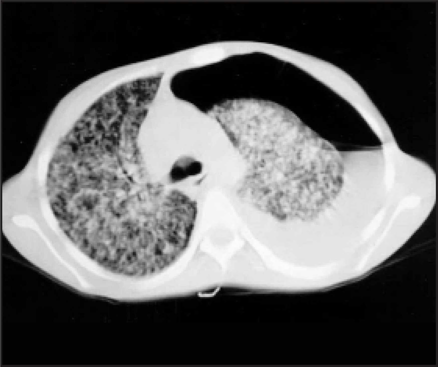
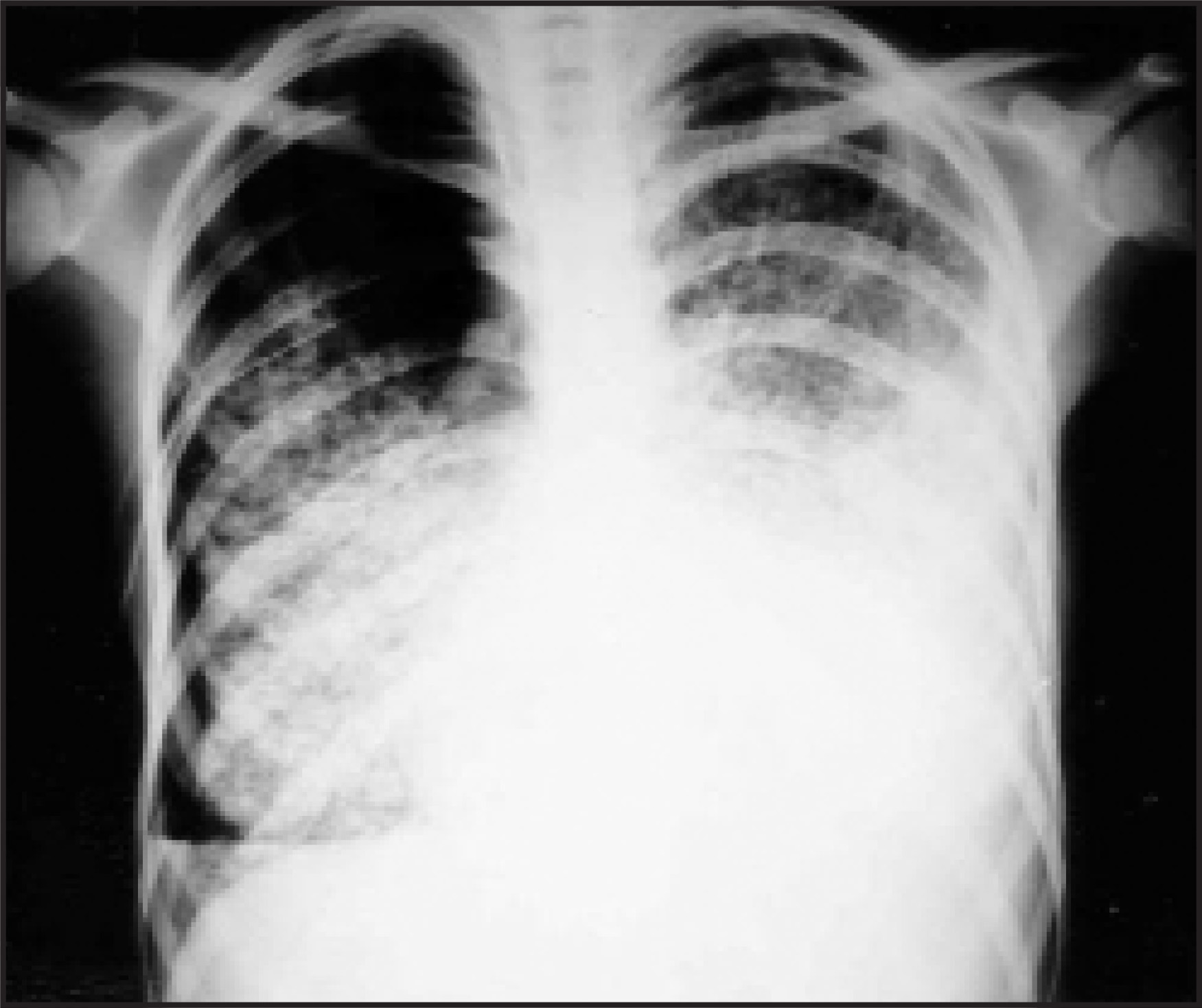
In addition to oxygen therapy, treatment with a combination of isoniazid, rifampicin, ethambutol, and pyrazinamide was started on the third day of hospitalization. On the fifth day of antituberculous treatment, her fever had not yet decreased, and methylprednisolone (1 mg/kg) was added to the therapy. Multiple blood cultures were negative. No pathogens were identified in aerobic cultures of sputum or urine. Wright and Rose Bengal agglutination tests for brucellosis were negative. The serum levels of CA-125 and CA-15.3 were high, but gynecological examination and thyroid and mammary ultrasonography showed no signs of malignancy. No fiberoptic bronchoscopy or other invasive procedures were performed because of respiratory failure. On the 38th day of hospitalization, she complained of pleuritic chest pain, and chest CT revealed a left-sided pneumothorax (Figure 2). A tube thoracostomy was placed, but after 1 week she developed a new pneumothorax on the other side (Figure 3). The patient was managed with bilateral tube thoracostomy, and both lungs were expanded. Her high temperature, which was occasionally subfebrile, continued for 2 months in spite of systemic corticotherapy. In the fifth month of therapy, the tumor markers (CA-125, CA-15.3) returned to normal levels. The treatment (anti-tuberculosis drugs and corticosteroids) was continued for 12 months, but the miliary shadows did not disappear. Her forced vital capacity (FVC) was 55%, her forced expiratory volume in 1 second (FEV1 ) was 58%, and her FEV1 /FVC was 93% in pulmonary function tests. Diffusion capacity was 65%, but the patient was not a candidate for an invasive diagnostic procedure at this time.
DIAGNOSIS
Bilateral spontaneous pneumothorax in a patient with miliary pattern tuberculosis
DISCUSSION
Miliary pattern and pneumothorax are rare radiological features in pulmonary tuberculosis. Their incidences are nearly 1.3% and 1.5%, respectively.1 However, the coincidence of pneumothorax on miliary tuberculosis is a very rare event. We found only 9 well-documented cases in the English literature.2-7 The age distribution of these patients showed adult predominance (Table 1), which supported the findings of other studies.8,9 Table 1 also illustrates that the male/female ratio was 4:6. However, this rate was 3:1 in all causes of secondary spontaneous pneumothorax.10 The pathogenesis of pneumothorax in miliary tuberculosis is unclear, but the following mechanisms can be considered: caseation or necrosis of subpleural miliary nodules and their subsequent rupture can cause pneumothorax. On the other hand, acute miliary dissemination may lead to emphysematous changes. This mechanism may explain the bilateral, simultaneous, and/or recurrent pneumothoraces.2,4 Despite this knowledge, it is not clear why a pneumothorax complicating miliary tuberculosis is commonly left-sided.
It has been noted that surgical pleurectomy should be attempted early in simultaneous bilateral secondary spontaneous pneumothorax.5 In miliary tuberculosis, open thoracotomy should not be considered until the patient has received antituberculous therapy for at least several weeks. The initial treatment for nearly every patient with a secondary spontaneous pneumothorax should be tube thoracostomy.
In this patient, at the end of the twelfth month of antituberculous therapy with corticotherapy, the miliary pattern was unchanged and her diffusion capacity was reduced, despite the presence of good performance status. These findings clearly suggested an interstitial lung disease, such as histiocytosis X or Langerhans cell histiocytosis. Pulmonary histiocytosis X is an uncommon, smoking-related, interstitial lung disease that primarily affects young adults. Spontaneous pneumothorax, which may be recurrent, is a recognized feature of this disease and likely results from the destruction of lung parenchyma with associated cysts or nodules (with or without cavitation). Prior to a biopsy procedure, a high-resolution chest CT can be helpful in the diagnostic evaluation. The natural history is variable, with some patients experiencing spontaneous remission of symptoms and others progressing to end-stage fibrotic lung disease. There is no clear benefit for either corticosteroids or cytotoxic agents.11,12
CONCLUSION
The possibility of a histiocytosis X should be considered in a young adult with a miliary pattern that is unresponsive to a long duration of antituberculosis drugs and corticosteroids, particularly in the absence of neurologic findings. Tuberculosis can accompany an interstitial lung disease that is characterized by a miliary pattern. In such a condition, fever may not respond to corticosteroid therapy. Finally, tumor markers may be used in the follow-up of tuberculosis.
References
- Aktogu S, Yorgancioglu A, Cirak K. Clinical spectrum of pulmonary and pleural tuberculosis: a report of 5,480 cases. Eur Respir J Eur Respir J.
- Peiken A, Lamberta F, Seriff N. Bilateral recurrent pneumothoraces: A rare complication of miliary tuberculosis. Am Rev Respir Dis. 1974;110:512-517.
- Narang R, Kumar S, Gupta A. Pneumothorax and pneumomediastinum complicating acute miliary tuberculosis. Tubercle. 1977;58:79-82.
- Chandra K, Prasad A, Prasad C. Recurrent pneumothoraces in miliary tuberculosis. Trop Geogr Med. 1988;40:347-349.
- Graf-Deuel E, Knoblauch A. Simultaneous bilateral spontaneous pneumothorax. Chest. 1994;105:1142-1146.
- Wammanda R, Ameh E, Ali F. Bilateral pneumothorax complicating miliary tuberculosis in children: Case report and review of the literature. Ann Trop Paediatr. 2003;23:149-152.
- Mert A, Bilir M, Akman C. Spontaneous pneumothorax: A rare complication of miliary tuberculosis. Ann Thorac Cardiovasc Surg. 2001;7:45-48.
- Kim J, Langston A, Gallis H. Miliary tuberculosis: Epidemiology, clinical manifestations, diagnosis, and outcome. Rev Infect Dis.. 1990;12:583-590.
- Maartens G, Willcox P, Benatar S. Miliary tuberculosis: Rapid diagnosis, hematologic abnormalities, and outcome in 109 treated adults. Am J Med. 1990;89:291-296.
- Melton L, Hepper N, Offord K. Incidence of spontaneous pneumothorax in Olmsted County, Minnesota: 1950 to 1974. Am Rev Respir Dis. 1979;120:1379-1382.
- Mendez J, Nadrous H, Vassallo R. Pneumothorax in pulmonary Langerhans cell histiocytosis. Chest. 2004;125:1028-1032.
- Vassallo R, Ryu J, Schroeder D. Clinical outcomes of pulmonary Langerhans’-cell histiocytosis in adults. N Engl J Med. 2002;346:484-490.
Citation
. RCOM RADIOLOGICAL CASE OF THE MONTH. Applied Radiology. 2007;36(4):38-40. doi:10.37549/AR1500.