RCOM RADIOLOGICAL CASE OF THE MONTH
Applied Radiology — Vol. 36 , Issue 4 , pp. 48E -48G
DOI: 10.37549/AR1504
Published: April 1, 2007
Categories
CASE SUMMARY
A 35-year-old man presented with a 2-year history of bilateral painless swelling at the angle of the jaw, which had increased in size over a 2-month period. He reported 3 days of fever, pain, and earache. There was no increase in size of the swelling with eating, no symptoms of dryness of the eyes or mouth, and no history of autoimmune diseases. The physical examination showed bilateral, fairly symmetric, nontender, nonfluctuant, soft tissue swelling at the angle of the jaw. The openings of Stensen’s ducts appeared unremarkable. The complete blood cell count laboratory values included a white blood cell count of 21.4 with 85 neutrophils. The CD4 count was 170.
DIAGNOSIS
Bilateral benign lymphoepithelial parotid cysts in diffuse infiltrative lymphocytic syndrome (DILS) in a patient with human immunodeficiency virus (HIV)
DIFFERENTIAL DIAGNOSIS
The presence of bilateral painless parotid gland swelling with no history or evidence of infection excludes acute parotitis. Lack of increased swelling or pain with food would exclude the possibility of calculus in the Stensen’s duct. Absence of xerostomia and xerophthalmia excludes Sjögren’s syndrome, which can present with benign lymphoepithelial hyperplasia. Secondary Sjögren’s syndrome in association with auto-immune diseases has a greater incidence of mucosa-associated lymphoid tissue or mucosa-associated lymphoid tissue lymphoma with a predilection for the parotid gland.1
IMAGING FINDINGS
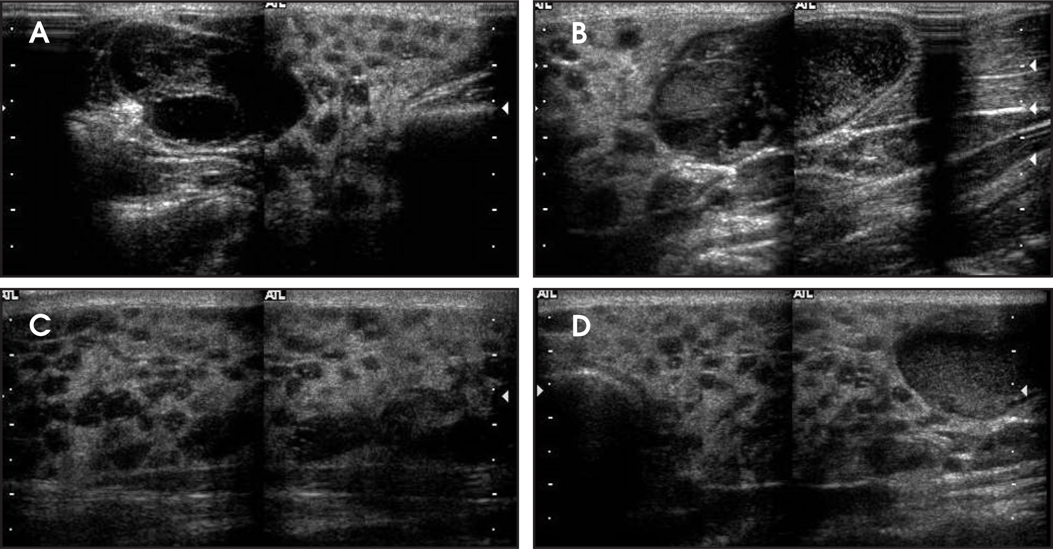
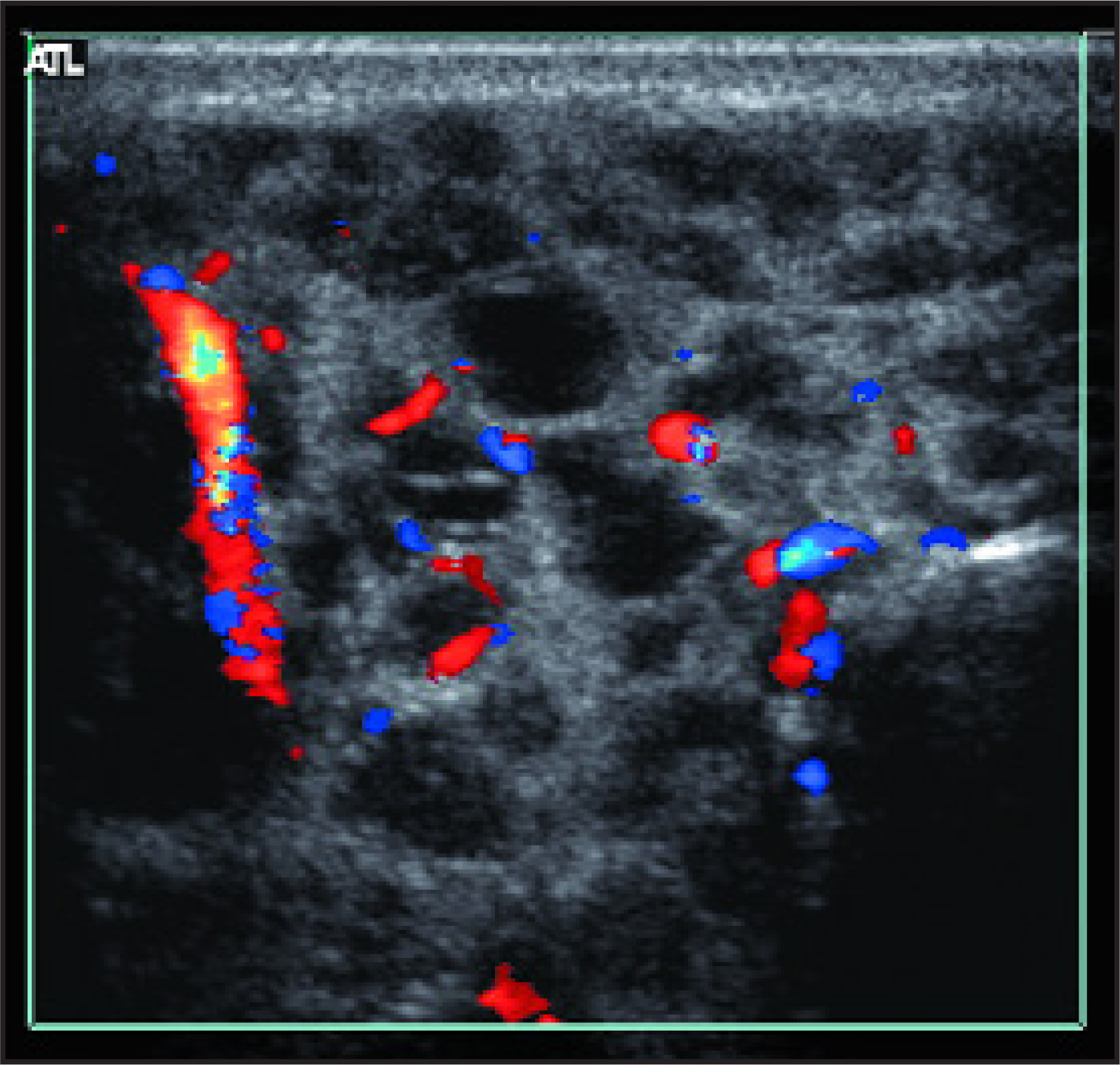
Diffusely enlarged parotid glands with multicystic involvement were seen on ultrasound (Figure 1). The cysts were noted to be thin-walled, fluid-filled structures with few internal septations noted. The color Doppler image confirms the cystic nature of the masses (Figure 2).
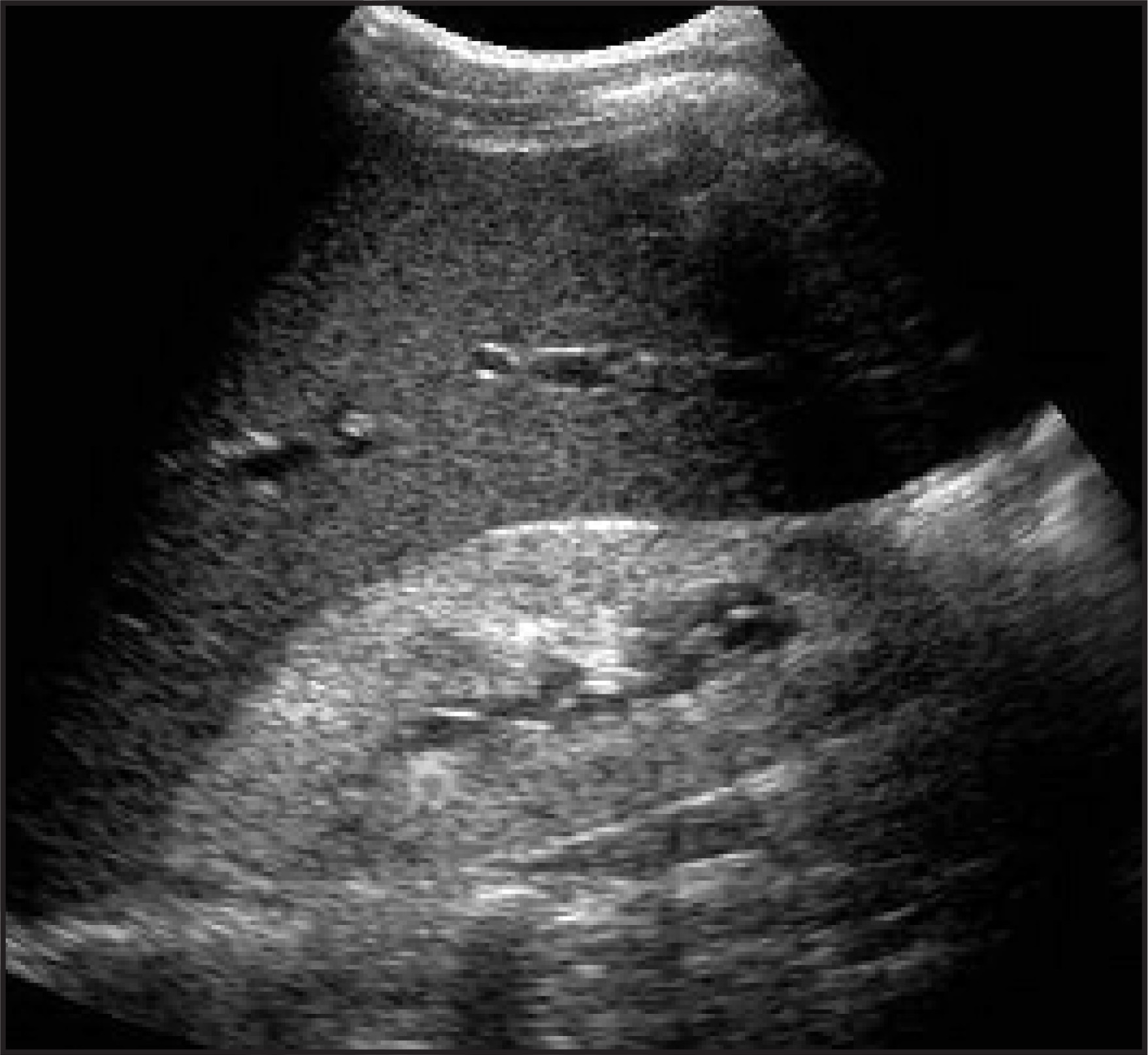
A longitudinal scan through the right upper quadrant shows increased cortical echogenicity of the right kidney in this patient with known HIV, reflecting the presence of HIV nephropathy (Figure 3).
PATHOLOGIC FINDINGS
Fine-needle aspiration of the right parotid gland showed polymorphs, abundant histiocytes, and foamy macrophages in a background of proteinaceous material. Stains for acid-fast bacilli and fungi were negative.
DISCUSSION
Diffuse infiltrative lymphocytic syndrome is a subset of HIV disease. Roughly 40% of patients will present with head- and neck-related symptoms, and parotid enlargement occurs in approximately 5% of this group. Diffuse infiltrative lymphocytic syndrome is associated with diffuse visceral infiltration, a persistent CD8 lymphocytosis, bilateral parotid swelling, and cervical lymphadenopathy. Cysts are thought to arise from intra- or periparotidal lymph nodes that may contain intrasalivary inclusions. These lymph nodes form a part of the gland during embryological development and may contain salivary gland acini and ducts. With replication of the HIV-1 virus, salivary gland hyperplasia and swelling appears. There is hyperplasia of the intraglandular lymphocytes and infiltration of the parotid from extraglandular lymphocytic proliferation. Soft and painless enlargement of the parotids involving the superficial lobes is present.2
CONCLUSION
With HIV nephropathy, lymphoepithelial cysts are a part of the spectrum of the HIV/acquired immunodeficiency syndrome (AIDS)-related complex syndrome. Development of parotid gland lymphoepithelial cysts in an HIV patient may be a precursor to AIDS.3 Differential diagnoses of parotid gland swelling include reactive lymphadenopathy, parotid cyst, and DILS. None of these lesions present bilaterally. Non-HIV causes of parotid enlargement should include Sjörgen’s syndrome. Ultrasound is useful to assess parotid masses and can differentiate intraglandular lesions from extraglandular lesions.4
References
- Mihovilovic Z, Mandel L. Sjögren’s syndrome and MALT lymphoma. Columbia Dent Rev. 2000;5:27-29.
- Hung C, Mandel L. HIV disease and parotid lymphoepithelial cysts. Columbia Dent Rev. 2000;5:25-26.
- Holliday R, Cohen W, Schinella R. Benign lymphoepithelial parotid cysts and hyperplastic cervical adenopathy in AIDS-risk patients: A new CT appearance. Radiology. 1988;168:439-441.
- Yousem D, Kraut M, Chalian A. Major salivary gland imaging. Radiology. 2000;216:19-29.
Citation
. RCOM RADIOLOGICAL CASE OF THE MONTH. Applied Radiology. 2007;36(4):48E-48G. doi:10.37549/AR1504.