RCOM RADIOLOGICAL CASE OF THE MONTH
Applied Radiology — Vol. 36 , Issue 9 , pp. 48A -48D
DOI: 10.37549/AR1540
Published: September 1, 2007
Categories
CASE SUMMARY
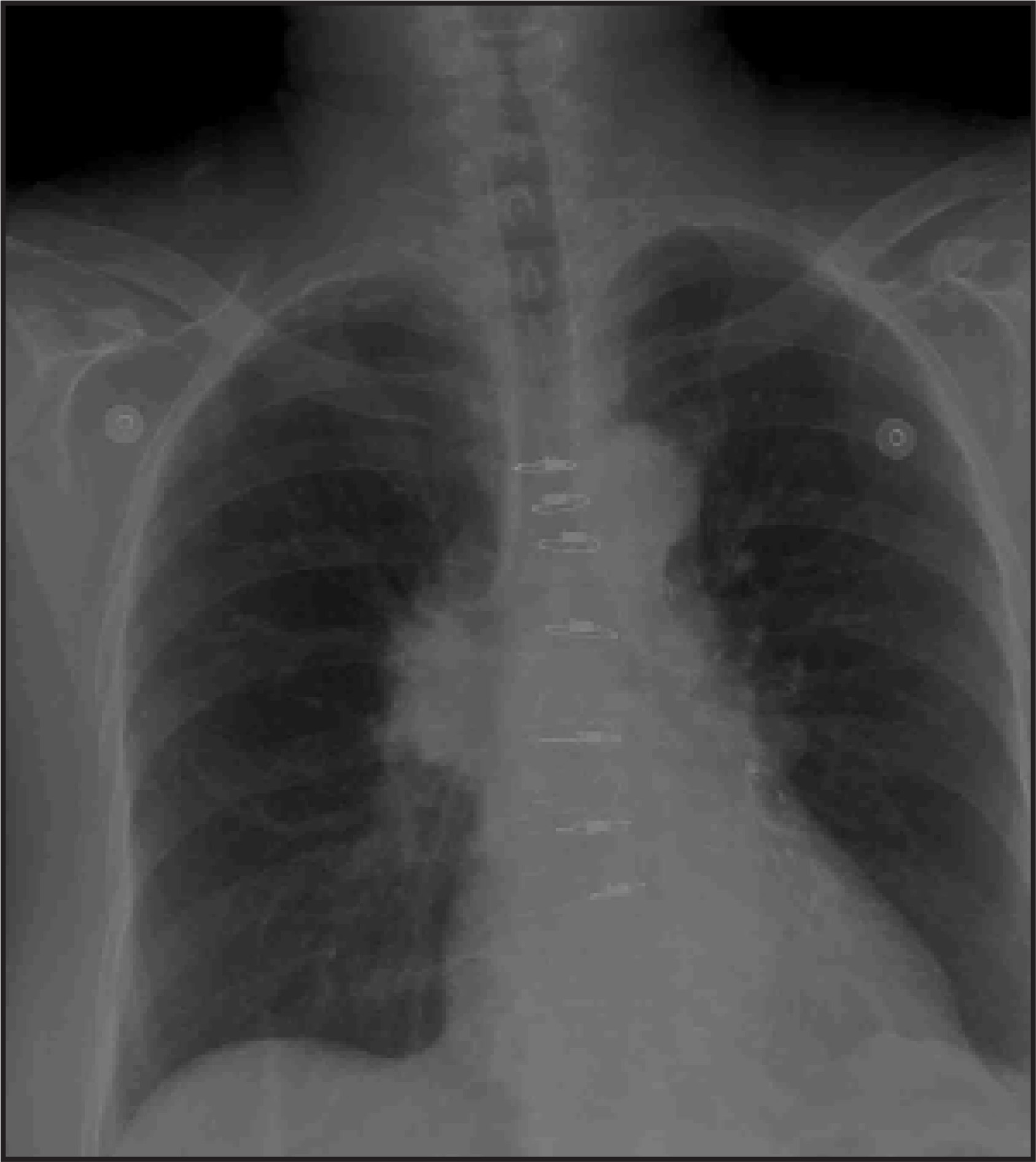
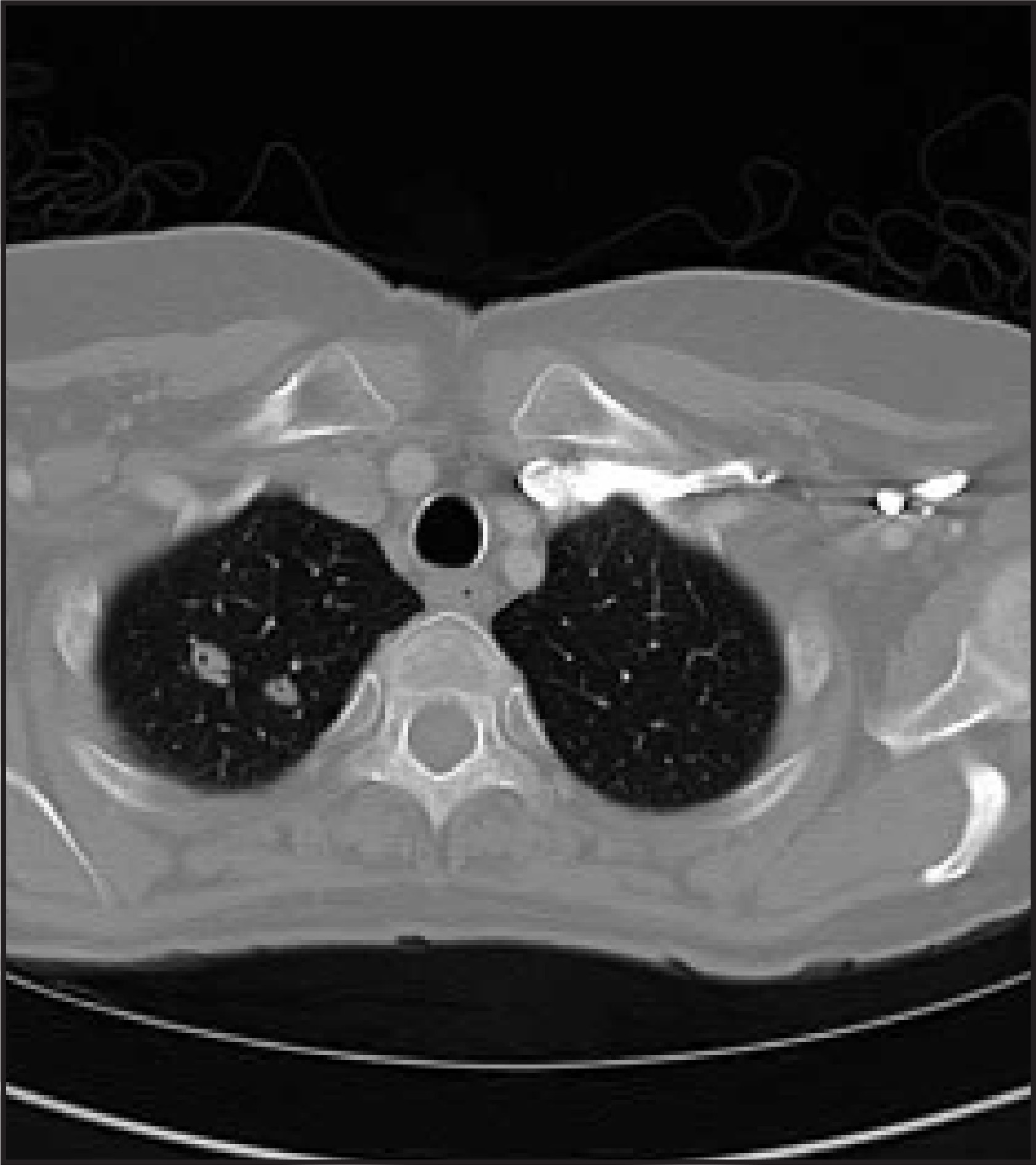
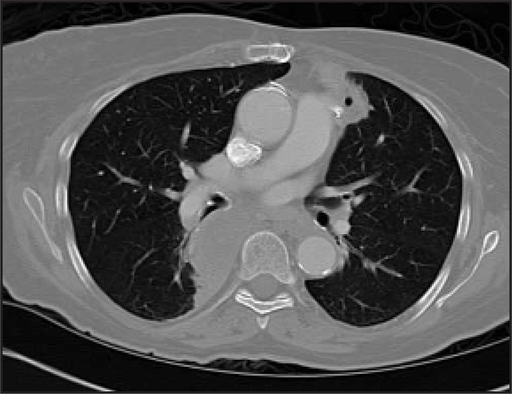
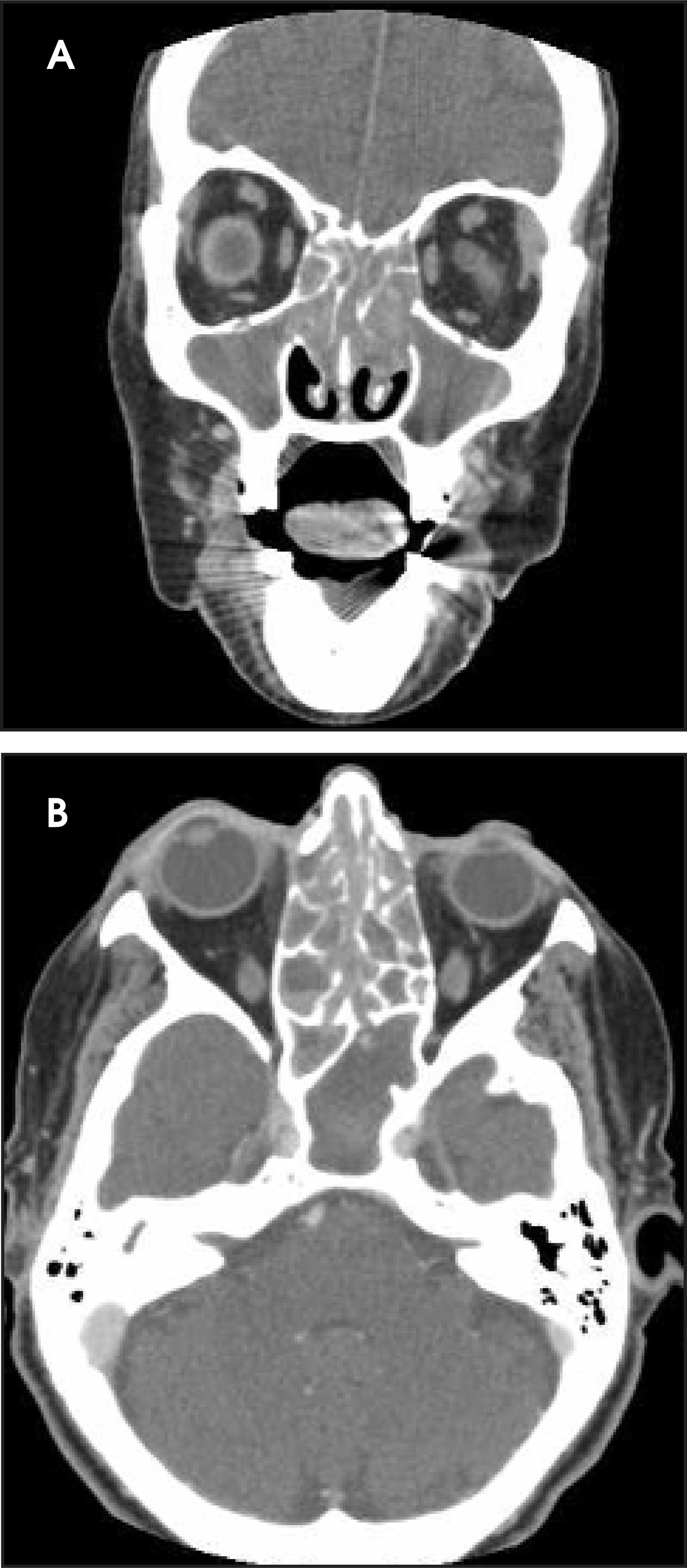
A 77-year-old woman presented with a 1-month history of productive cough and hemoptysis. Additional complaints included fever, chills, night sweats, and generalized weakness of the same duration. Recently, she had experienced unilateral hearing loss. Relevant surgical history included oral surgery for recurrent jaw pain. A physical examination revealed expiratory wheezes over the left lung base. Her neurological examination was nonfocal except for decreased hearing in her right ear. Laboratory analysis showed elevations in C-reactive protein and antiproteinase-3 antibody (anti-PR3). Blood cultures were negative. Chest radiography (Figure 1) and computed tomography (CT) of the chest (Figures 2 and 3) and sinuses (Figure 4) were performed, followed by video-assisted thoracoscopy with lung biopsies.
IMAGING FINDINGS
The initial radiographs that were obtained at the time of the patient’s presentation showed a large right hilar mass along with smaller nodules at the right lung apex (Figure 1). Subsequently, a chest CT was obtained to better characterize the plain film findings. An axial image at the level of the lung apices demonstrated 2 cavitary nodules (Figure 2). An image at the level of the hila showed a masslike area of consolidation with another cavitary nodule seen in the lingula (Figure 3). The attenuation of the hilar “mass” approached that of soft tissue and was thought to represent advanced focal consolidation.
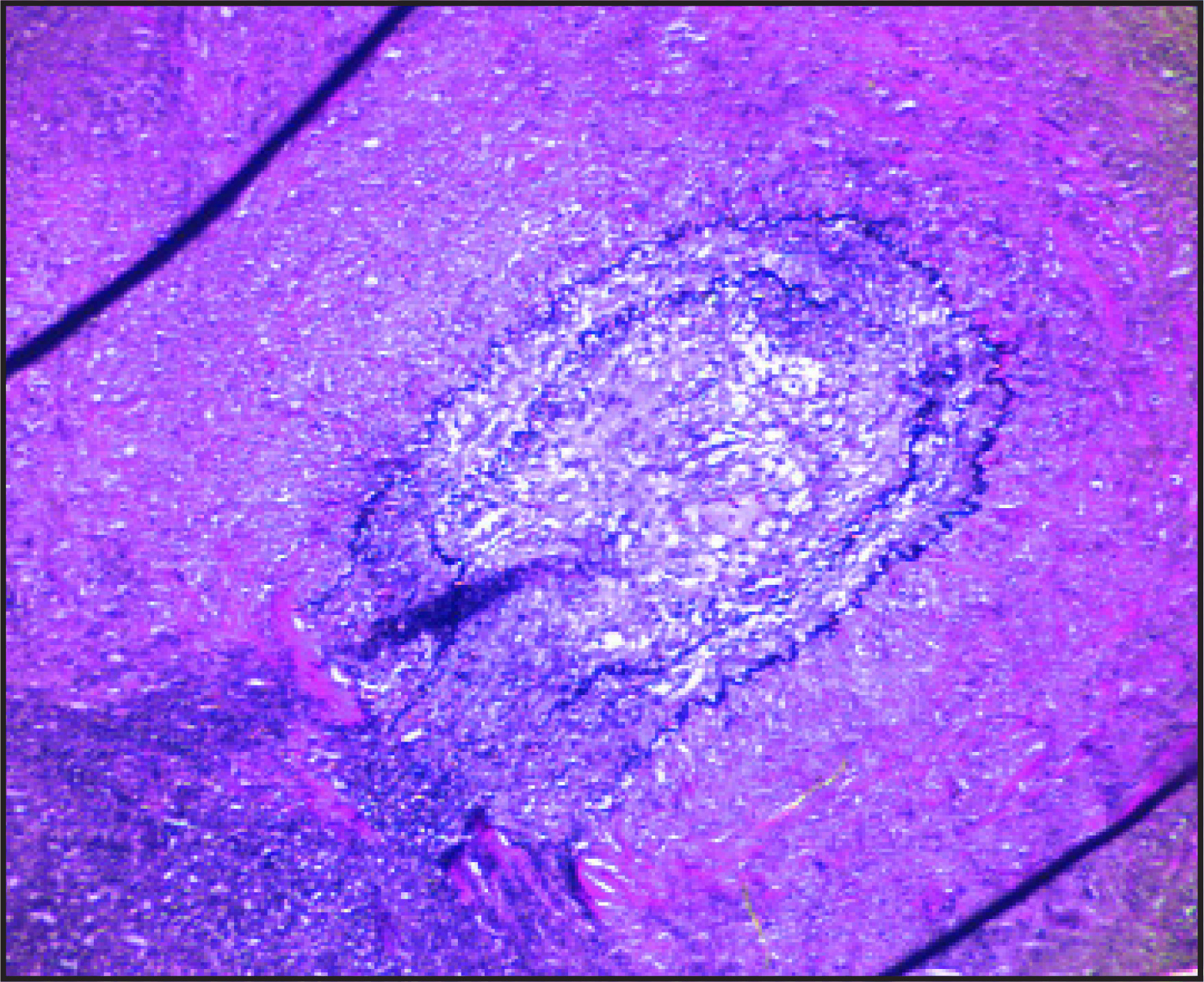
Sinus CT was then obtained and revealed soft tissue density opacification of the maxillary, ethmoid, sphenoid, and frontal sinuses (Figure 4). The imaging workup was followed by a lung biopsy. Histologic examination revealed characteristic findings of Wegener’s granulomatosis (WG) (Figure 5).
DIAGNOSIS
Wegener’s granulomatosis
DISCUSSION
Wegener’s granulomatosis is an infrequent chronic vasculitis involving the upper and lower respiratory tracts and, in most cases, the kidneys. Specific laboratory findings include an elevated anti-PR3, which corresponds to the cytoplasmic pattern of antineutrophil cytoplasmic antibodies (c-ANCA).1 The prevalence is reported at 3.0 per 100,000. Reports on gender distribution yield conflicting results; the National Institutes of Health reports suggest that WG affects both sexes equally, while others indicate a male predominance.2,3
Upper airway disease is the most frequent presenting feature of WG. It is seen in >70% of patients at onset and ultimately in >90% of cases. Otologic manifestations may be part of the initial presentation in approximately 25% of patients. Significant degrees of hearing loss (usually conductive) may be seen in 14% to 42% of patients. Sinusitis is a component of the initial presentation in 50% to 66% of patients and is seen in 85% of cases at some point in the disease process.
Pulmonary involvement is one of the cardinal features of WG, with cough, hemoptysis, and pleuritis being the most common pulmonary symptoms. It is also important to note that up to one third of patients with radiographically visible pulmonary lesions may not have lower airway symptoms.2
Until 1990, WG was considered a lethal disease with a mortality of 90% within 2 years. Improvement in the prognosis is primarily related to earlier diagnosis and improved chemotherapeutic treatment strategies that generally include a cytotoxic drug (eg, cyclophosphamide) and glucocorticoids. Even though no specific radiologic criteria exist, given the appropriate clinical setting, imaging may suggest the diagnosis at an early stage.4
Chest CT abnormalities in WG have been well-characterized. Typical findings include multiple nodules, air-space opacification, and tracheobroncial disease. Also reported are interstitial fibrosis, pleural effusions, and mediastinal lymphadenopathy. Multiple nodules or masses are the most common pulmonary finding. They are seen in 70% of patients. The nodules range in size from a few millimeters to 10 cm. The nodules are usually multiple and tend to increase in size and number as the disease progresses, but they rarely exceed 10 in total. Cavitation is common, being seen in approximately 50% of cases and in most nodules >2 cm. Air-fluid levels and calcification are uncommon.
Areas of air-space opacification, either consolidation or ground-glass opacification, are also common, occurring in 50% of patients. The opacification is variable in terms of size, density, and distribution. It may occur with or without the presence of accompanying nodules. Several patterns have been described, including wedge-shaped areas of peripheral consolidation abutting the pleura and mimicking pulmonary infarcts, peribronchoarterial distribution of consolidation, focal consolidation (with or without cavitation), parenchymal bands, and diffuse bilateral areas of ground-glass opacification, which represent diffuse pulmonary hemorrhage.5
The patterns of recurrent WG are as varied as those seen at the initial presentation. Wegener’s granuloma may recur as multifocal or diffuse ground-glass opacities or consolidation, multiple cavitary nodules, and multifocal or long-segment airway stenosis (with or without resultant atelectasis). The recurrent pattern has been reported to be different from the initial pattern; however, according to the study by Lee et al,6 both the same and different patterns of recurrence were seen.
Several studies have sought to characterize the findings of sinus CT in WG patients. Some typical bony changes that are indicative of WG include bony obliteration of a sinus, neo-osteogenesis within a sinus, and punctuate bony erosions, primarily at the septum and turbinates. Periantral soft tissue infiltration and nodular mucosal thickening are also often seen. The maxillary sinus is the most frequently involved, followed by the frontal and sphenoid sinuses. The bony changes are believed to represent chronic periostitis from the underlying vasculitis. In the appropriate clinical setting, the above changes seen on sinus CT scans may provide radiologic evidence of WG, which can help to expedite diagnosis and improve prognosis.1,3,4
Treated WG leaves substantial residual damage in the lungs, and high-resolution CT can assist in the distinction between active and inactive lesions. Ground-glass opacification, cavitating nodules/masses, and lesions that measure >3 cm typically represent active disease. Noncavitary small nodules and septal or nonseptal linear densities can be either active or cicatricial, residual lesions; however, patients with residual nodules or lines tend to have no more relapses than patients with completely cleared lungs. In the study by Komocsi et al,7 all masses resolved in response to treatment, whereas smaller nodules either resolved or remained unchanged. Ground-glass opacification is a feature of active disease and typically resolves completely with clinically effective treatment. Cavitation is another prognostically helpful feature, as it typically portends resolution with treatment. Linear densities are impossible to classify prospectively into active or inactive lesions and, therefore, may or may not show response to therapy. Tracheobronchial lesions, such as segments of stenosis as well as intra- and extraluminal soft tissue masses, are also reported to not show improvement with drug treatment.7
The histologic diagnosis of WG requires identification of necrotizing granulomatous vasculitis. The predominant vessels affected are the medium-sized muscular arteries. The necrosis and obliteration of these vessels may result in the radiographic appearances of pulmonary infarcts. However, small-vessel vasculitis sited mainly in alveolar septal capillaries and small arterioles can also be encountered. Capillary involvement is usually found at the edges of the more typical granulomatous vasculitis.8
Although WG has a well-characterized “classic” presentation, the disease may also have atypical presentations. There have been case reports of WG causing a vasculitis of the main pulmonary arteries and presenting with findings identical to chronic pulmonary thromboembolic disease.9 There is also a rare, “limited” form of WG, in which lung and kidney involvement may be absent. In an even more exceptional presentation, limited WG may appear as Tolosa-Hunt syndrome (painful ophthalmoplegia due to inflammation of the cavernous sinus or superior orbital fissure), meningeal and cerebral parenchymal granulomatous inflammation, chronic meningitis, and cranial neuropathies.10
CONCLUSION
Wegener’s granulomatosis is a relatively infrequent vasculitis that can cause significant morbidity and even mortality. Prompt diagnosis may be possible if its characteristic imaging findings are recognized in the appropriate clinical setting, and may allow for earlier treatment and improved prognosis.
References
- Gubbels S, Barkhuizen A, Hwang P. Head and neck manifestations of Wegener’s granulomatosis. Otolaryngol Clin North Am. 2003;36:685-705.
- Calabrese L, Duna G, Harris E, Budd R, Firestein G. Kelley’s Textbook of Rheumatology. 2005:1357-1378.
- Yang C, Talbot J, Hwang P. Bony abnormalities of the paranasal sinuses in patients with Wegener’s granulomatosis. Am J Rhinol. 2001;15:121-125.
- Benoudiba F, Marsot-Dupuch K, Rabia M. Sinonasal Wegener’s granulomatosis: CT characteristics. Neuroradiology. 2003;45:95-99.
- Sheehan R, Flint J, Muller N. Computed tomography features of the thoracic manifestations of Wegener’s granulomatosis. J Thoracic Imaging. 2003;18:34-41.
- Lee K, Kim T, Fujimoto K. Thoracic manifestations of Wegener’s granulomatosis: CT findings in 30 patients. Eur Radiol. 2003;13:43-51.
- Komocsi A, Reuter M, Heller M. Active disease and residual damage in treated Wegener’s granulomatosis: An observational study using pulmonary high-resolution CT. Eur Radiol. 2003;13:36-42.
- Hansell D. Small-vessel diseases of the lung: CT-pathologic correlates. Radiology. 2002;225:639-653.
- Doyle D, Fanning N, Silke C. Wegener’s granulomatosis of the main pulmonary arteries: Imaging findings. Clin Radiol. 2003;58:329-331.
- Thajeb P, Tsai J. Cerebral and oculorhinal manifestations of a limited form of Wegener’s granulomatosis with c-ANCA-associated vasculitis. J Neuroimaging. 2001;11:59-63.
Citation
. RCOM RADIOLOGICAL CASE OF THE MONTH. Applied Radiology. 2007;36(9):48A-48D. doi:10.37549/AR1540.