RCOM RADIOLOGICAL CASE OF THE MONTH
Applied Radiology — Vol. 36 , Issue 9 , pp. 43 -45
DOI: 10.37549/AR1542
Published: September 1, 2007
Categories
CASE SUMMARY
A 34-year-old woman presented to the clinic complaining of chronic right shoulder pain. The patient was an otherwise healthy person, denying any previous shoulder trauma or history of athletic activity involving overhead arm motions. The physical examination was unremarkable, revealing intact sensory and motor function as well as full range of motion. Radiography of the shoulder was obtained, and further evaluation was later performed with computed tomographic (CT) examination, skeletal scintigraphy, and magnetic resonance (MR) imaging.
IMAGING FINDINGS
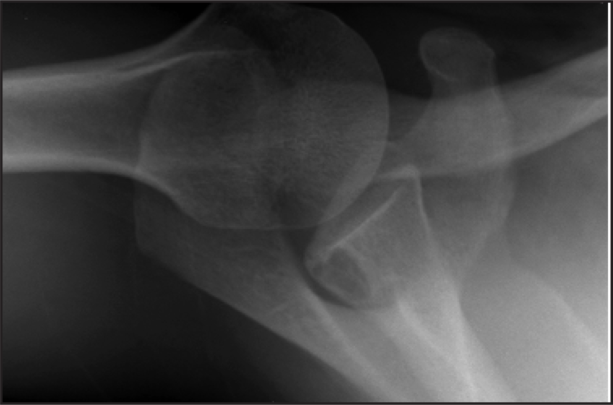
Radiographic evaluation of the shoulder showed a well-circumscribed subchondral lytic lesion of the glenoid. The lesion exhibited lobulation and sclerosis of its margins, without evidence of periostitis or cortical disruption (Figure 1).
Noncontrast CT of the shoulder (not shown) verified the well-circumscribed lytic appearance of the lesion without internal matrix calcification or ossification.
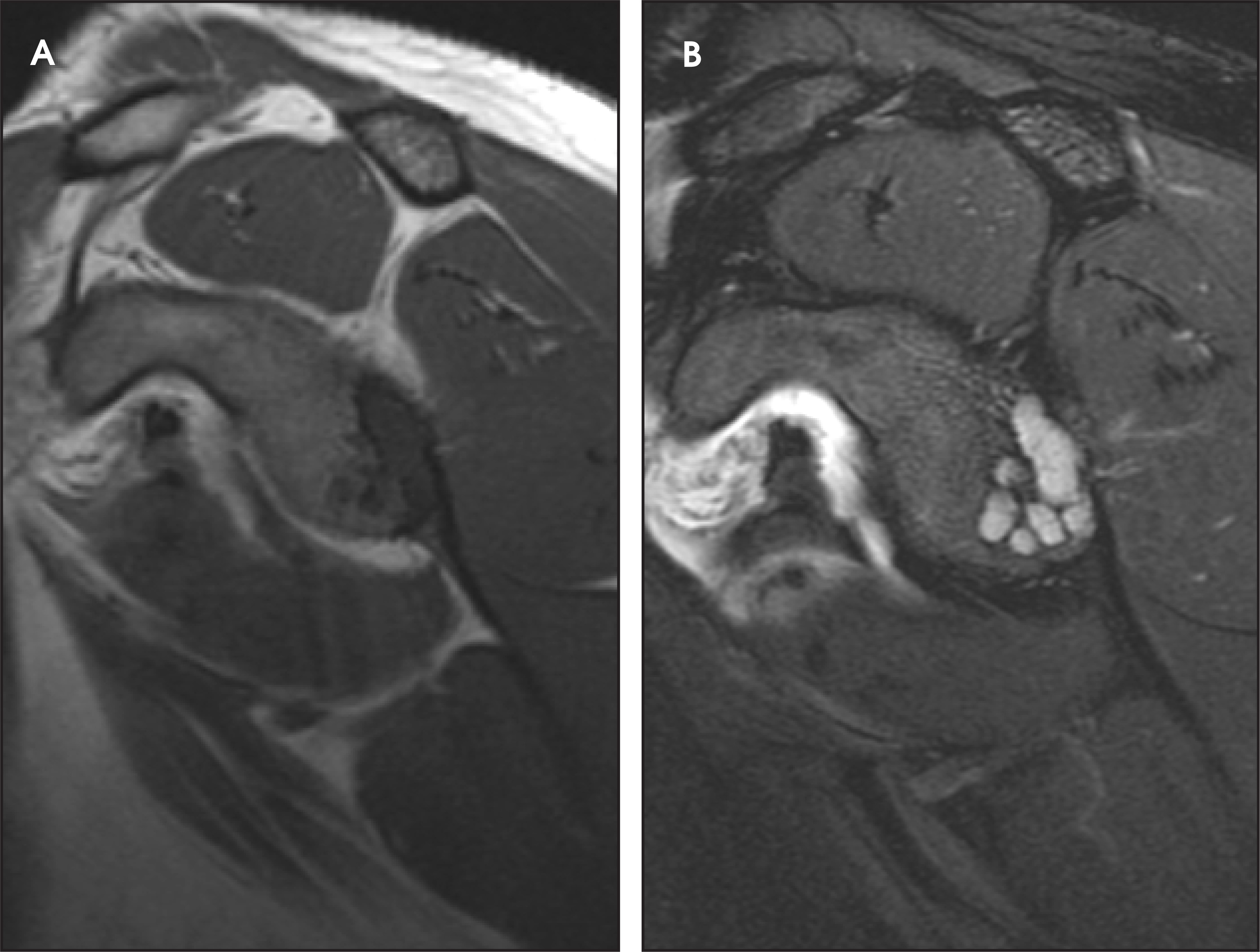
MR imaging shows the presence of a T1 hypointense (Figure 2A), T2 hyperintense lobulated lesion involving the glenoid (Figure 2B). The surrounding cortical bone appears intact, and there are no associated inflammatory changes or evidence of soft tissue mass.
DIAGNOSIS
Intraosseous ganglion of the glenoid (which was proven surgically)
DISCUSSION
Intraosseous ganglion is a rare, but benign, tumor of bone that is considered in the differential diagnosis of epiphyseal lesions. It is characterized as a loculated, fibrous, and cystic lesion lined by mucin-secreting synovial-like histiocytes with histologic features that are identical to those of its soft tissue counterparts.1 The origin of this lesion is unclear, but most experts agree that they may arise de novo through a sequence of intramedullary metaplasia involving a nonspecific mesenchymal precursor cell that eventually leads to the formation of fibroblasts; these fibroblasts will undergo proliferation and mucin secretion. These changes are thought to lead to pressure atrophy of the surrounding trabecula, resulting in cyst formation.2
Intraosseous ganglia are usually encountered incidentally in asymptomatic young adults and middle-aged individuals. When symptomatic, patients generally complain of a long-standing aching discomfort that is associated with activity but is relieved by rest.3 Lesions are generally between 2 and 7 cm in size and are most frequently seen in the region of the hip (acetabulum and femoral head), comprising 40% of all lesions in 1 large series.2 The ankle/hindfoot (distal tibia, talus, and calcaneus) is a close second in terms of location; rarely, there is involvement of the upper extremity as well.2 Intraosseous ganglia of the glenoid is one of the most rare locations encountered, with only 12 cases reported in the literature.1
Radiographically, intraosseous ganglia are characterized as well-defined, subchondral lytic lesions that contain a sclerotic margin.4,6 They exhibit benign features and typically are without periosteal reaction, cortical disruption, deformity, joint involvement, or associated soft tissue mass. Moreover, intralesional calcifications are almost never seen.2 The lesion usually shows an absence of significant radioactivity on skeletal scintigraphy. CT and MR imaging typically show benign, cystic findings. This constellation of imaging features is helpful in differentiating an intraosseous ganglion from other radiolucent epiphyseal lesions such as chondroblastoma, giant cell tumor, simple bone cyst, clear cell chondrosarcoma (in older patients), and subchondral cyst or geode, which occurs in the setting of arthritis.
Treatment of intraosseous ganglia is according to symptomatology. Asymptomatic individuals may be observed; the presence of pain requires treatment by curettage and bone grafting.3
CONCLUSION
Intraosseous ganglia represent an infrequent and benign cause of cystic bone lesions. They may be considered in the differential diagnosis when a benign-appearing cystic lesion is encountered in the epiphysis. They are generally asymptomatic but, as in this case, may be a source of chronic pain requiring surgical management with curettage and packing.
References
- Kligman M, Roffman M. Intraosseous ganglia of glenoid. J South Orthop Assoc. 2000;9:216-218.
- Feldman F, Johnston A. Ganglia of bone: Theories, manifestations, and presentations. CRC Crit Rev Clin Radiol Nucl Med. 1973;4:303-332.
- Willems D, Mulier J, Martens M, Verhelst M. Ganglion cysts of bone: Report of two cases and review of the literature. Acta Orthop Scand. 1973;44:655-662.
- Yamato M, Saotome K, Tamai K, Yamaguchi T. Case report 783. Intraosseous ganglion of scapula. Skeletal Radiol. 1993;22:227-228.
- Parman L, Murphey M. Alphabet soup: Cystic lesions of bone. Semin Musculoskelet Radiol. 2000;4(1):89-101.
- Bauer T, Dorfman H. Intraosseous ganglion: A clinicopathologic study of 11 cases. Am J Surg Pathol. 1982;6:207-213.
Citation
. RCOM RADIOLOGICAL CASE OF THE MONTH. Applied Radiology. 2007;36(9):43-45. doi:10.37549/AR1542.