RCOM RADIOLOGICAL CASE OF THE MONTH
Applied Radiology — Vol. 36 , Issue 9 , pp. 34 -36
DOI: 10.37549/AR1543
Published: September 1, 2007
Categories
CASE SUMMARY
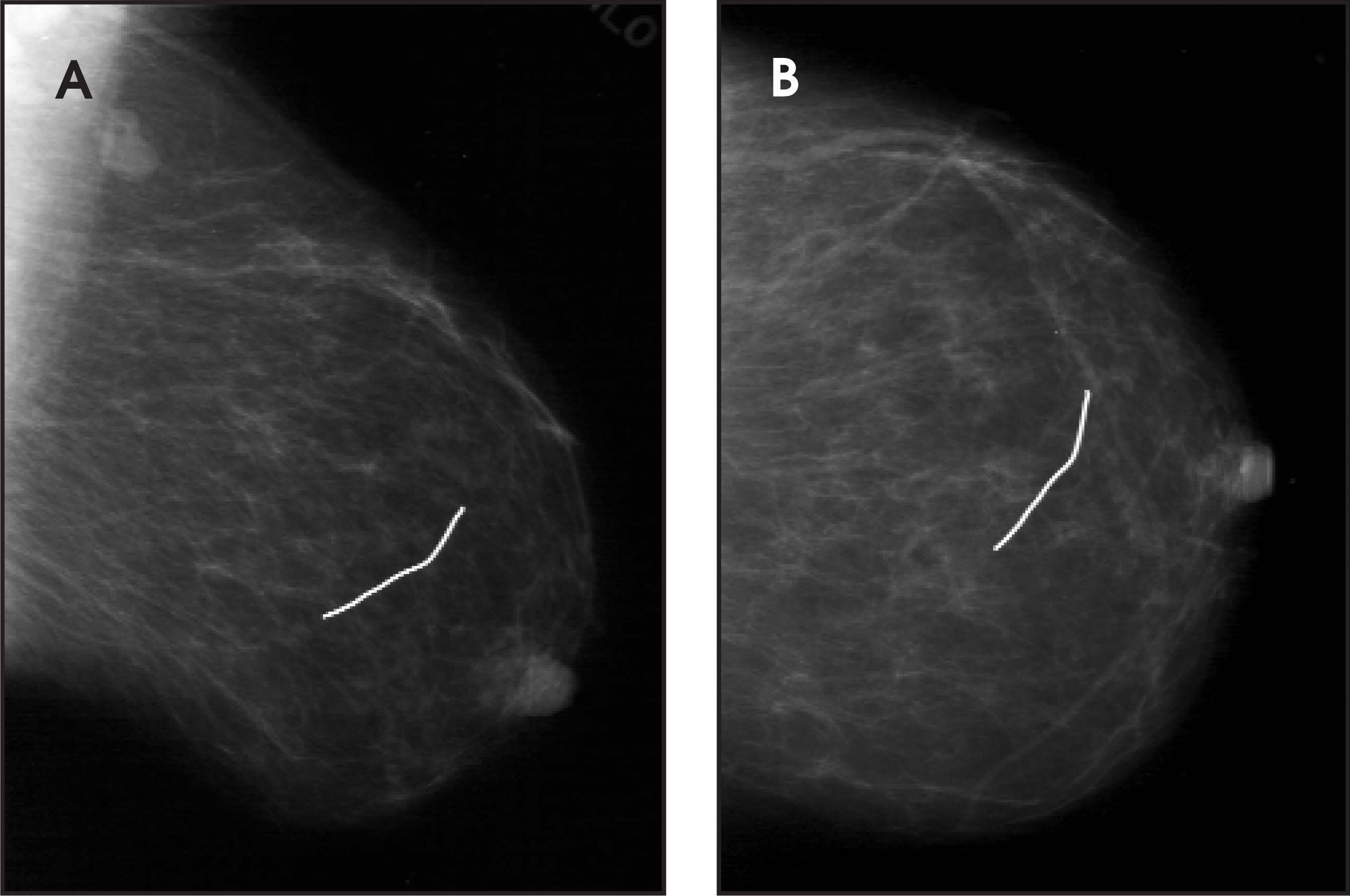
A 61-year-old woman presented to our mammography department without symptoms for a routine screening examination. We discovered a 3-cm wire in the images of the left breast (Figure 1).
IMAGING FINDINGS
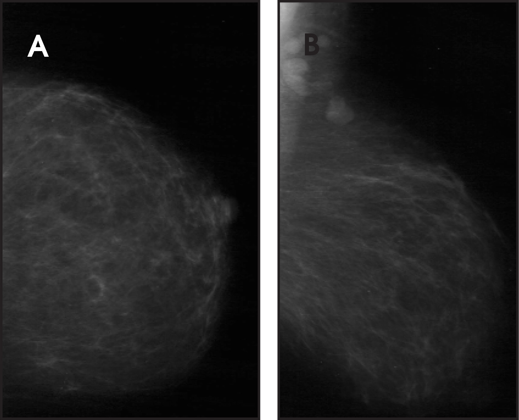
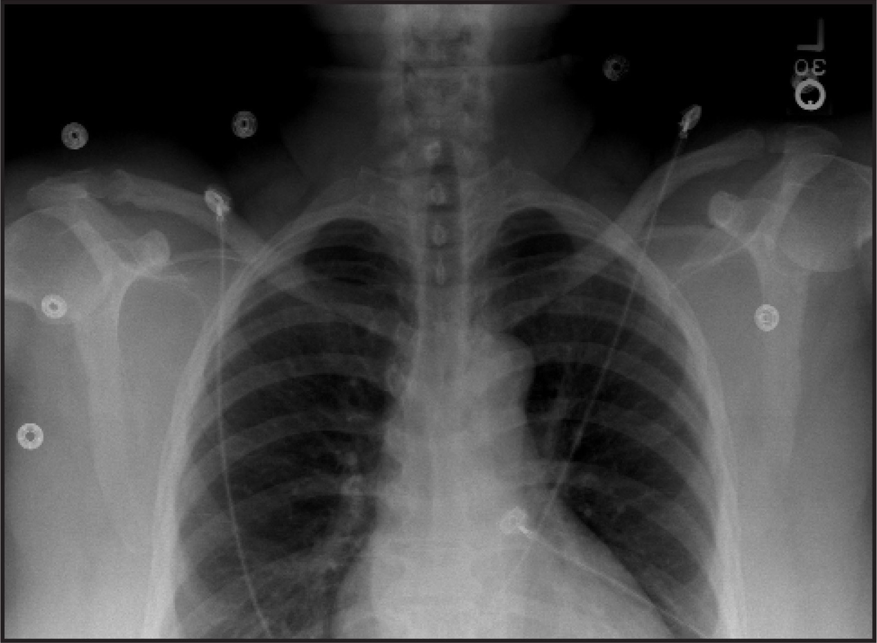
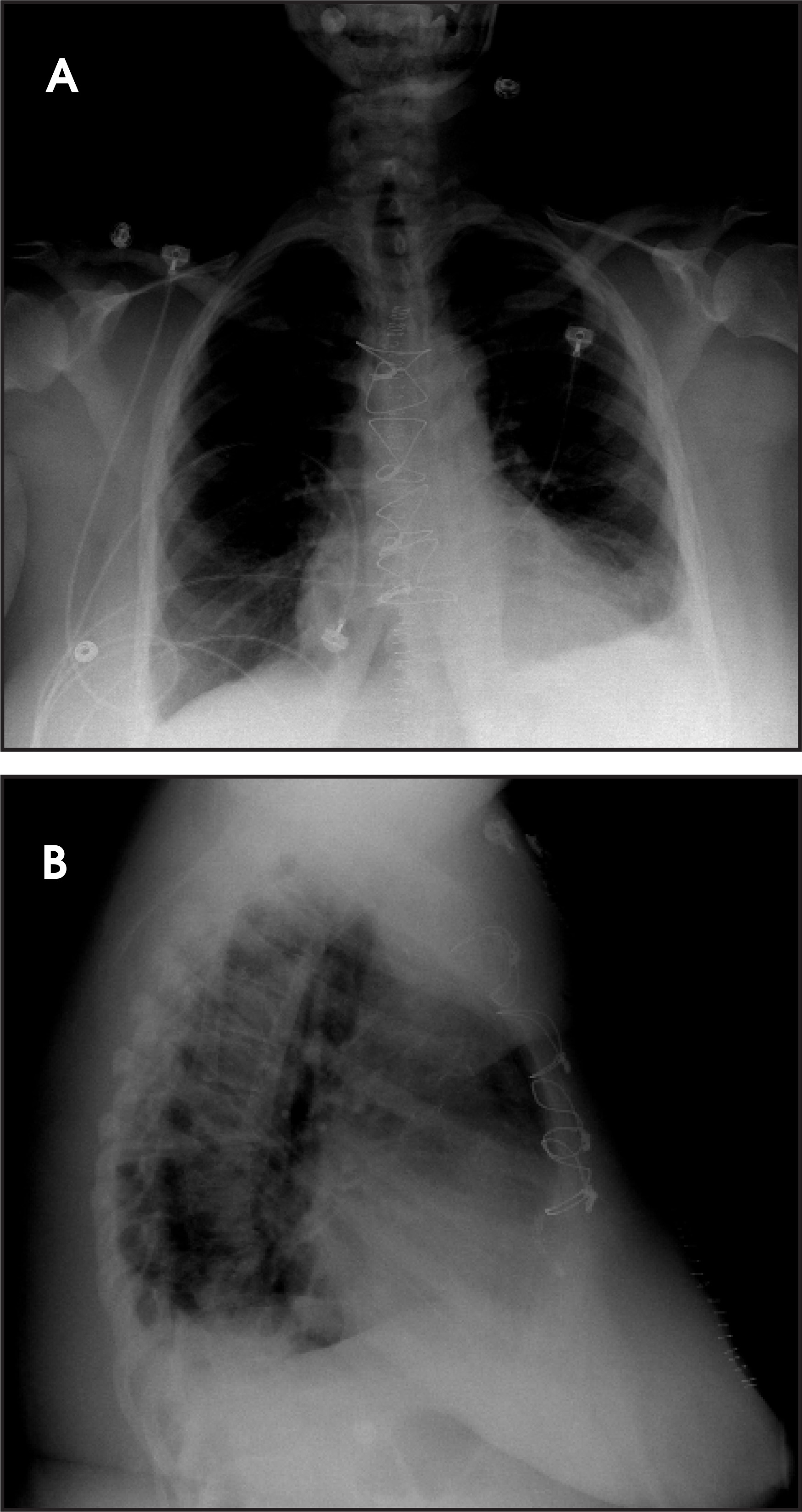
No wire markers were described on the technician’s standard mammographic questionnaire. On questioning the technician, the absence of wire markers was confirmed. The technician did, however, report a long mid-line scar over the sternum. The patient was then contacted to ask about any history of chest trauma and/or surgeries. The patient denied any history of injury to her chest. She also denied any history of left breast pain. She did report a relatively recent coronary artery bypass graft surgery after which she recovered with minimal complications. On further evaluation of the left craniocaucal and left mediolateral oblique images, it was determined that the wire was in the central middle third of the left breast. No associated architectural distortion or masses were noted. The patient’s prior chest X-rays, mammograms, and chest computed tomograms were then reviewed. A 2001 screening mammogram (Figure 2) and a preoperative chest X-ray (Figure 3) revealed no such metallic density in the region of the left breast. Postoperative chest X-rays (Figure 4) showed 4 unremarkable sternal wires and a small left pleural effusion.
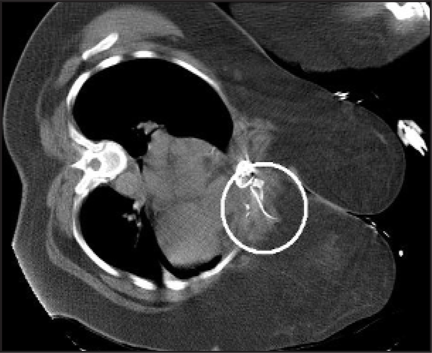
Seven months later, the patient’s effusion had increased and a CT-guided percutaneous drainage was required. On the images from this procedure, the inferior-most wire was noted to be fractured posteriorly (Figure 5). A fragment was identified that extended antero-laterally toward the left breast.
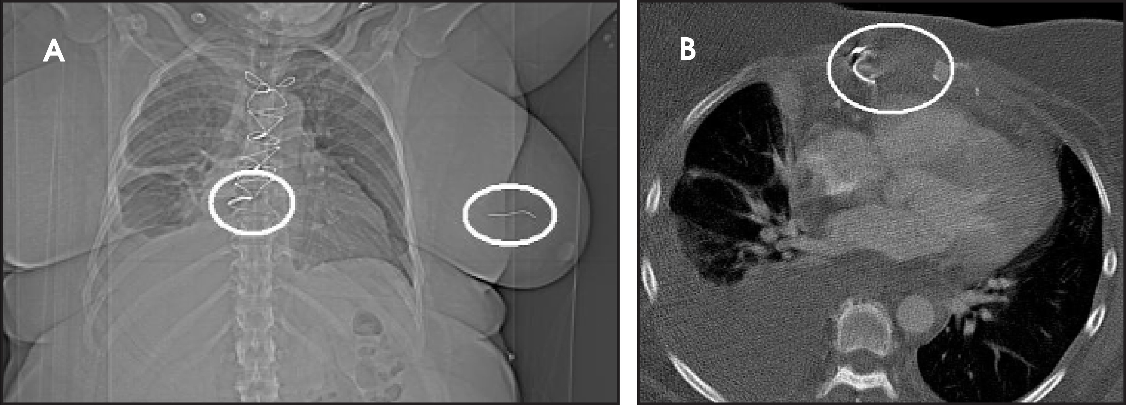
Approximately 1 month later, a follow-up CT examination of the chest was performed (Figure 6). On the scout image (Figure 6A), the wire could be seen in the left breast. However, this was not noted on the initial examination, as it was not in the field of view of the axial images.
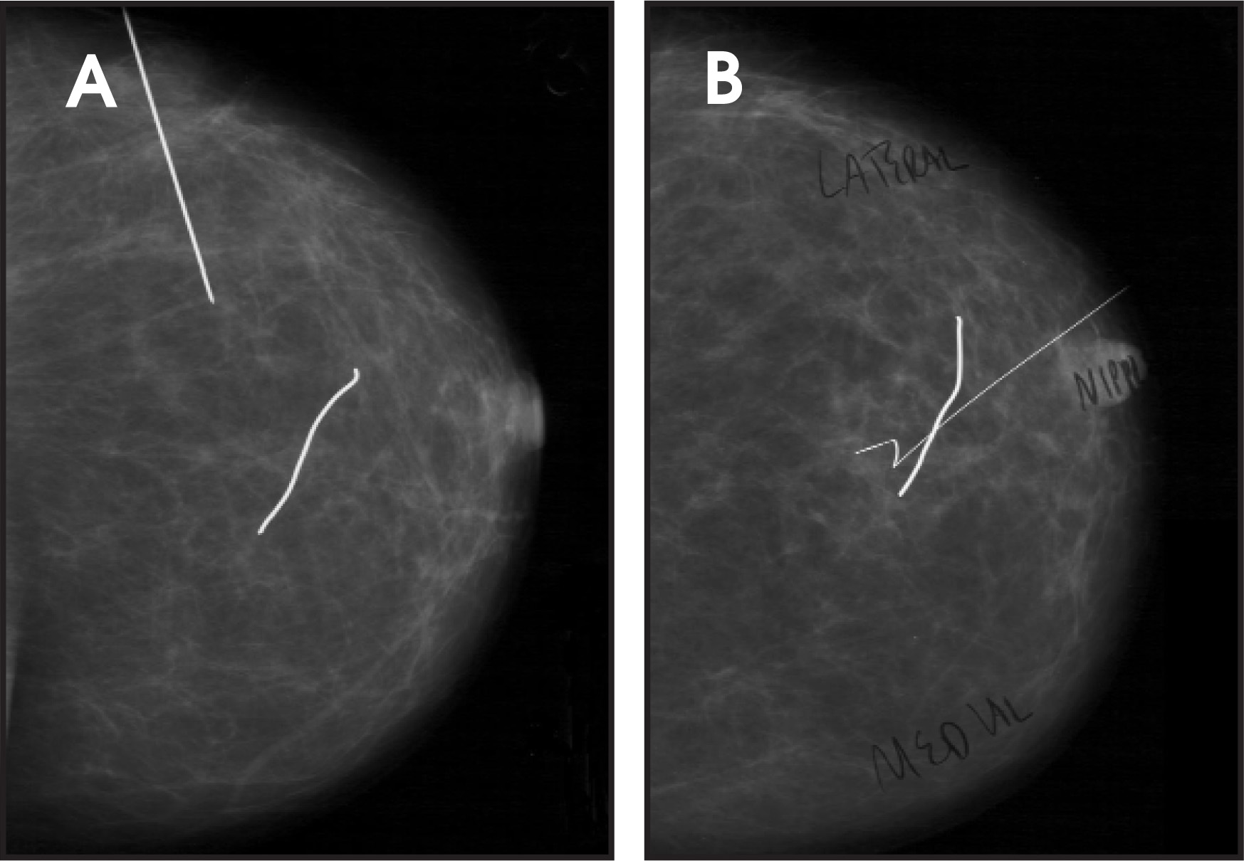
At this point, the cardiothoracic surgery service was contacted for further evaluation of the patient. The cardiothoracic surgeon reviewed the case and sent the patient to the breast surgery clinic. The breast surgeons recontacted our department to set up needle localization for excision of the wire. Six months after the follow-up CT study, we localized the wire using a standard technique. At surgery, a 3.2-cm wire was removed without incident (Figure 7).
DIAGNOSIS
Ectopic position of a fractured sternal wire fragment
DISCUSSION
In recent medical literature, there have been many diverse cases of migration of fractured sternal and other surgical wires in the chest.1,2 These were usually described as traversing an internally directed course, such as those that eventually impinged on and/or penetrated various cardiac chambers,3 great vessels,4 or the pleural cavity.5 The case reported here appears unique in this respect.
CONCLUSION
In this case, the fractured sternal wire was migrating peripherally into the breast, where it was removed without complication. However, a unidirectional migration path does not encompass all wayward wires. Thus, removal at the time of detection appears most prudent.
References
- Kopans D. Breast Imaging. 1998:369.
- Cardenosa G. The Core Curriculum: Breast Imaging. 2004:352.
- Schreffler A, Rumisek J. Intravascular migration of fractured sternal wire presenting with hemoptysis. Ann Thorac Surg. 2001;71:1682-1684.
- Kao C, Chang J. Aortic graft pseudoaneurysm secondary to fracture of sternal wires. Tex Heart Inst J. 2003;30:240-242.
- Radich G, Altinok D, Silva J. Marked migration of sternotomy wires: A case report. J Thoracic Imaging. 2004;19:117-119.
Citation
. RCOM RADIOLOGICAL CASE OF THE MONTH. Applied Radiology. 2007;36(9):34-36. doi:10.37549/AR1543.