RCOM RADIOLOGICAL CASE OF THE MONTH
Applied Radiology — Vol. 36 , Issue 9 , pp. 37 -39
DOI: 10.37549/AR1548
Published: September 1, 2007
Categories
CASE SUMMARY
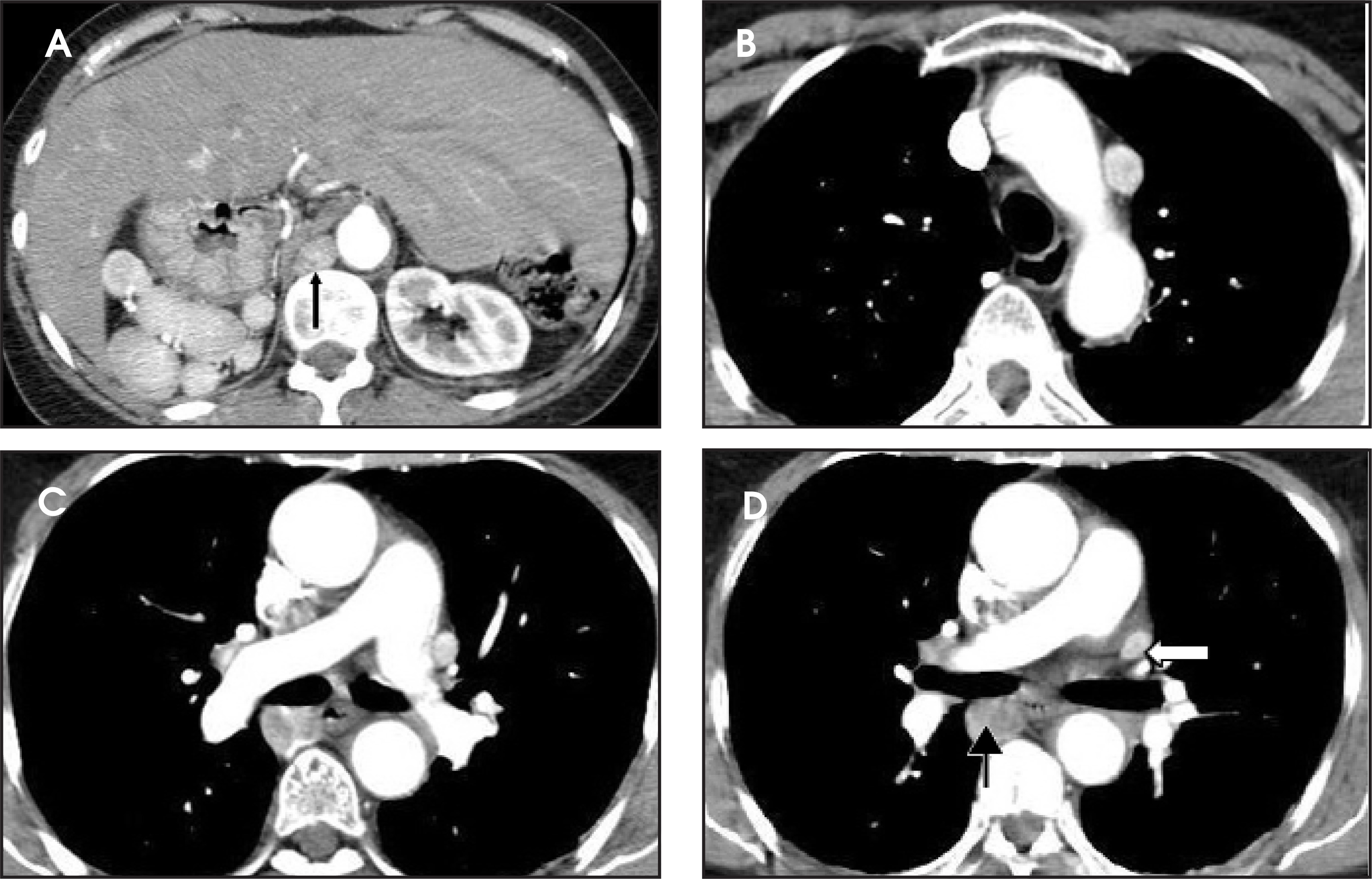
A 62-year-old white woman with chronic atrial fibrillation and severe aortic regurgitation underwent elective transcatheter radiofrequency ablation. The patient experienced postprocedure chest pain, and contrast-enhanced computed tomography (CT) scan of the chest was performed utilizing the protocol for the detection of pulmonary emboli (Figure 1). A physical examination revealed atrial fibrillation, a diastolic murmur in the aortic valve region, and a benign abdomen. Laboratory values were unremarkable.
IMAGING FINDINGS
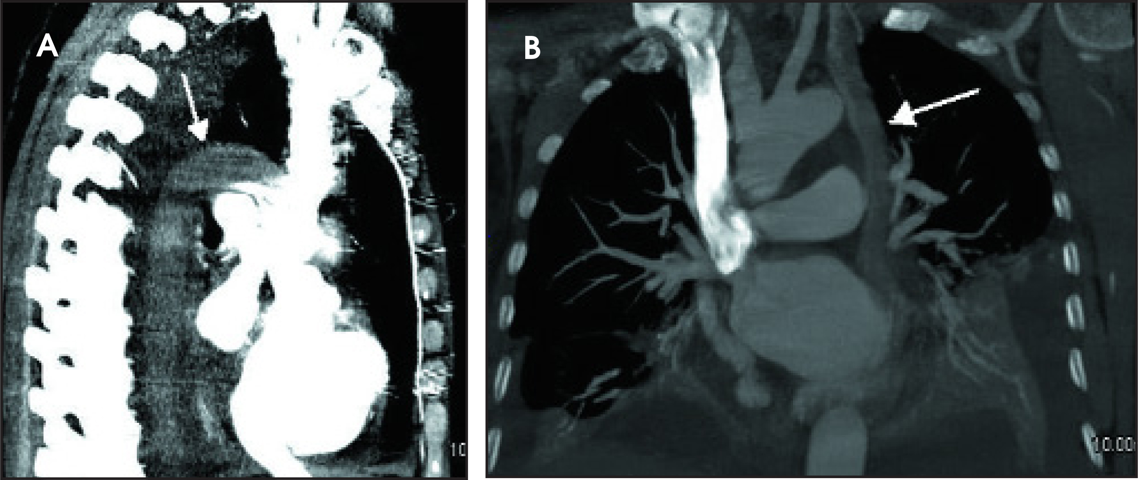
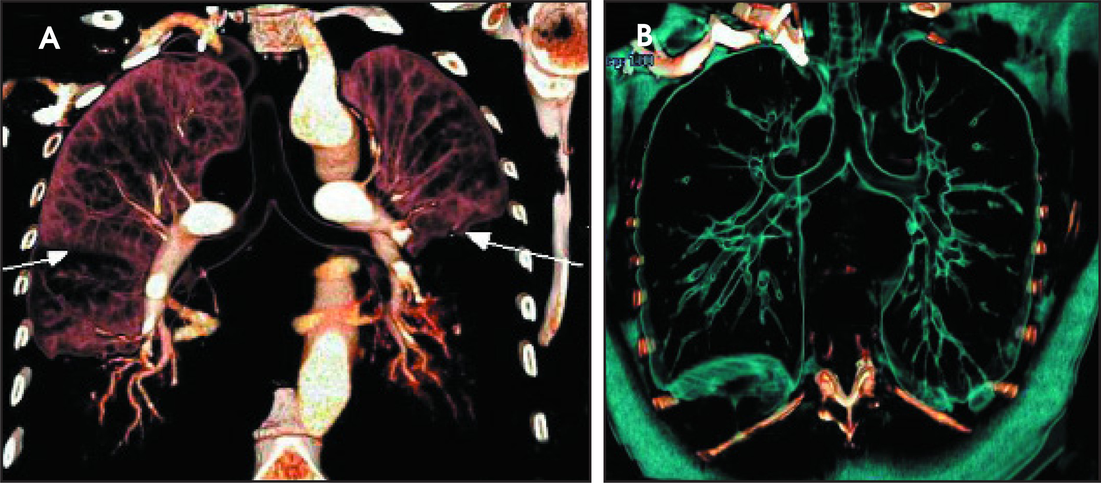
Multiple visceral anomalies were incidentally discovered on the CT of the chest and the upper abdomen, which was performed for chest pain. There was a midline liver, right-sided stomach, polysplenia in the right upper quadrant posterior to the stomach, and inferior vena cava (IVC) interruption with azygos continuation (Figures 1 and 2A). Each spleen ranged in size from 2 to 3 cm. No spleen was noted in the left upper quadrant. A dilated azygos vein drained into the superior vena cava (SVC). There was a persistent left SVC (Figures 1B, 1C, and 2B) that emptied into the coronary sinus (Figure 2B). Both main pulmonary trunks coursed over the main stem bronchi (Figures 1C, 1D, and 3A), representing the characteristic bilateral hyparterial bronchial pattern seen in this syndrome. Both lungs were bilobed. Each main stem bronchus branched in a left-sided pattern into an upper- and lower-lobe bronchus (Figure 3B).
DIAGNOSIS
Heterotaxy syndrome with polysplenia
DISCUSSION
Heterotaxia, or situs ambiguous, indicates visceral dysmorphism. This is an abnormal arrangement of organs and vessels in the chest and abdomen.1 The 2 main subtypes of situs ambiguous are situs ambiguous with polysplenia and situs ambiguous with asplenia.2
Heterotaxia with polysplenia, also known as left isomerism or bilateral left-sidedness, is characterized by a midline or ambiguous location of the majority of chest and abdominal viscera, multiple spleens, and an absence of some right-sided structures, such as the IVC.2 Although there is great variability in the spectrum of abnormalities and no single anomaly that is pathognomonic for this disorder, IVC interruption with azygos continuation is the most common finding.1 The syndrome is characterized by bilateral bilobed left-sided lungs and hyparterial bronchial pattern, intrahepatic IVC interruption with continuation of the azygos vein, a centrally located transverse liver, a stomach in an indeterminate position, and multiple spleens, all of which were present in our patient. The spleens vary in size and number, but are always on the same side as the stomach.1 While 99% to 100% of the patients with situs ambiguous and asplenia have congenital heart diseases, cardiac anomalies are less common in polysplenic patients (50% to 90%).3 They are also often not as complex, which explains their survival into adulthood.1,2 In our patient, the only cardiac abnormalities were a persistent left SVC draining into the coronary sinus, a persistent foramen ovale, and a bicuspid aortic valve (not shown).
Other gastrointestinal abnormalities associated with polysplenia include biliary atresia, intestinal malrotation, and truncated pancreas (only the pancreatic head or body is present). Neither abdominal heterotaxy nor multiple spleens need not always be present. The stomach and cardiac apex are discordant in roughly 50% of cases.2 The clinical presentation, morbidity, and mortality are usually related to the presence and severity of cardiac defects, biliary atresia, or bowel malrotation.
Although heterotaxy and polysplenia in most adult patients is asymptomatic and is detected incidentally, the role of radiologists in recognizing the complex anatomy is crucial in the clinical setting. The presentation of an acute abdomen in these patients can be confusing because the pain from acute cholecystitis, appendicitis, or other pathology is often not at the usual location.2 Similarly, the imaging appearance can be perplexing if the radiologist is not aware of the spectrum of findings associated with these syndromes.
CONCLUSION
Heterotaxia or situs ambiguous with polysplenia is a rare spectrum of congenital abnormalities involving the visceral organs and cardiovascular structures in the chest and abdomen. The knowledge and recognition of the complex and often confusing anatomy of these patients is important in diagnosing pathology and planning appropriate surgical, diagnostic, and interventional procedures.
References
- Applegate K, Goske M, Pierce G, Murphy D. Situs revisited: Imaging of the heterotaxy syndrome. RadioGraphics. 1999;19.
- Fulcher A, Turner M. Abdominal manifestations of situs anomalies in adults. RadioGraphics. 2002;22:1439-1456.
- Tonkin I, Friedman W, Higgins C. Pediatric Cardiac Imaging. 1983:157-187.
Citation
. RCOM RADIOLOGICAL CASE OF THE MONTH. Applied Radiology. 2007;36(9):37-39. doi:10.37549/AR1548.