RCOM RADIOLOGICAL CASE OF THE MONTH
Applied Radiology — Vol. 36 , Issue 10 , pp. 40 -41
DOI: 10.37549/AR1553
Published: October 1, 2007
Categories
CASE SUMMARY
The patient was a 52-year-old African-American man with a history of insulin-dependent diabetes mellitus, chronic pancreatitis, and a Whipple procedure several years previously. He presented with complaints of 2 weeks of vague chills, a body temperature of 102°F, several days of black stools, a hemoglobin level of 6.8 g/dL, and weight loss.
IMAGING FINDINGS
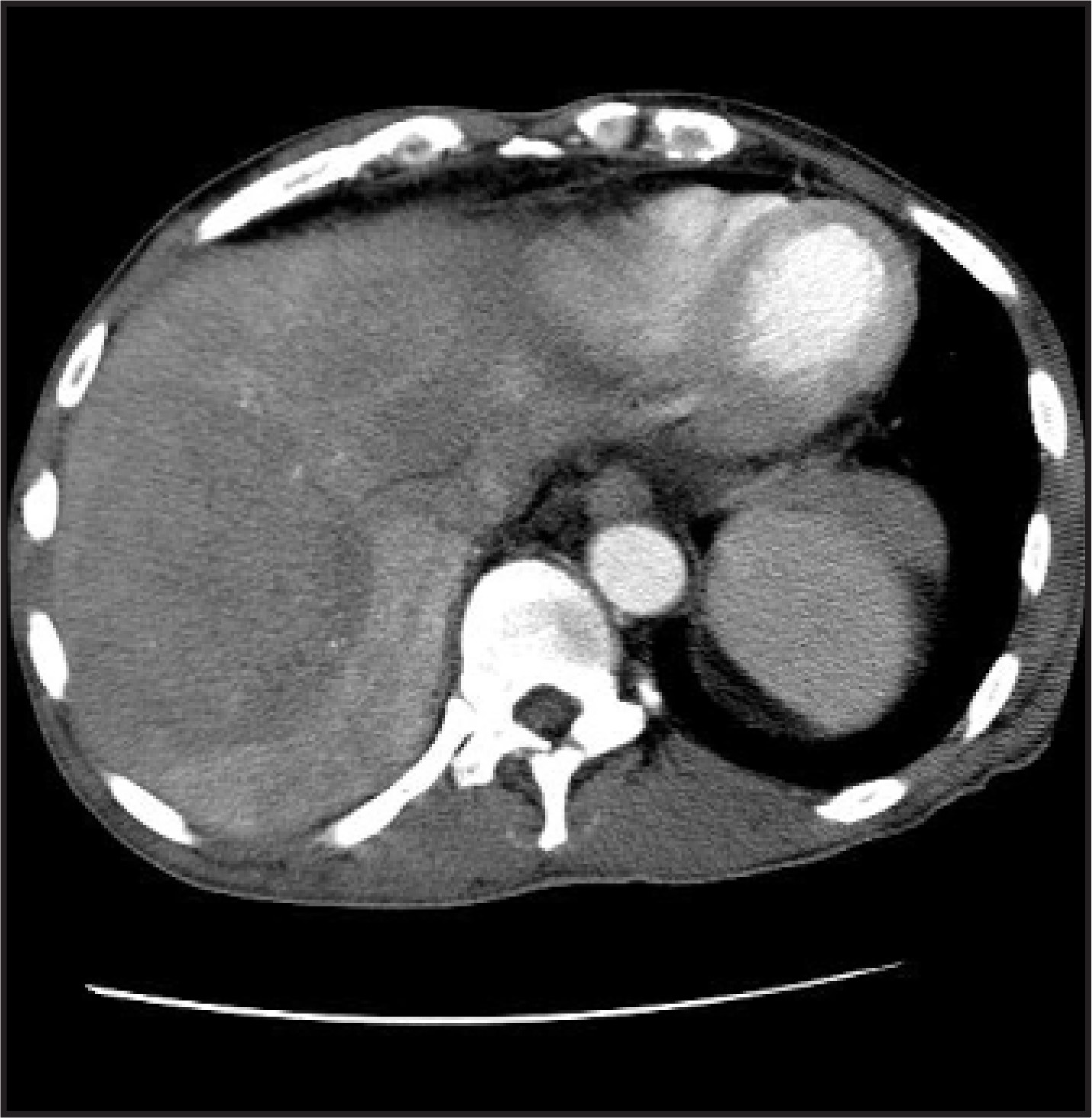
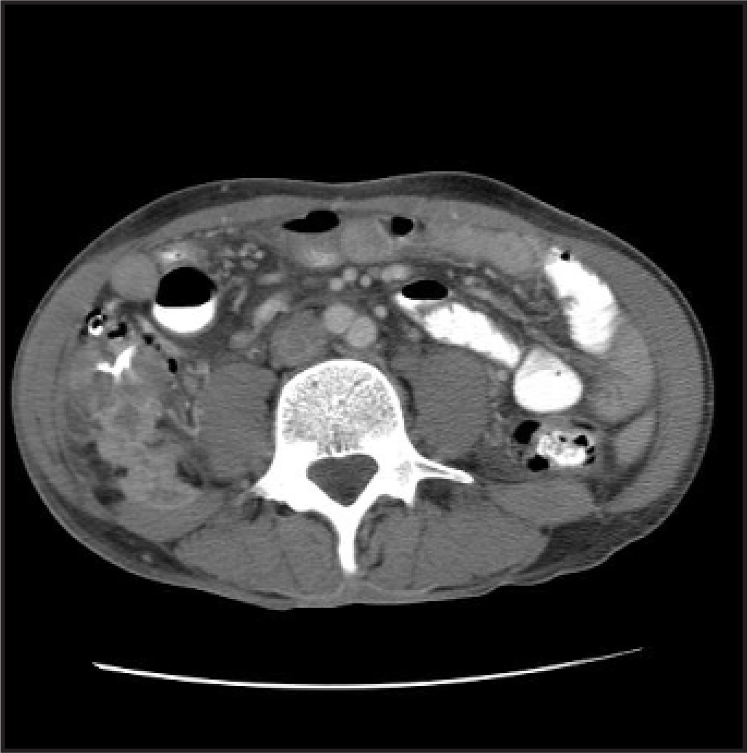
An abdominal computed tomography (CT) scan was obtained, which revealed large hepatic masses (Figure 1) with extension to the surrounding soft tissues (Figure 2). This finding prompted a CT-guided biopsy of the largest lesion. The pathologic examination determined the diagnosis of hepatic actinomycosis.
DIAGNOSIS
Hepatic actinomycosis
CASE FOLLOW-UP
Based on the pathologic results, intravenous treatment was instituted. Unfortunately, the patient died from his disease during his hospital stay.
DISCUSSION
Actinomycosis is a chronic suppurative bacterial infection that is characterized by a granulomatous inflammatory reaction, which results in the formation of multiple abscesses and sinus tracts. It is caused by a non–acid-fast, non–spore-forming Gram-positive filamentous bacteria. The bacteria are slow growing in anaerobic-to-microaerophilic conditions.
An abdominal infection may result from intestinal perforation, most often a ruptured appendix, diverticulitis, foreign body, peptic ulcer, or gastrectomy. Appendicitis, especially with perforation, has been reported to be the most common culprit.1 Abdominal and pelvic actinomycosis accounts for approximately 10% to 20% of reported cases.
Hepatic involvement is rare, occurring in approximately 5% of all patients with actinomycosis and in roughly 15% of abdominal actinomycosis cases. Affected patients are typically immunocompetent. A history of prior surgical procedures or manipulations is often uncovered2,3; these allow the organism to be introduced into the deep tissues. Classically, it will present as one or more slow-growing hepatic masses, and their appearance is often confused with a hepatic neoplasm. However, direct extension to adjacent organs and the abdominal wall can ensue, with the possibility of draining sinuses formation. Its presence should suggest the diagnosis to the radiologist.
Presenting symptoms may include fever, abdominal pain, anorexia, and weight loss. Physical examination may reveal fever, abdominal tenderness, and hepatomegaly. Chronic anemia, leukocytosis with a left shift, elevated serum erythrocyte sedimentation rate, and an elevated level of alkaline phosphatase are often present on laboratory analysis. CT is the preferred imaging modality. The most common radiographic appearance encountered is a single low-attenuation mass or multiple low-attenuation masses (abscesses), which are found in approximately 68% of cases. These “masses” often exhibit enhancement after contrast administration. As was the case with our patient (Figure 2), extension to adjacent soft tissues is found in approximately 33.3% of reported cases.2 The presence of lymphadenopathy is uncommon.
In most cases, tissue biopsy with isolation of the organism from a clinical specimen or from sulfur granules is necessary for a conclusive diagnosis. Antibiotic treatment suffices for most patients, although surgery can be an adjunct treatment in selected cases.4,5 High-dose penicillin G is most commonly used.
Even with appropriate treatment, hepatic actinomycosis has been reported to recur after several years. Therefore, careful follow-up is warranted in such patients.6
CONCLUSION
Hepatic actinomycosis is a rare condition. Clinical signs are often nonspecific, and imaging findings can be misleading, often resulting in a delay in diagnosis and proper treatment. However, knowledge of the entity and its imaging appearance should alert the radiologist to the diagnosis.
References
- Deshmukh N, Heaney S. Actinomycosis at multiple colonic sites. Am J Gastroenterol. 1986;81:1212-1214.
- Sharma M, Briski L, Khatib R. Hepatic actinomycosis: An overview of salient features and outcome of therapy. Scand J Infect Dis. 2002;34:386-391.
- Vargas C, González C, Pagani W. Hepatic actinomycosis presenting as liver mass: Case report and review of the literature. PR Health Sci J. 1992;11(1):19-21.
- Goldberg N, Anthony W, Yen M, Hashmi S. Hepatic actinomycosis. Md Med J. 1989;38:751-752.
- Meade R. Primary hepatic actinomycosis. Gastroenterology. 1980;78:355-359.
- Tambay R, Côté J, Bourgault A, Villeneuve J. An unusual case of hepatic abscess. Can J Gastroent. 2001;15:615-617.
Citation
. RCOM RADIOLOGICAL CASE OF THE MONTH. Applied Radiology. 2007;36(10):40-41. doi:10.37549/AR1553.