RCOM RADIOLOGICAL CASE OF THE MONTH
Applied Radiology — Vol. 36 , Issue 10 , pp. 45 -47
DOI: 10.37549/AR1558
Published: October 1, 2007
Categories
CASE SUMMARY
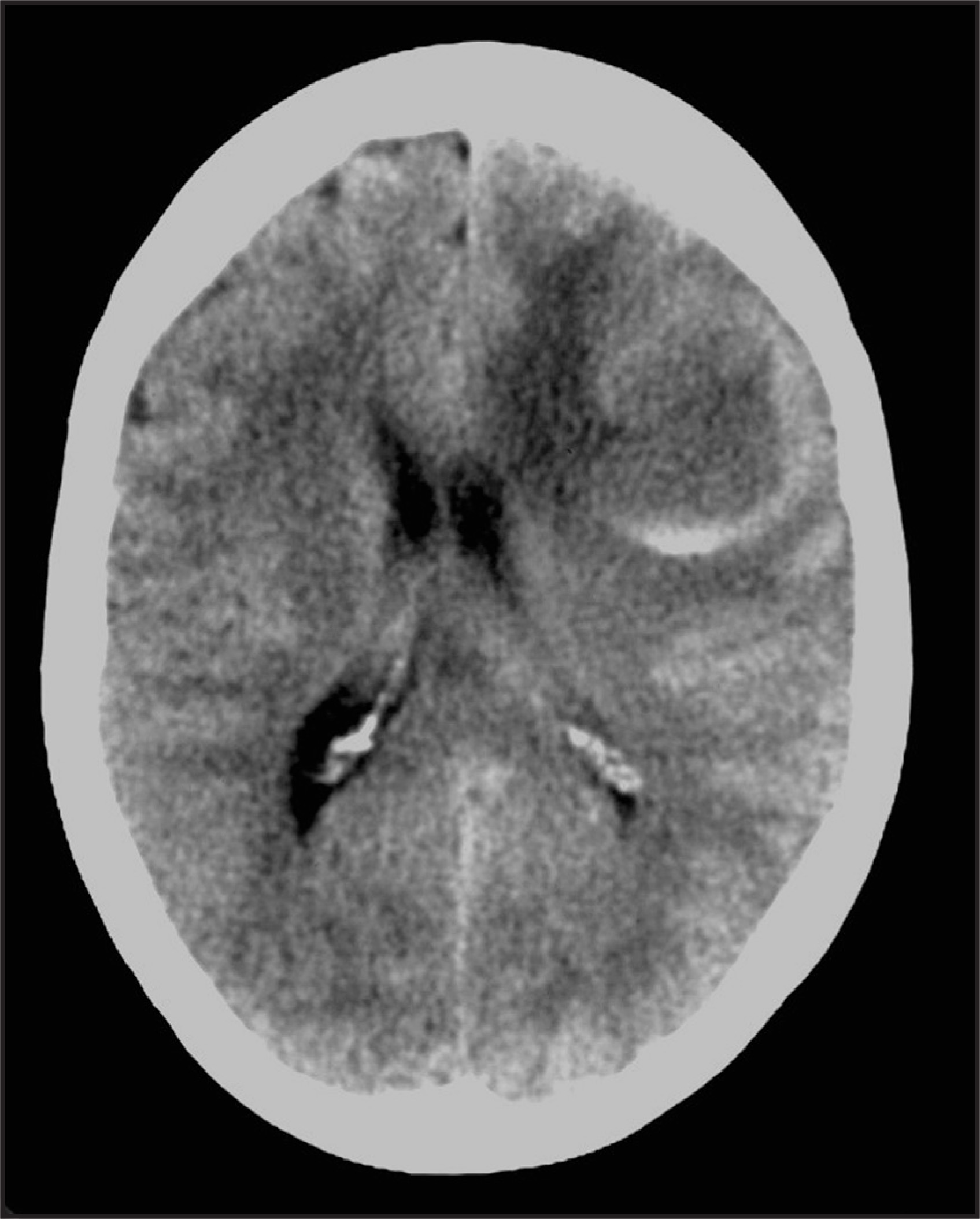
A 62-year-old woman experienced symptoms of intermittent forgetfulness and aphasia that had worsened over a 2-week period. Her medical history was significant for smoking 2 packs of cigarettes per day for many years and for a right hemicolectomy as a result of long-standing Crohn’s disease. A noncontrast computed tomography (CT) of the brain was performed (Figure 1).
IMAGING FINDINGS
Noncontrast CT of the brain revealed what appeared to be an intra-axial mass of predominantly low attenuation, in the left frontal lobe (Figure 1). Layering debris levels were present. Vasogenic edema was evident, as was mass effect with compression of the left lateral ventricle and small subfalcine herniation. The overlying calvarium was normal, with no evidence of hyperostosis or erosion.
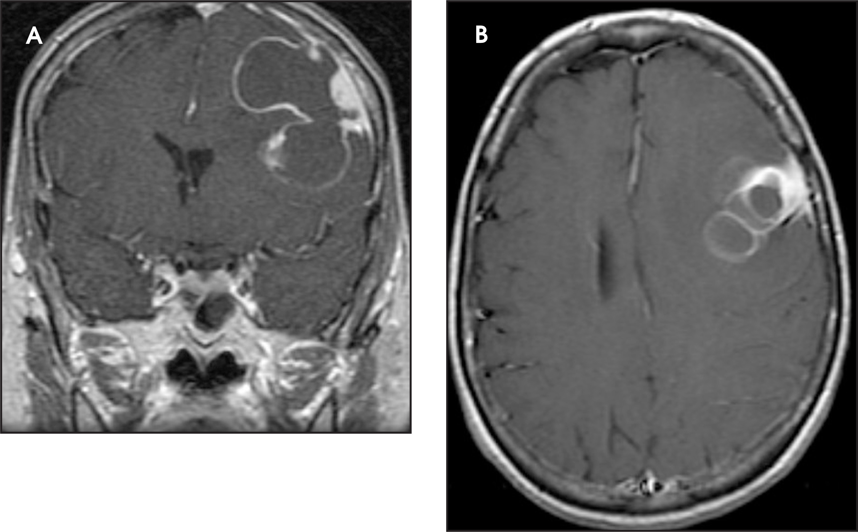
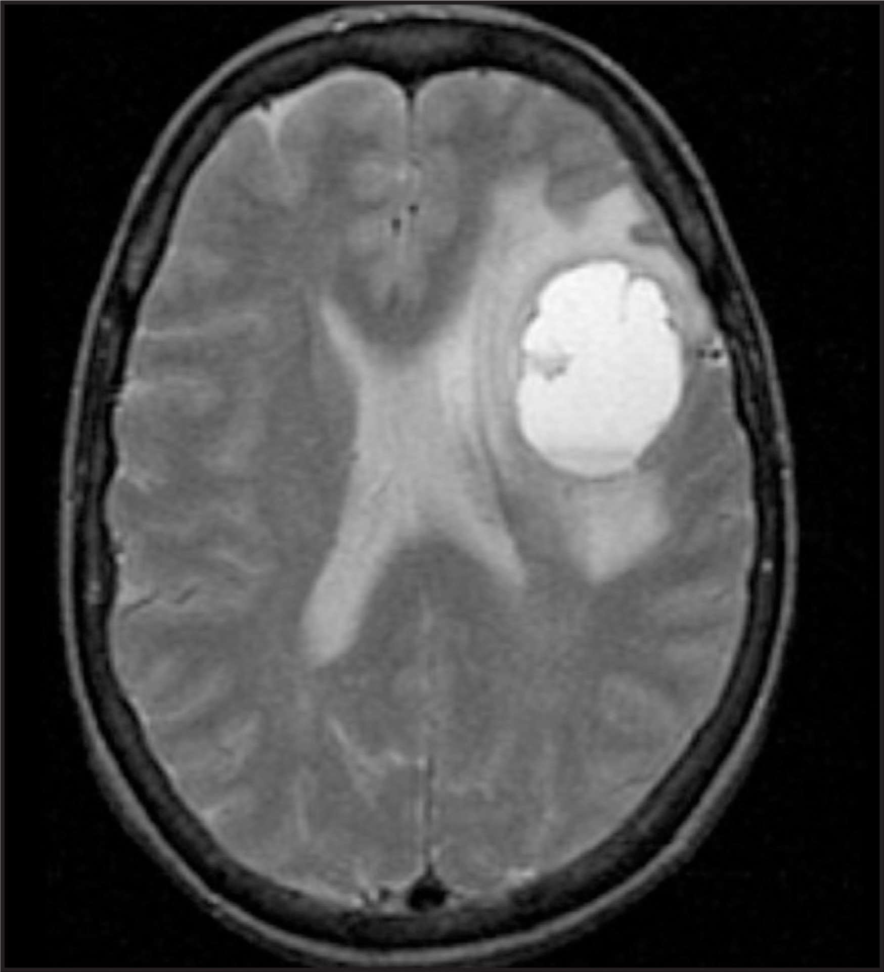
Based upon these findings, magnetic resonance (MR) imaging was performed and revealed a multilobular, multiseptate cystic mass with an enhancing mural nodule and enhancement of the cyst wall. A minimal dural tail was seen (Figure 2). Layering debris was confirmed. Vasogenic edema and mass effect were again present. (Figure 3). Several large flow voids were seen at the periphery of the mass. Chief diagnostic considerations were a primary central nervous system neoplasm such as astrocytoma, hemangioblastoma, or ganglioglioma, rather than metastatic disease.
CT of the chest, abdomen, and pelvis and a bone scan were all negative (not shown).
The diagnosis of typical benign meningioma (nodule and cyst wall) was established at surgery. The cyst contained xanthochromic fluid.
DIAGNOSIS
Cystic meningioma
DISCUSSION
This case provides an opportunity to review both the common and unusual imaging features of meningiomas. The typical meningioma will be obviously extra-axial, buckling brain cortex. It is unilobular, dense on noncontrast CT, and nearly isointense to the brain on T1- and T2-weighted MR images. Contrast enhancement is homogeneous.1
Some findings, such as vasogenic edema and dural attachment, are common but nonspecific.2 The etiology of vasogenic edema associated with meningioma is uncertain and debated. Gross hemorrhage is uncommon.3 Meningiomas are usually described as solid tumors. Large or multilobular cysts are uncommon, as is ring enhancement. Cyst formation may be the result of hemorrhage or necrosis, fluid secretion from the meningioma, or loculation of cerebrospinal fluid (CSF). Intratumoral and peritumoral cysts have been described. The intratumoral cysts are presumably formed from hemorrhage, necrosis, or fluid secretion within the tumor. Peritumoral cysts are thought by some to be formed by CSF entrapment as in an arachnoid cyst. Ring enhancement suggests an intratumoral cyst.1 The atypical features can easily lead to confusion with glial neoplasms, metastases, or even abscesses.
CONCLUSION
Meningiomas are common intracranial neoplasms. Atypical features have been described and radiologic diagnosis requires familiarity with these features. When confronted with any lesion, one should consider the uncommon presentation of a common lesion, including the possibility that several atypical features are present within the same lesion. When this occurs, even an experienced imager could be misled.
References
- Buetow M, Buetow P, Smirniotopoulos J. Typical, atypical and misleading features in meningioma. RadioGraphics. 1991;11:1087-1106.
- Wasenko J, Hochhauser L, Stopa E, Winfield J. Cystic meningiomas: MR characteristics and surgical correlations. AJNR Am J Neuroradiol. 1994;15:1959-1965.
- Osborn A, Tong K. Handbook of Neuroradiology: Brain and Skull. 1996:289-301.
Citation
. RCOM RADIOLOGICAL CASE OF THE MONTH. Applied Radiology. 2007;36(10):45-47. doi:10.37549/AR1558.