RCOM RADIOLOGICAL CASE OF THE MONTH
Applied Radiology — Vol. 36 , Issue 11 , pp. 66 -70
DOI: 10.37549/AR1564
Published: November 1, 2007
Categories
CASE SUMMARY
A 43-year-old woman presented to an outside hospital with a 10-month history of progressive headaches, nausea, and vomiting. On physical examination, mild bilateral nystagmus on lateral gaze and rotatory nystagmus were present. Mild ataxia was present.
Magnetic resonance imaging (MRI) of the brain obtained at the outside hospital reported a large posterior fossa tumor within the fourth ventricle as well as obstructive hydrocephalus. The patient underwent a second MRI after being transferred to another hospital for neurosurgical removal of the tumor. The differential considerations for both studies included ependymoma, subependymoma, neurocytoma, giant cell astrocytoma, and metastasis. The patient was scheduled for a craniotomy and surgical excision. In the operating room, the patient was noted to have a large “pulsatile mass,” which was determined to be an aneurysm. The aneurysm was successfully clipped. Neither examination was reviewed by a neuroradiologist until an intraoperative consult was given, at which time a correct diagnosis of giant posterior-inferior cerebellar artery (PICA) aneurysm was made.
IMAGING FINDINGS
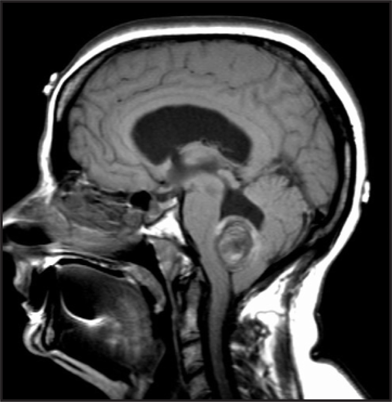
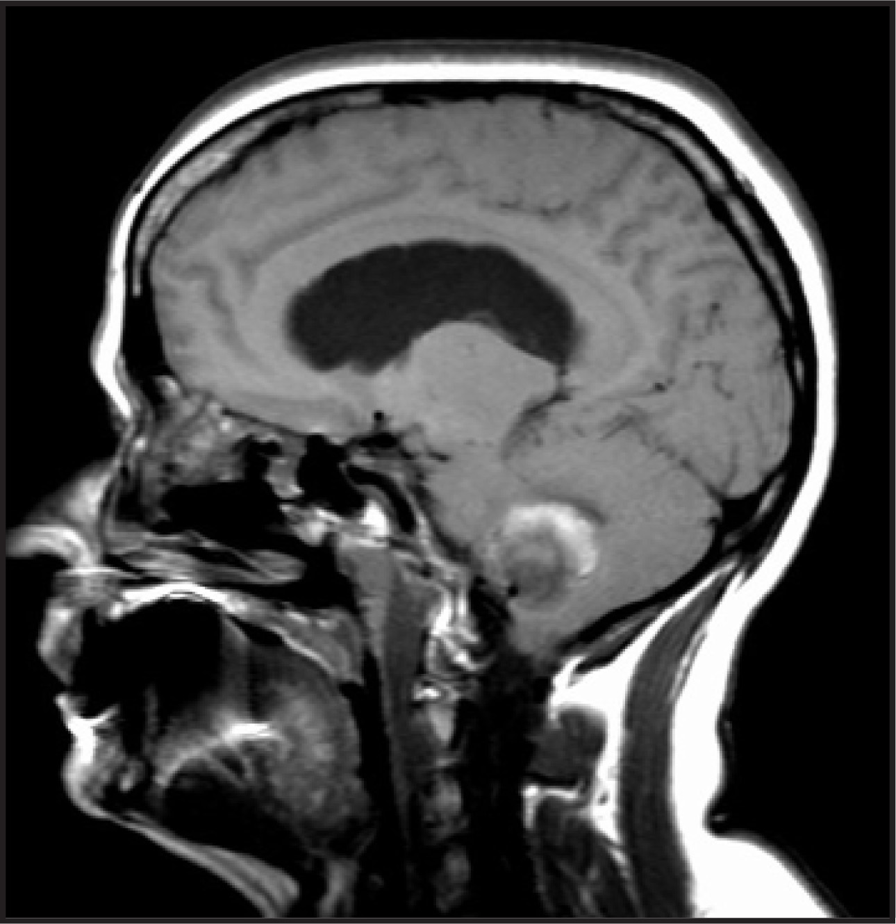
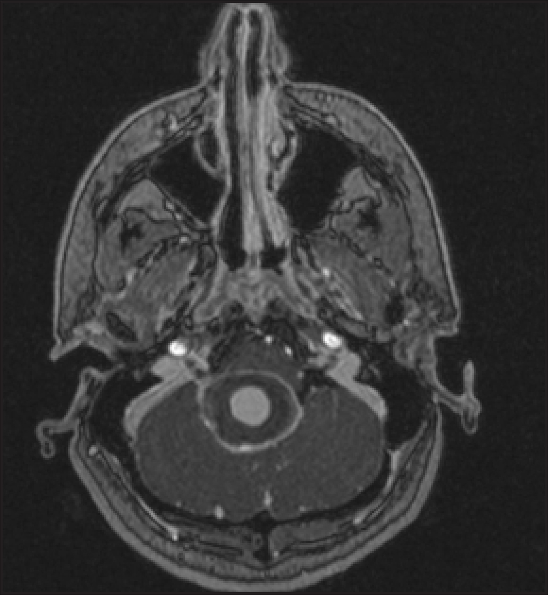
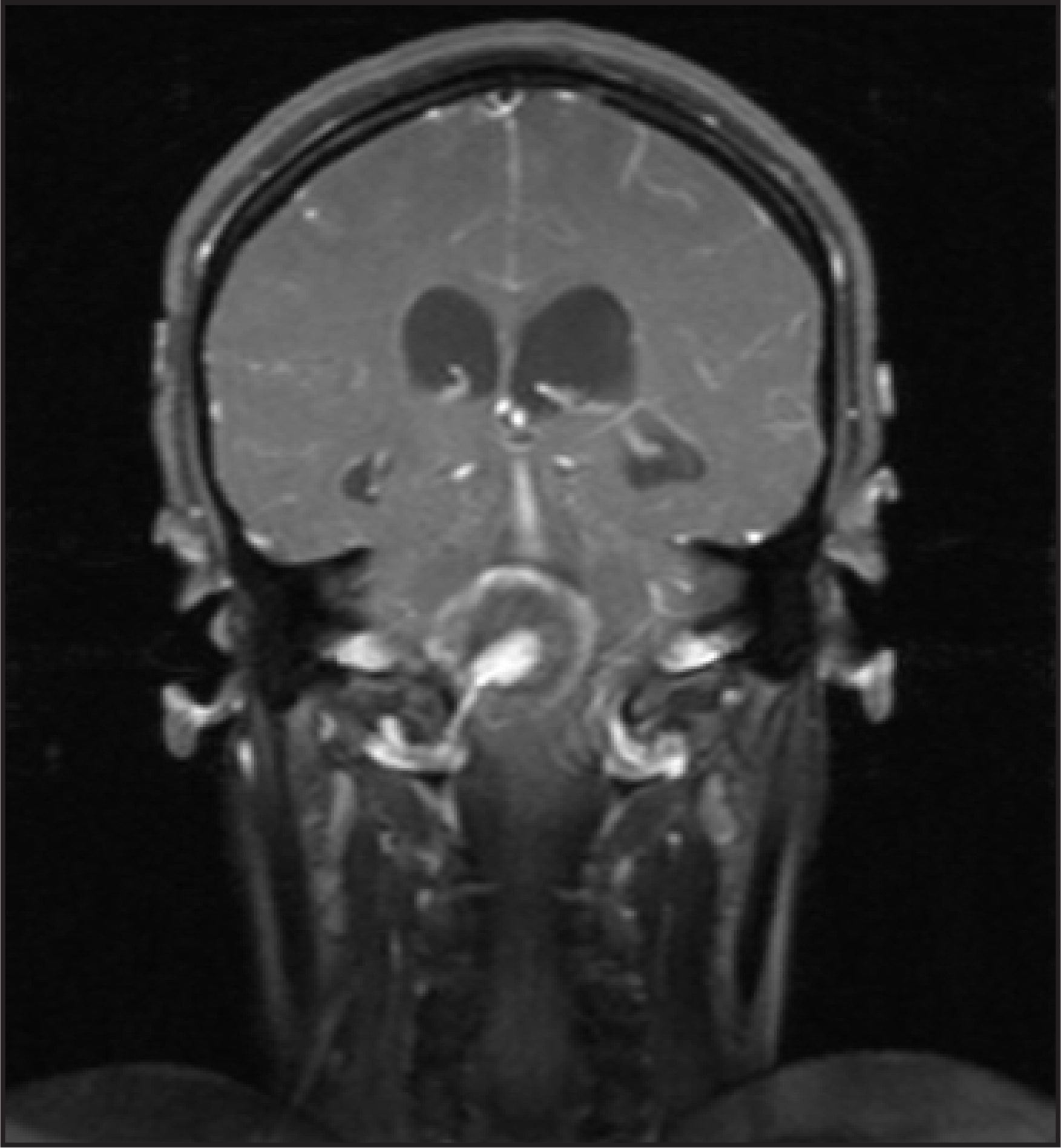
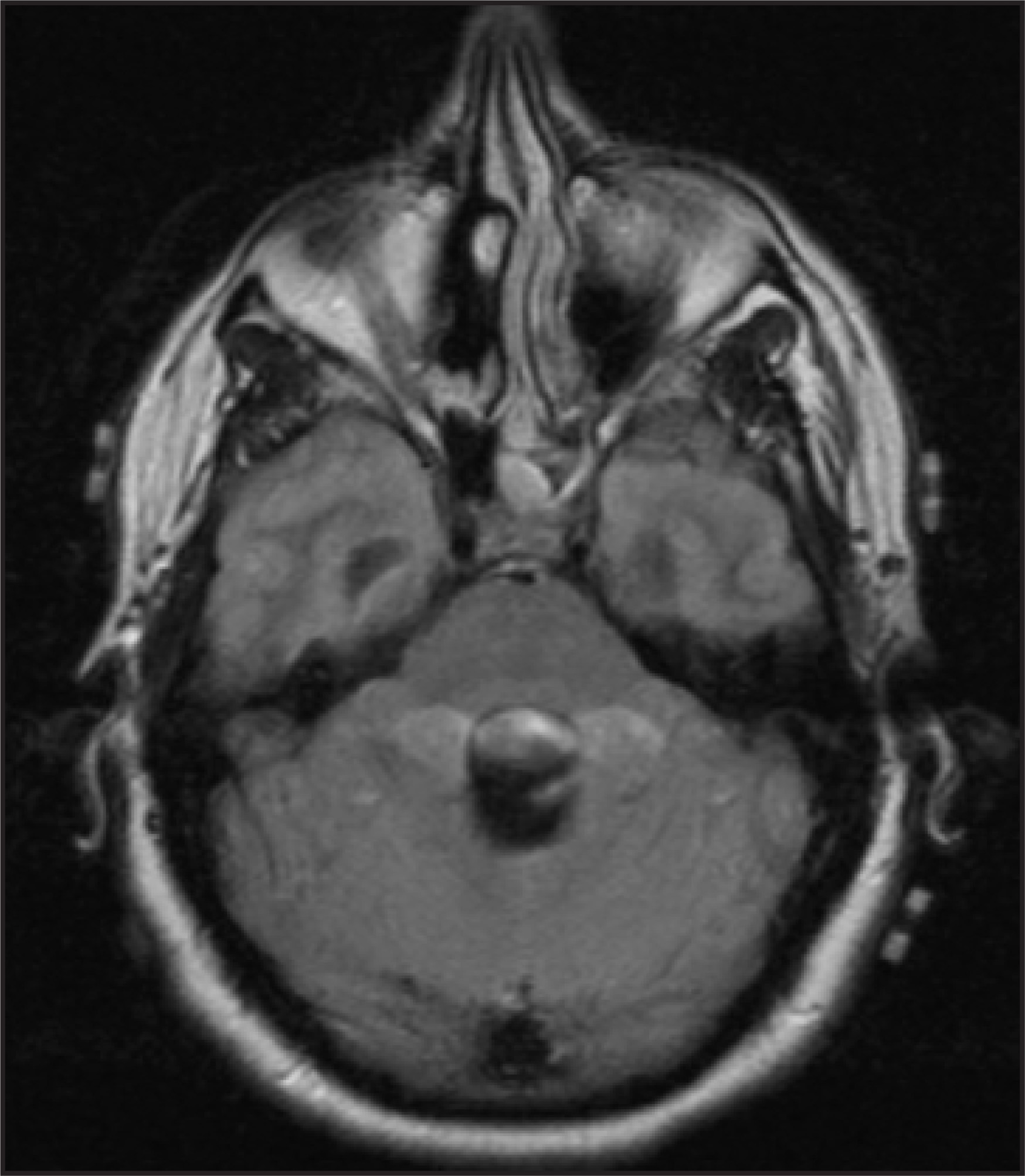
MRI of the brain with and without gadolinium enhancement showed a 3.4 × 2.6 × 3.0-cm lesion situated within the region of the fourth ventricle. It had a concentric multilaminated appearance (Figure 1). A flow void originating from the right PICA was present on midline sagittal T1-weighted images (Figure 2). Gadolinium injection revealed central enhancement of the mass (Figure 3) with enhancement of the PICA that was seen directly feeding into it (Figure 4). Pulsation ghosting artifact was also present (Figure 5).
DIAGNOSIS
Giant PICA aneurysm
DISCUSSION
An aneurysm that measures >2.5 cm in diameter is termed a giant intracranial aneurysm.1-4 They reportedly represent approximately 2.5% to 13% of all intracranial aneurysms1,3 and occur more commonly in women.1,4 Aneurysms that originate from the PICA are especially rare.5 Giant intracranial aneurysms more commonly present secondary to mass effect rather than to subarachnoid hemorrhage, as is the case in the smaller versions.1,2,4 Examples of previously reported presentations include cranial nerve palsies,5,6 dysphagia,7 homonymous hemianopsia,8 seizures,9,10 and obstructive hydrocephalus,2,11-13 as in the current case.
Our patient presented with symptoms related to obstructive hydrocephalus and underwent 2 separate MRIs. On both occasions, the studies were interpreted as a fourth ventricular mass causing the hydrocephalus. Differential diagnoses that were given included those mentioned previously. The patient subsequently underwent surgery to remove this mass. Aneurysm was never considered in the diagnosis prior to surgery.
Since most giant aneurysms are associated with the extradural internal carotid artery, the middle cerebral artery, or the basilar artery,4 “masses” occurring within these regions should include aneurysm in the differential diagnosis. Imaging features and characteristics of giant intracranial aneurysms have been well described. These were present in the current case and should be investigated when a mass is identified within the head, especially within regions in which giant aneurysms are more commonly found.
Giant intracranial aneurysms that are partially thrombosed exhibit both a multilayered thrombosed portion and a patent vessel lumen.1,4 The thrombosed portion of the aneurysm is characterized by mixed heterogeneous signal on MRI. Layers of clot of differing ages result in a multilaminated appearance (Figure 1). A patent lumen with rapidly flowing blood will result in a signal void that may be visualized on T1- or T2-weighted images1,2 (Figure 2). Flow void occurs because protons do not remain within the selected slice long enough to produce a signal when 90° and 180° degree pulses are given.3 Slow flow or turbulence with the vessel can occur and can result in heterogeneous signal.2
With the administration of gadolinium, the patent lumen of the aneurysm along with the parent vessel can be visualized (Figures 3 and 4). Gadolinium can also help to identify a patent lumen with slow flow. An extremely helpful clue to the vascular etiology of these lesions is the phase-ghosting phenomenon.6 Phase-ghosting artifact occurs secondary to the pulsation present within the vessel and should indicate the vascular nature of the mass. Although it was not performed in the current case, MR angiography can further characterize the lesion. MR angiography may, however, fail to adequately characterize the size and flow of the aneurysm secondary to slow intraluminal flow.3,4
CONCLUSION
Giant aneurysms represent a minority of intracranial aneurysms, and those involving the PICA are especially rare. More commonly, they present secondary to the mass effects imparted, such as hydrocephalus, rather than to subarachnoid hemorrhage. The large size and heterogeneity of appearance would lead to a differential diagnosis that includes various tumors.
When encountering an intracranial mass, especially within the regions in which giant aneurysms commonly occur, it is important to include aneurysm in the differential diagnosis. Key characteristics that help to determine the etiology include a flow void or contrast enhancement within the mass or patent vessel leading into the mass, a multilaminated appearance, and artifacts that indicate that flowing blood is present, such as phase ghosting. Searching for these clues and providing a correct differential diagnosis could lead to a better overall outcome. In this case, although the PICA is an unusual location for giant aneurysms to occur, we believe a correct preoperative diagnosis could have been made based on the imaging features discussed above.
References
- Atlas S, Grossman R, Goldberg H. Partially thrombosed giant intracranial aneurysms: Correlation of MR and pathologic findings. Radiology. 1987;162:111-114.
- Smith K, Kraus G, Johnson B. Giant posterior communicating artery aneurysm presenting as third ventricle mass with obstructive hydrocephalus. Case report. J Neurosurg. 1994;81:299-303.
- Biondi A, Scialfa G, Scotti G. Intracranial aneurysms: MR imaging. Neuroradiology. 1988;30:214-218.
- Atlas S, Atlas S, Do H. Magnetic Resonance Imaging of the Brain and Spine. 2002:899-905.
- O’Dell K, Gordon R. Intracavernous carotid aneurysm: An unusual cause of isolated abducens nerve palsy. Ann Emerg Med. 1990;19:1063-1065.
- Sarwar M. Abducens nerve paralysis due to giant aneurysm in the medial carotid canal. Case report. J Neurosurg. 1977;46:121-123.
- Massey C, El Gammal T, Brooks B. Giant posterior inferior cerebellar artery aneurysm with dysphagia. Surg Neurol. 1984;22:467-471.
- Versavel M, Witmer J, Matricali B. Giant aneurysm arising from the anterior cerebral artery and causing an isolated homonymous hemianopsia. Neurosurgery. 1988;22:560-563.
- Pasqualin A, Da Pian R, Colamaria V, Bardin P. Giant unruptured aneurysm of the middle cerebral artery manifesting with epilepsy: Successful surgical treatment. J Neurosurg Sci. 1979;23:303-310.
- Whittle I, Allsop J, Halmagyi G. Focal seizures: An unusual presentation of giant intracranial aneurysms. A report of four cases with comments on the natural history and treatment. Surg Neurol. 1985;24:533-540.
- Morota N, Ohtsuka A, Kameyama S. Obstructive hydrocephalus due to a giant aneurysm of the internal carotid bifurcation. Surg Neurol. 1988;29:227-231.
- Bose B, Northrup B, Osterholm J. Giant basilar artery aneurysm presenting as a third ventricular tumor. Neurosurgery. 1983;13:699-702.
- Piek J, Lim D, Bock W. Obstructive hydrocephalus caused by a growing, giant aneurysm on the upper basilar artery. Surg Neurol. 1983;20:288-290.
Citation
. RCOM RADIOLOGICAL CASE OF THE MONTH. Applied Radiology. 2007;36(11):66-70. doi:10.37549/AR1564.