RCOM RADIOLOGICAL CASE OF THE MONTH
Applied Radiology — Vol. 36 , Issue 11 , pp. 72 -78
DOI: 10.37549/AR1566
Published: November 1, 2007
Categories
CASE SUMMARY
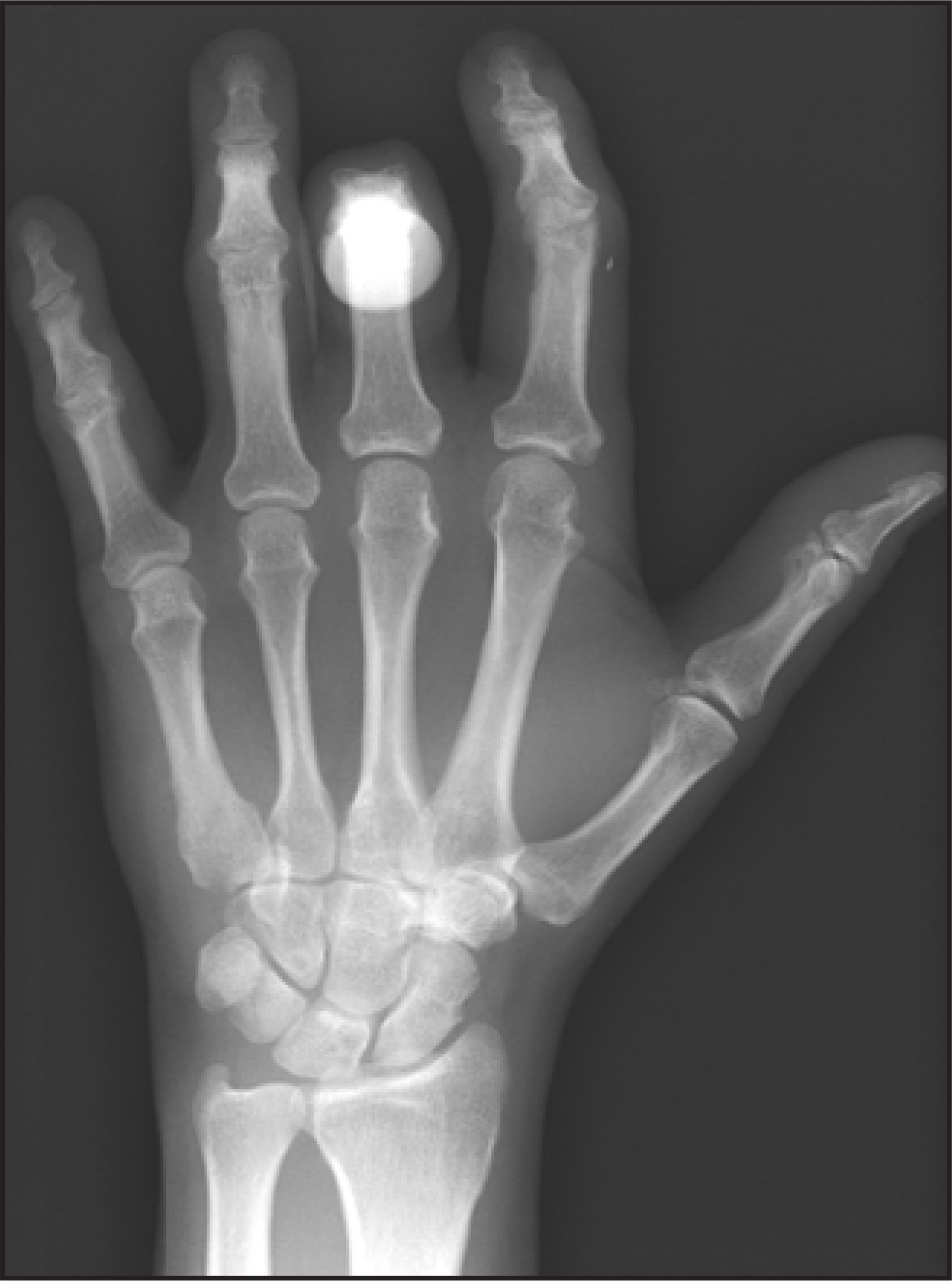
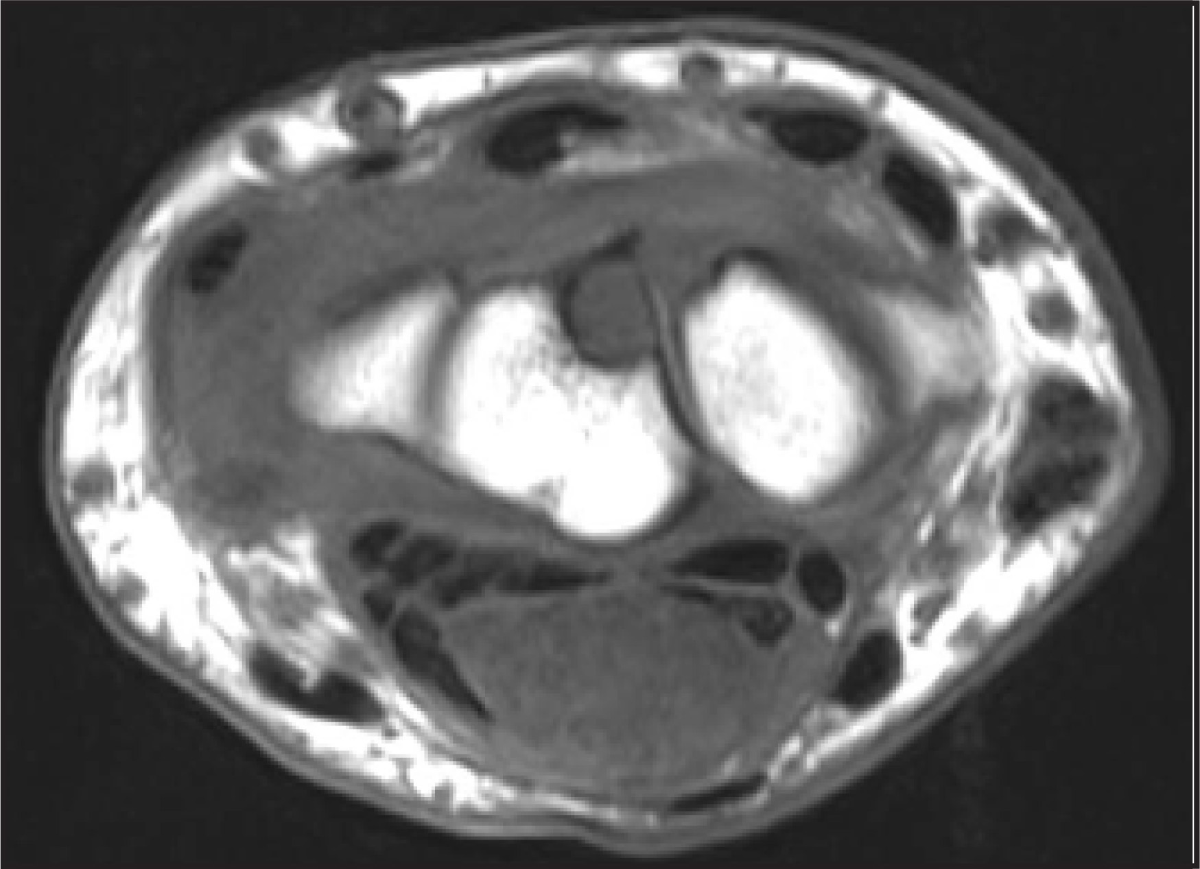
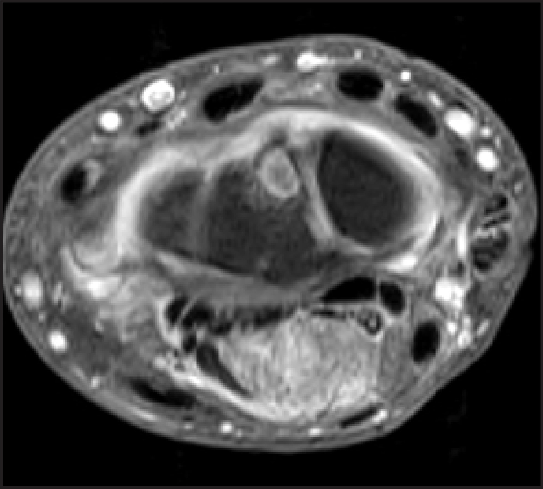
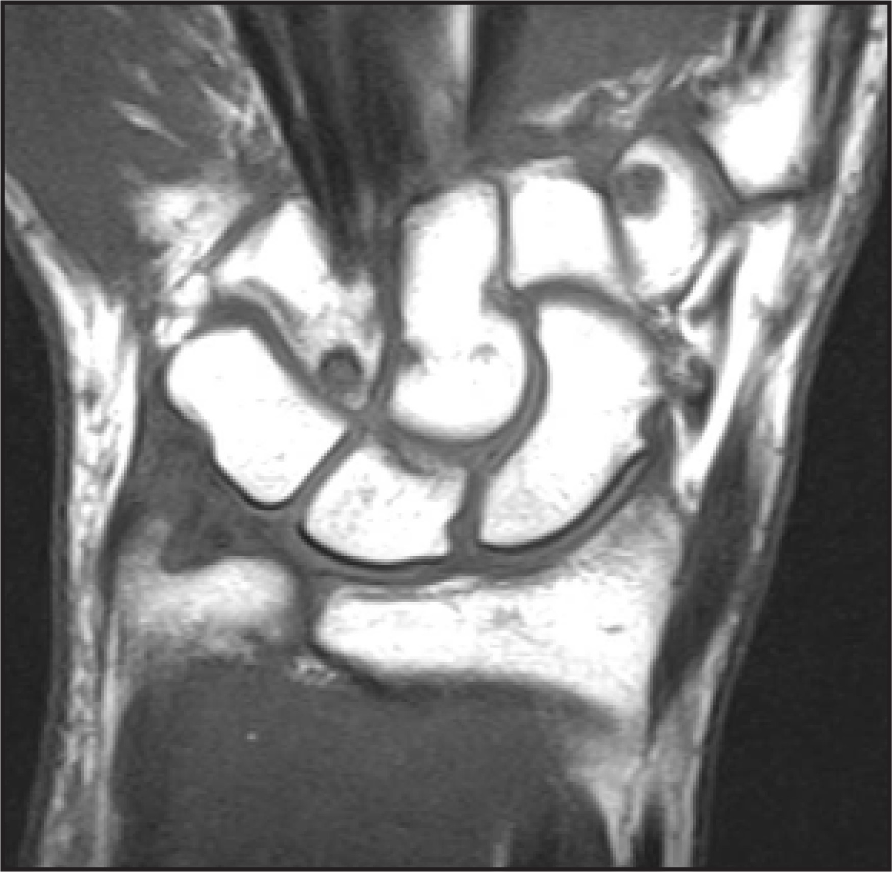
A 44-year-old man presented with a 5-month history of progressive contracture of the left middle finger and a mass that had been increasing in size in the volar aspect of the left wrist. The patient denied any history of trauma or infection in this area. On physical examination, he had tight flexure contracture of the proximal interphalangeal joint (PIP) of the third finger of the left hand and a 2 × 2.5-cm soft tissue cystlike mass on the volar aspect of the left wrist. He had a mild Tinel sign with radiation to the second and third finger and also mild thenar atrophy. Radiography of the left hand (Figure 1) and magnetic resonance imaging (MRI) of the left wrist (Figures 2 through 6) were performed.
IMAGING FINDINGS
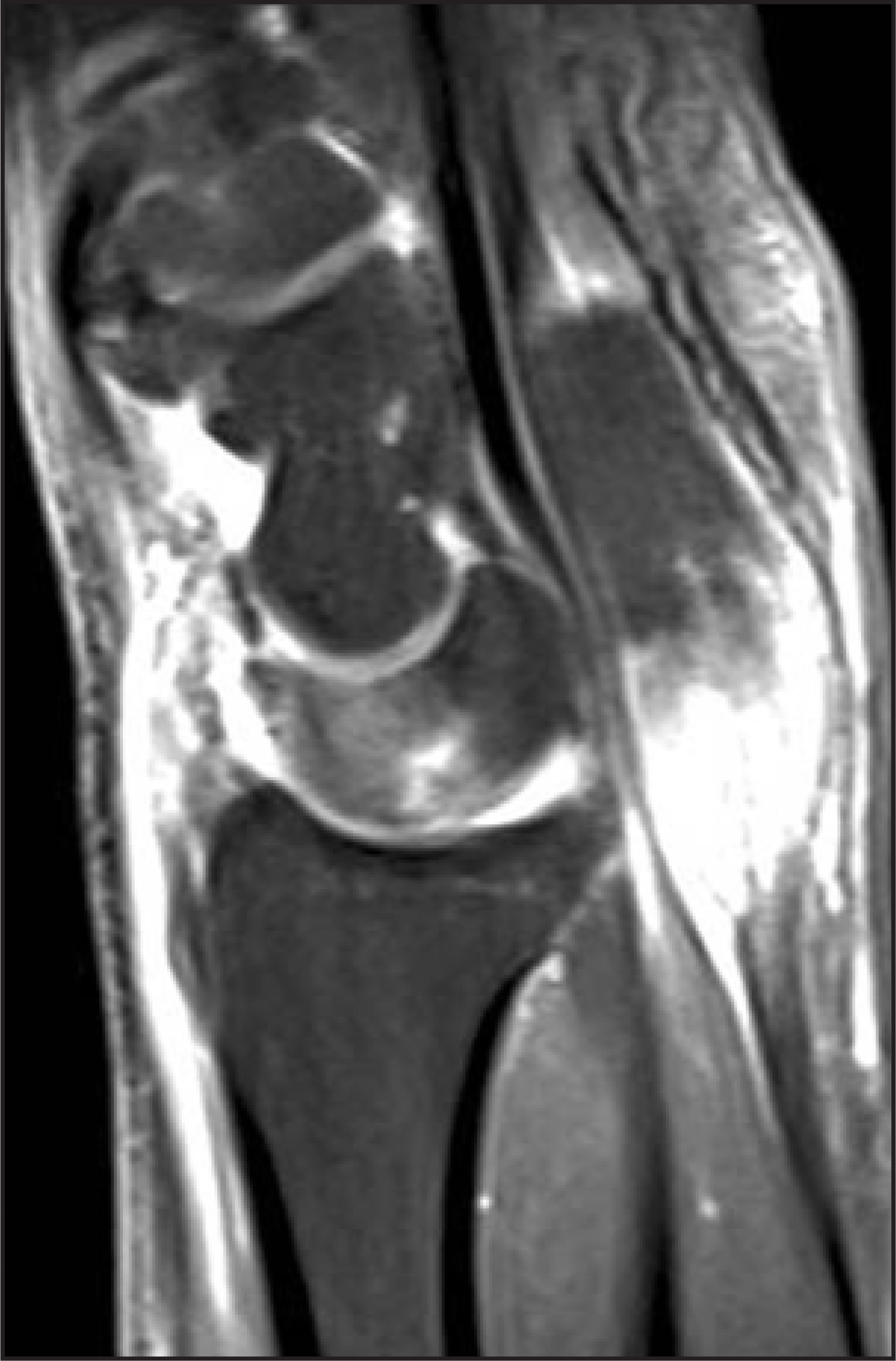
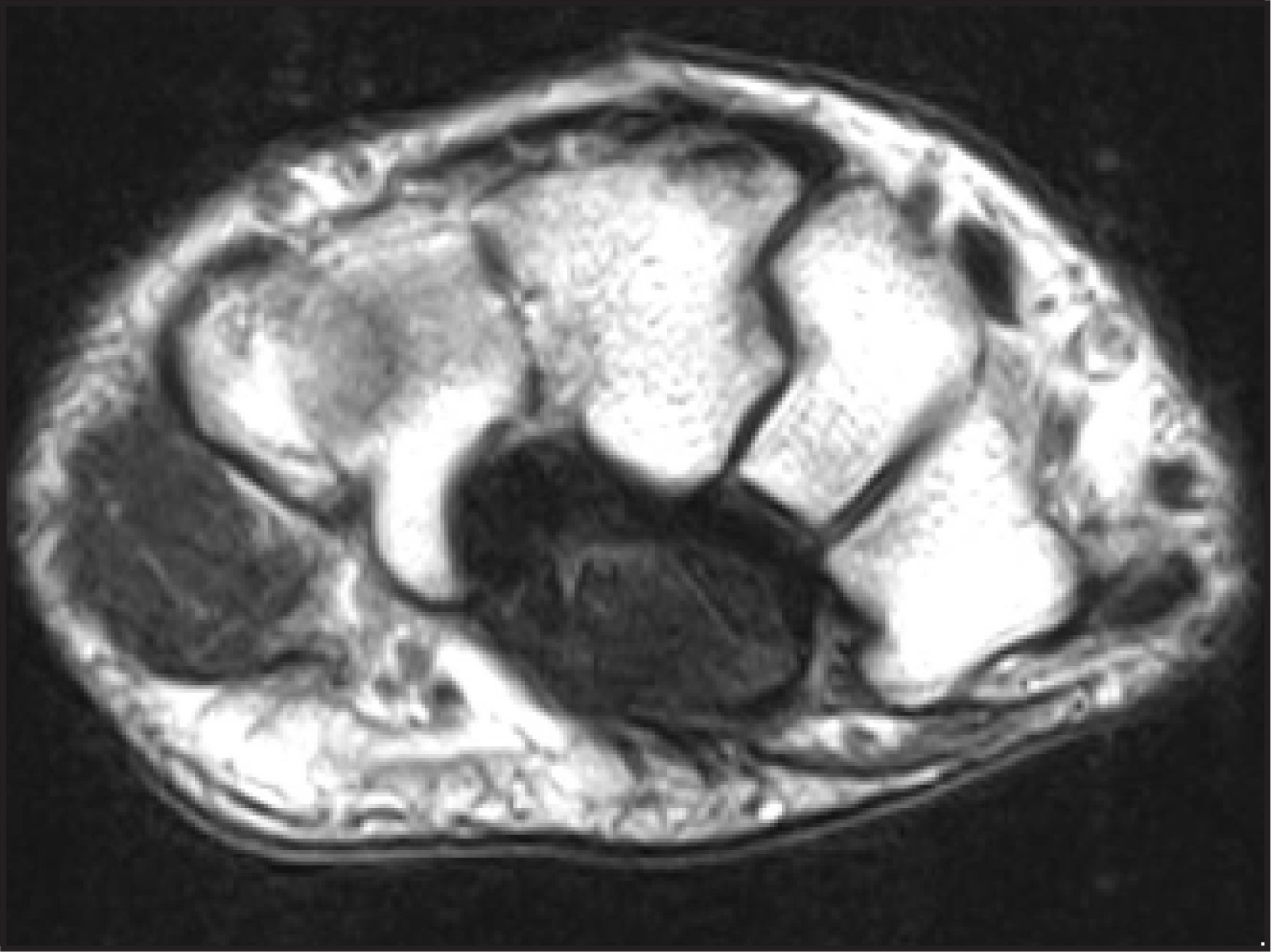
The initial radiograph of the left hand revealed contracture of the PIP of the third finger (Figure 1) with erosion of the metacarpophalangeal joint (MCP) of the second finger and a small cyst in the lunate. MRI (Figures 2 through 6) revealed a large mass measuring 3.9 × 2.9 × 1.5 cm that involved the flexor tendons in the area of the carpal tunnel. This mass showed low signal on T1-weighted (T1W) images (Figures 2 and 4) and intermediate-to-low signal on T2-weighted (T2W) images (Figure 6). There was postcontrast enhancement only in the proximal half (Figures 3 and 5). Multiple erosions that were seen as focal areas of low signal intensity on T1W imaging and contrast enhancement in the trapezium, hamate, lunate, capitate, and scaphoid bones were noted (Figures 3 and 5). There was evidence of synovitis in the intercarpal joints and tenosynovitis of the extensor pollicis brevis (Figure 3) and the abductor pollicis longus.
The rest of the bone marrow signal was normal. The differential diagnosis included inflammatory arthritidies (such as rheumatoid arthritis or psoriatic arthritis), gouty arthritis, amyloidosis, pigmented villonodular synovitis, and xanthomatosis. Other causes of carpal tunnel syndrome (such as congestive heart failure, myxedema, and trauma) did not match this patient’s clinical and imaging findings.
SURGICAL FINDINGS
The patient subsequently underwent an open biopsy. Intraoperative frozen sections of the biopsied specimen were consistent with gout. The mass was very firm, it involved and encased the flexor digitorum superficialis tendon of the third and possibly fourth fingers, and it had very thickened surrounding synovium. The median nerve was very flattened and hyperemic.
PATHOLOGIC FINDINGS
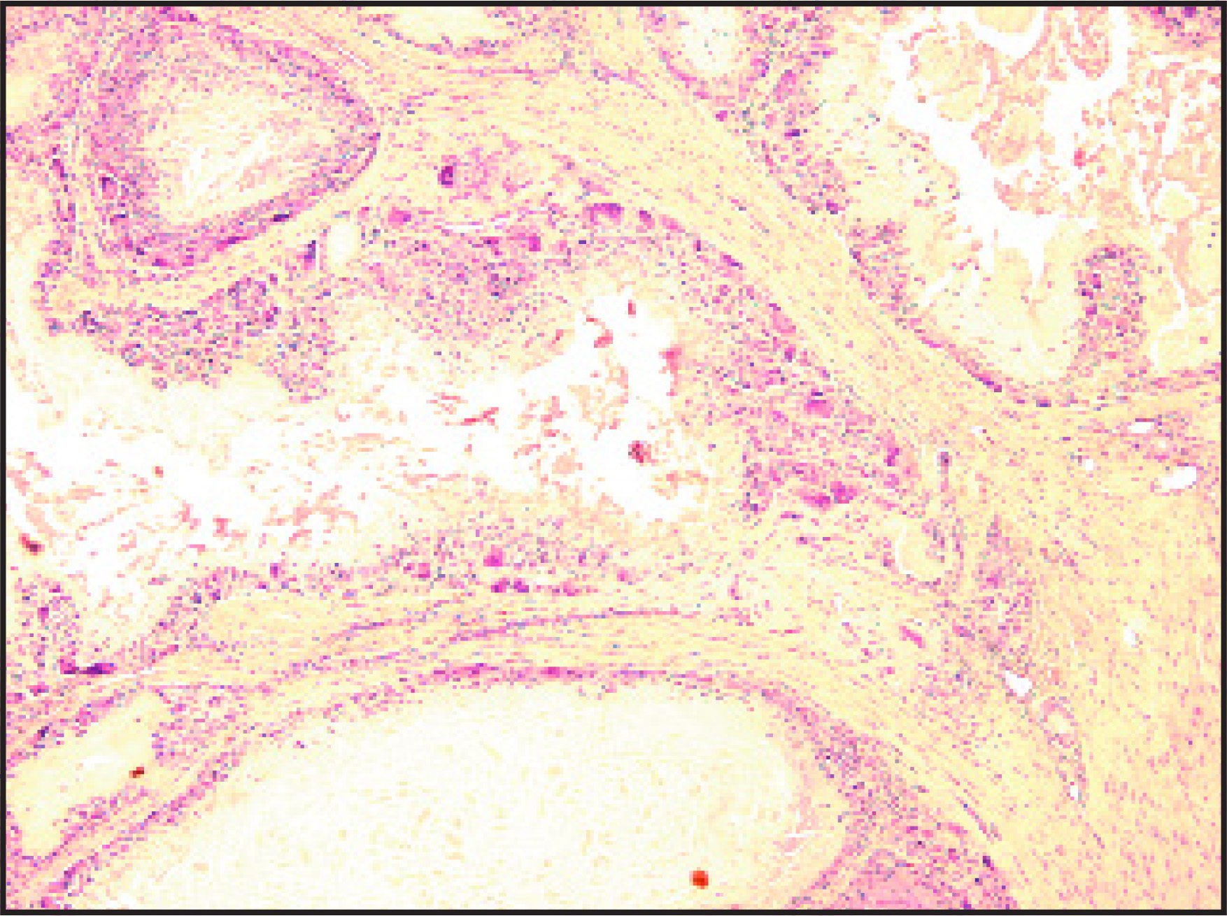
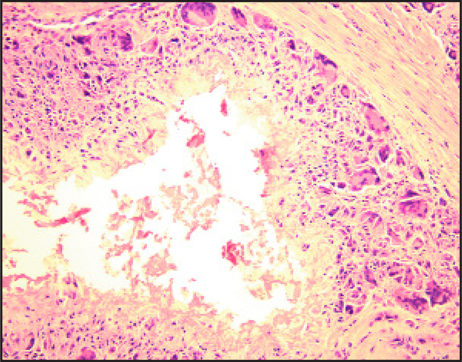
The gross specimen was a chalky white, gritty tubular tissue measuring 4.5 × 2.2 × 1.2 cm and labeled as “left wrist tendon.” A low-power microscopic view showed tophi consisting of nodules of dissolved urate crystals during formalin fixation surrounded by large multinucleated giant cells (Figure 7). A high-magnification view of the specimen showed tophi surrounded by histiocytes and multinucleated giant cells (Figure 8).
DIAGNOSIS
Gout of the hand and wrist, with carpal tunnel syndrome
DISCUSSION
The typical upper-extremity lesions of gout are tophi within the subcutaneous tissues, more commonly around the extensor surface of the elbow joint1 and PIP joints of the hand, followed, in order, by the MCP and distal interphalangeal joints.2-4 Gouty deposits may also manifest themselves with tenosynovitis5 or bony erosions (as in our patient); the tophi were located in the synovium and eroded and entrapped the flexor tendons. Even tendon rupture may occur in some cases.2,4
MRI is the modality of choice for the early detection of bony erosions. These erosions were readily detected on MRI in the carpal bones and on radiography in the MCP joint of the index finger.
Nerve entrapment may be another manifestation of gout in the upper extremity. Carpal tunnel syndrome related to tophaceous flexor tenosynovitis has been reported earlier.2,6,7 Compression of the ulnar nerve due to large gouty deposits within the elbow cubital tunnel has also been observed.2
MRI features of gouty tophi include homogeneous signal intensity on T1W images that is generally isointense to muscle. However, T2W images are more variable and may have homogeneous high signal intensity or low signal intensity. The most commonly reported signal intensity characteristic of tophi on T2W images has been heterogeneous deposits. The hyperintense signal intensity seen on T2W spin-echo images may reflect the high protein content in the amorphous center of the tophus, while the decreased signal intensity may indicate regions of calcification within the tophus, fibrous tissue and crystals, hemosiderin deposition, or proton immobility.3
The reported patterns of enhancement have been inconsistent in the literature, with some descriptions indicating homogeneous and intense enhancement and others showing heterogeneous and peripheral enhancement.3,8 Furthermore, the proliferative synovitis that is seen in gouty arthritis may be accompanied by enhancement of a tophus, reflecting hypervascularity of the affected synovium.3,8 In our case, the tophi showed low signal intensity in T1W images and intermediate-to-low signal in T2W images, with postcontrast enhancement in the proximal segment of the lesion.
Although radiographic findings of gout can sometimes be very characteristic, when pathologic confirmation is needed, one should be aware that monosodium urate crystals dissolve in an aqueous solution and that specimen loss occurs in culture and transport media, formalin fixative, and even during the hematoxilin-and-eosin–staining process. Thus, clinical information for pathologists is helpful to ensure that the specimen is preserved in 100% alcohol for fixation when the material is scanty. When crystals are abundant, such as in the present case, incomplete dissolution results in amorphous cloudy material (Figure 7). In cases in which crystals are completely dissolved, one can attempt to polarize unstained sections to prevent loss during the staining process. Under polarization, urate crystals demonstrate negative birefringence. When urate crystals are not seen, the surrounding histiocytic reaction (Figure 8) resembles granulomatous inflammation, especially tuberculosis. Fungal and acid-fast bacilli stains can be performed in these cases to rule out microorganisms. Fine-needle aspiration biopsy with 21-gauge needles can also provide a cost-effective diagnostic method. In the current case, the frozen section showed needlelike crystals that were consistent with gout.
CONCLUSION
MRI is the modality of choice for the early detection of erosions in the hand and wrist. Although these erosions may appear as common changes in arthritis, rarely gout may manifest with carpal tunnel syndrome as a presenting sign of the disease.
References
- Weniger F, Davison S, Risin M. Gouty flexor tenosynovitis of the digits: Report of three cases. J Hand Surg [Am]. 2003;28:669-672.
- Schuind F, Clermont D, Stallenberg B. Gouty involvement of flexor tendons. Chir Main. 2003;22:46-50.
- Chen C, Chung C, Yeh L. Carpal tunnel syndrome caused by tophaceous gout: CT and MR imaging features in 20 patients. AJR Am J Roentgenol. 2000;175:655-659.
- Moore J, Weiland A. Gouty tenosynovitis in the hand. J Hand Sur Am. 1985;10:291-295.
- Primm D, Allen J. Gouty involvement of flexor tendon in the hand. J Hand Surg Am. 1983;8:863-865.
- Tan G, Chew W, Lai C. Carpal tunnel syndrome due to gouty infiltration of lumbrical muscles and flexor tendon. Hand Surg. 2003;8:121-125.
- Mockford B, Kincaid R, Mackay I. Carpal tunnel syndrome secondary to intratendinous infiltration by tophaceous gout. Scand J Plast Surg Hand Surg. 2003;37:186-187.
- Yu J, Chung C, Recht M. MR imaging of tophaceous gout. AJR Am J Roentgenol. 1997;168:523-527.
Citation
. RCOM RADIOLOGICAL CASE OF THE MONTH. Applied Radiology. 2007;36(11):72-78. doi:10.37549/AR1566.