RCOM RADIOLOGICAL CASE OF THE MONTH
Applied Radiology — Vol. 36 , Issue 11 , pp. 58 -61
DOI: 10.37549/AR1567
Published: November 1, 2007
Categories
CASE SUMMARY
A 51-year-old woman developed sudden-onset neck and right shoulder pain and numbness in the right upper extremity and both lower extremities. The following morning, she also developed chills and fever. She was seen in the clinic and was prescribed muscle relaxants, anti-inflammatory medications, and steroids for a presumed muscle strain. However, progressive weakness prompted the patient to go to the emergency room on the very same evening.
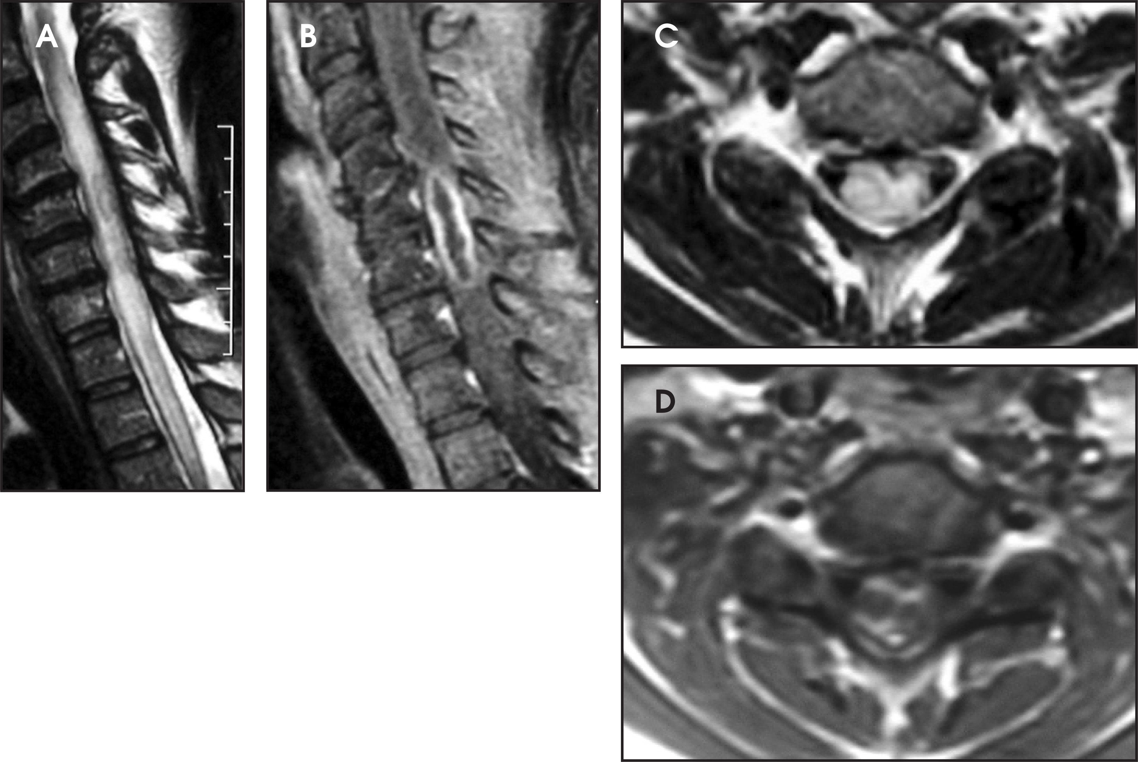
Computed tomography (CT) scans of the head and cervical spine that were performed on admission were negative. The following day, she underwent magnetic resonance imaging (MRI) of the neck, which revealed a large cervical spinal cord lesion with thickening of the cord and a rim of enhancement. This was interpreted as an intramedullary lesion, most likely a neoplasm (Figure 1).
IMAGING FINDINGS
MRI of the cervical spine without and with intravenous contrast shows a large lesion in the cervical spinal cord extending from the C4-C5 to the C6-C7 level with significant expansion of the cord at these levels. The lesion manifested a thick rim of enhancement on postcontrast T1-weighted images. There was also marked hyperintensity involving the entire cervical and upper thoracic spinal cord on T2-weighted images (Figure 1). This was interpreted as an intramedullary lesion, most likely a neoplasm.
The patient was placed on steroids, with plans for elective surgical intervention, which was later performed urgently due to progressive neurologic compromise. A myelotomy was performed and frank pus was identified. The abscess was drained. Intraoperative Gram stain showed Gram-positive cocci. Intravenous antibiotics (including vancomycin, nafcillin, and metronidazole) were started; the first dose was administered in the operating room.
After surgery, the patient was found to be quadriplegic. She continued antibiotics, rehabilitation, and wound care, including a trial of hyperbaric therapy. After approximately 2 weeks of continuous therapy, she was transferred to the cord trauma institute with persistent neurologic deficits, including marked proximal and distal right-sided weakness and hyperesthesia. She showed some improvement of the left side and achieved minor control of bowels.
DIAGNOSIS
Cervical spinal cord intramedullary abscess caused by Streptococcus intermedius
DISCUSSION
Intramedullary abscess is a well-known but relatively rare pathology. Since its first description by Hart 1 in 1830, only approximately 100 cases have been reported. Despite advances in microbial therapy, it still carries significant morbidity and mortality.1,2 Neuroradiologists and neurosurgeons should be very aware of this entity, since it is one of the few treatable causes of paraparesis.1 In this report, we present a case of an abscess in the cervical spinal cord caused by Streptococcus intermedius that developed acutely in an otherwise healthy middle-aged woman.
Only a few reports have described spontaneous spinal cord abscesses.3 In our case, the symptoms had been present for only 2 days prior to hospital admission. No anatomic defects, metabolic abnormalities, injuries, or sources of infection were identified.
Intramedullary abscess of the spinal cord is a rare infection of the central nervous system, occurring in 0.2 to 2.2 per 10,000 patients admitted to tertiary care hospitals.4 These infections have been associated with high mortality and neurologic morbidity. In a review by Menezes et al,5 a mortality rate of 24% for spinal cord abscesses was reported for cases between 1944 and 1977. Only timely surgical drainage and aggressive intravenous antimicrobial therapy with agents that have appropriate spectrum and good penetration into the central nervous system can offer a favorable outcome. The reported male-to-female ratio is roughly 3 to 1. The age of patients varies widely, and females are affected at a younger age.6
Mechanisms of acquiring infection include 1) hematogenous spread, 2) contiguous spread from an adjacent focus of infection, and 3) direct seeding (penetrating trauma, postsurgery, etc.). In the modern era, approximately 10% of cases result from hematogenous spread of infection as opposed to almost 50% of cases in the preantibiotic era. Roughly 25% of cases result from contiguous spread of infection associated with a dermal sinus tract. This mode of infection did not show a noticeable change when compared to the preantibiotic era.7 The majority of cases in recent times are idiopathic, as it was in our case. In children, more than half of patients have underlying anatomic spinal defects.8 Neuroectodermal defects or congenital dermal sinus are commonly present in children.
Patients usually present to the physician with mixed neurologic deficits, fever, and pain symptoms. Neurorologic deficits, especially sensory and sphincter disturbances, are common.6 Children usually present with fever and an inability to walk.8
Common findings on gadolinium-enhanced MRI, the gold standard for this diagnosis, include increased signal intensity on T2-weighted imaging and central iso- to low-signal-intensity lesions with a peripheral enhancing rim on T1-weighted imaging. Generalized thickening of the cord and diffuse hyperintensity have also been reported. MRI is also valuable in assessing any accompanying anatomic defects and in surgical planning. Follow-up examinations are important in the detection of potential recurrences.
CT findings are variable and range from normal (as in our case) to a thickened or deformed spinal cord. Since the lesions involve predominantly soft tissues, CT is inferior to MRI in obtaining detailed tissue characteristics. Myelography may show obstruction of cerebrospinal fluid flow secondary to thickening of the spinal cord, but it is an invasive technique and is used less commonly.
The only form of treatment that offers a chance of favorable outcome is early surgical drainage followed by appropriate antimicrobial therapy. Cases of intramedullary abscesses from the preantibiotic era and a few later cases in which patients did not receive proper antimicrobial agents noted 100% mortality. This is in contrast to a surgically treated group with only 13.6% mortality.6 Laminectomy with myelotomy and drainage of abscess is the most common surgical technique. Any coexisting anatomic abnormalities (such as dermal sinuses, dermoids, or meningoceles) should also be corrected during the surgery if no contraindications are present.8
Antibiotics should be administered as soon as a preliminary diagnosis is made. Intravenous agents with a wide antimicrobial spectrum and good penetration to the central nervous system (such as vancomycin, metronidazole, or cefotaxime) should be used. Multiagent therapy is preferred. Appropriate adjustments should be made when culture and sensitivity results are available. Postoperative duration of therapy varies and should be tailored to the individual patient, but most authors report at least 6 weeks of treatment.
CONCLUSION
Intramedullary abscesses are rare pathologies with potentially good outcomes, providing that an early diagnosis is made and appropriate therapy is instituted immediately. The radiologist should be aware of this diagnosis and raise the possibility of this entity in order to initiate the proper treatment and avoid a grave outcome. MRI is the most effective diagnostic tool. Surgical drainage followed by aggressive antimicrobial therapy offers a good chance of survival and minimizes neurologic deficits.
References
- Hart J. Case of encysted abscess in the center of the spinal cord. Dublin Hospital Report. 1830;5:522-524.
- Artzt P. Abscess within the spinal cord: Review of the literature and report of three cases. Arch Neurol Psych. 1944;51:533-543.
- Blacklock J, Hood T, Maxwell R. Intramedullary spinal cord abscess. J Neurosurg. 1982;57:270-273.
- Tacconi L, Arulampalam T, Johnston F, Thomas D. Intramedullary spinal cord abscess: Case report. Neurosurgery. 1995;37:817-819.
- Menezes A, Graf C, Perret G. Spinal cord abscess: A review. Surg Neurol. 1977;8:461-467.
- Bartels R, Gonera E, Van der Spek J. Intramedullary spinal cord abscess: A case report. Spine. 1995;20:1199-1204.
- Chan C, Gold W. Intramedullary abscess of the spinal cord in the antibiotic era: Clinical features, microbial etiologies, trends in pathogenesis, and outcomes. Clin Infect Dis. 1998;27:619-626.
- Simon J, Lazareff J, Diament M, Kennedy W. Intramedullary abscess of the spinal cord in children: A case report and review of the literature. Pediatr Infect Dis J. 2003;22:186-192.
Citation
. RCOM RADIOLOGICAL CASE OF THE MONTH. Applied Radiology. 2007;36(11):58-61. doi:10.37549/AR1567.