RCOM RADIOLOGICAL CASE OF THE MONTH
Applied Radiology — Vol. 36 , Issue 12 , pp. 37 -40
DOI: 10.37549/AR1571
Published: December 1, 2007
Categories
CASE SUMMARY
A 36-year-old man presented to the emergency department with a history of sudden-onset neck pain during his usual weight-lifting workout. He also reported transient paresthesias in his arms. Clinically, he had diffuse tenderness of the cervical spine. The neurological examination was unremarkable.
Conventional plain-film radiography followed by computed tomography (CT) of the cervical spine was requested. The patient was managed conservatively.
IMAGING FINDINGS
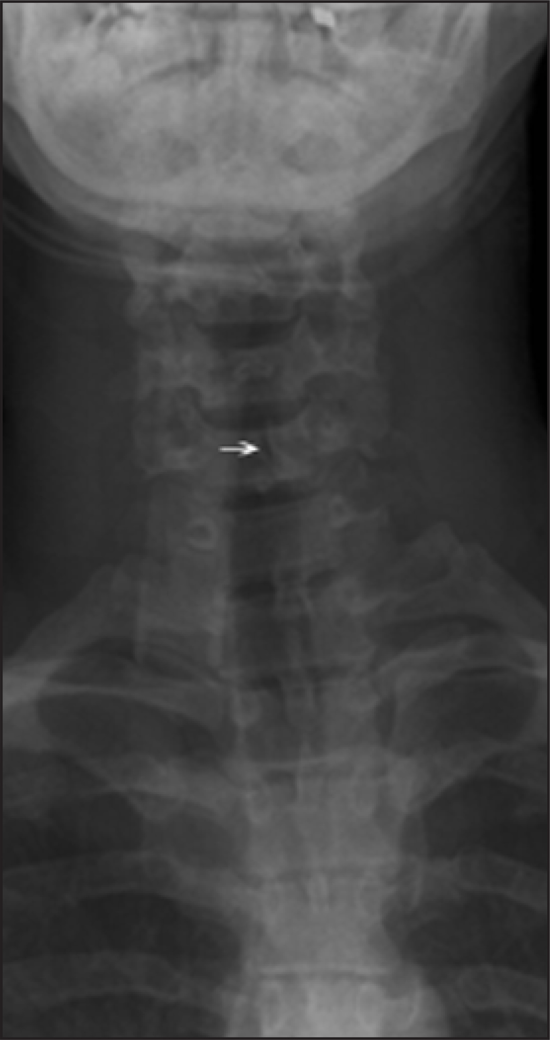
The lateral radiograph of the cervical spine showed the presence of a defect of the pars interarticularis at C6. Grade-1 (<25%) spondylolisthesis of C6 anteriorly with respect to C7 was also noted on the lateral view. There was no increase in the prevertebral soft tissue density anterior to C6. Flexion and extension views did not reveal instability of the cervical spine (Figure 1). Additionally, spina bifida occulta was observed on the anteroposterior view (Figure 2).
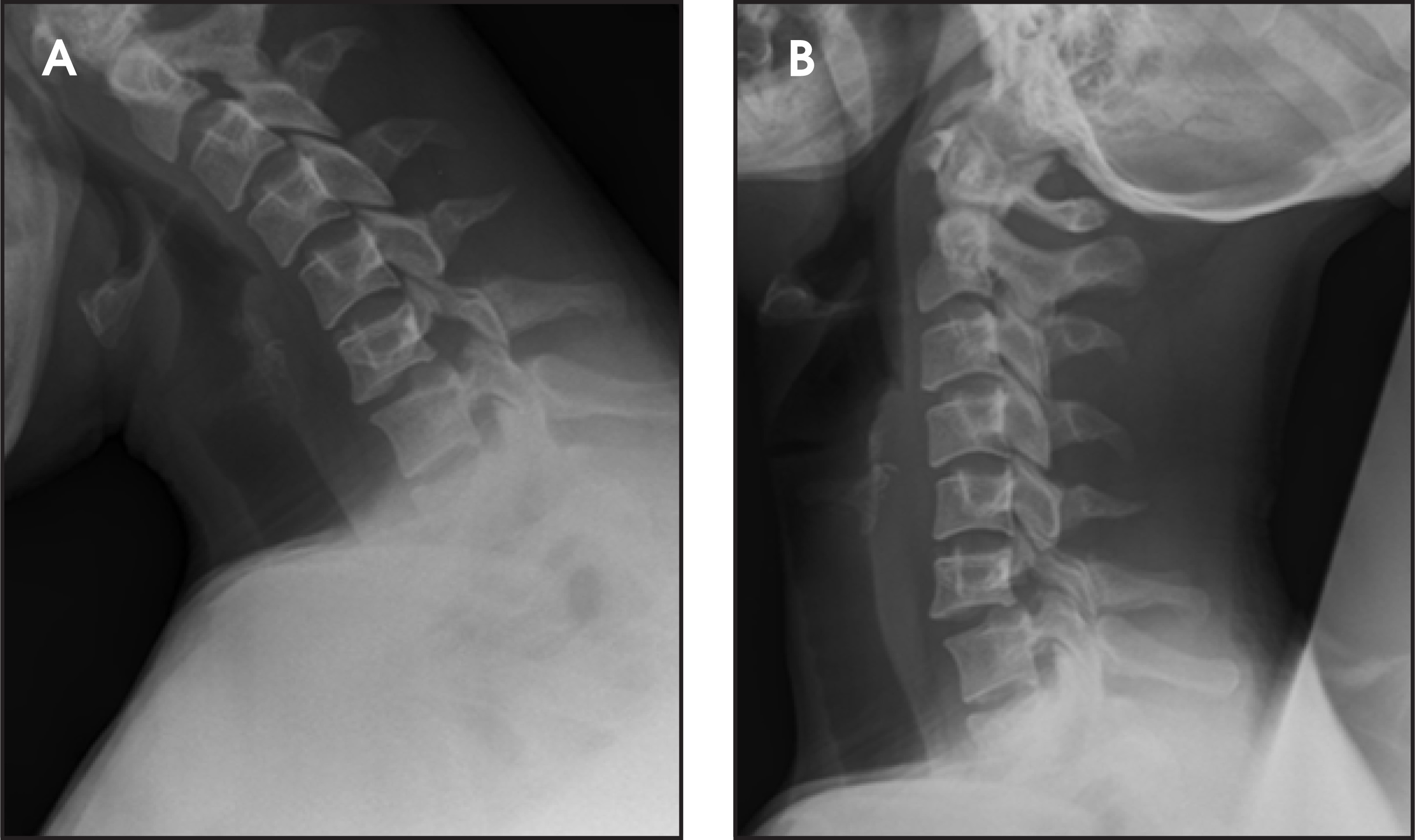
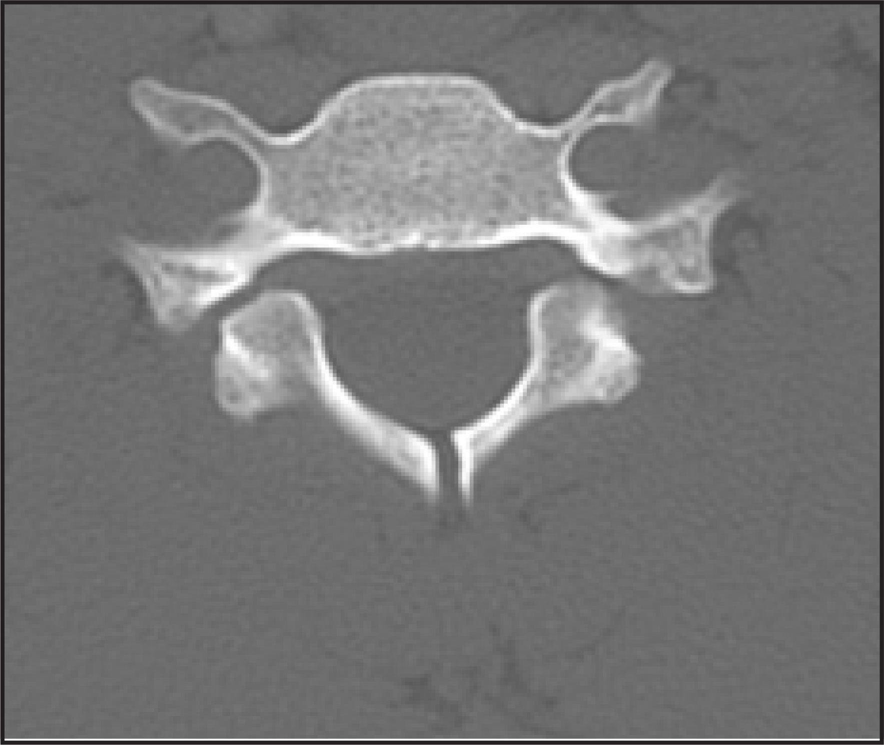
Axial noncontrast CT at the C6 level showed bilateral well-corticated defects of the pars interarticularis. It also confirmed the presence of spina bifida occulta and medial displacement of the laminae. Again, soft tissue swelling indicative of acute trauma was not detected (Figure 3).
DIAGNOSIS
Bilateral spondylolysis of the sixth cervical vertebra
DISCUSSION
Spondylolysis with spondylolisthesis is a common condition in the lumbar spine. Cervical spondylolysis is, however, much more rare than its lumbar counterpart and is described as a well-corticated cleft between the superior and inferior articular facets of the articular pillar of the cervical spine.1,2
It is usually discovered as an incidental radiologic finding in patients who are undergoing cervical spine imaging for neck pain or trauma.2,3 Associated neurological deficit may be detected, although spinal cord compression is rare.4 This condition mostly affects the sixth cervical vertebra, although involvement of other cervical vertebrae has been reported.5
Plain-film radiography and CT are the principal diagnostic modalities that help in differentiating cervical spondylolysis from articular pillar fracture or dislocation. In addition to the distinctive pars interarticularis defect, other imaging findings described in cervical spondylolysis are hyposplasia of the pedicles, dysplastic laminae, and spina bifida. Medial displacement of inferior facets and laminae, compensatory hypertrophic changes of the articular process in the adjacent vertebrae, and spondylolisthesis are also seen.2,6,7 The pars interarticularis defect is difficult to identify on magnetic resonance imaging because of poor bone resolution; however, the absence of the spinous process on sagittal sequences should raise the suspicion of this abnormality.2,8
The exact etiology of cervical spondylolysis remains unknown. The detection of articular dysplasia and spina bifida occulta on imaging studies suggests a congenital origin of this condition. A dysplasia mechanism with stress fractures of a weakened pars interarticularis is another possibility.3 Patient management is usually conservative, with surgical treatment reserved for patients with a neurologic deficit.1,2
CONCLUSION
Cervical spondylolysis is a rare condition. It is usually discovered incidentally and should be differentiated from traumatic spondylolysis. Plain-film radiography and CT are the main diagnostic imaging modalities.
References
- Poggi J, Martinez S, Hardaker W, Richardson W. Cervical spondylolysis. J Spinal Disord. 1992;5:349-356.
- Forsberg D, Martinez S, Vogler J, Wiener M. Cervical spondylolysis: Imaging findings in 12 patients. AJR Am J Roentgenol. 1990;154:751-755.
- Fessy M, Durand J, Gunepin F. An unusual anomaly: Cervical spondylolysis in an adult. Rev Chir Orthop Reparatrice Appar Mot. 1999;85:174-777.
- Hinton M, Harris M, King A. Cervical spondylolysis. Report of two cases. Spine. 1993;18:1369-1372.
- Jeyapalan K, Chavda S. Case report 868. Congenital bilateral spondylolysis and spondylolisthesis of the fourth cervical vertebra. Skeletal Radiol. 1994;23:580-582.
- Jones D. Sage MR Bilateral spondylolysis and associated dysplasia of C6. Australas Radiol. 1992;36:260-261.
- Schwartz J. Case 36: Bilateral cervical spondylolysis of C6. Radiology. 2001;220:191-194.
- Redla S, Sikdar T, Saifuddin A, Taylor B. Imaging features of cervical spondylolysis with emphasis on MR appearances. Clin Radiol. 1999;54:815-820.
Citation
. RCOM RADIOLOGICAL CASE OF THE MONTH. Applied Radiology. 2007;36(12):37-40. doi:10.37549/AR1571.