RCOM RADIOLOGICAL CASE OF THE MONTH
Applied Radiology — Vol. 36 , Issue 12 , pp. 48A -48C
DOI: 10.37549/AR1573
Published: December 1, 2007
Categories
CASE SUMMARY
A 91-year-old woman presented with progressive dyspnea on exertion, paroxysmal nocturnal dyspnea, orthopnea, and increasing lower extremity edema despite compliance with her antihypertensive and diuretic medications. Her medical history was significant for recently diagnosed congestive heart failure after presenting with a cerebral vascular accident that resulted in mild left hemiplegia. Her other comorbidities included mild chronic renal insufficiency, hypertension, urge incontinence, osteoarthritis, and previous resection of a basal cell carcinoma. Her social history was unremarkable, and her family history was significant for osteoarthritis. She was admitted and treated for congestive heart failure exacerbation. The rheumatology service was consulted for chronic, painful, deforming bilateral hand arthritis.
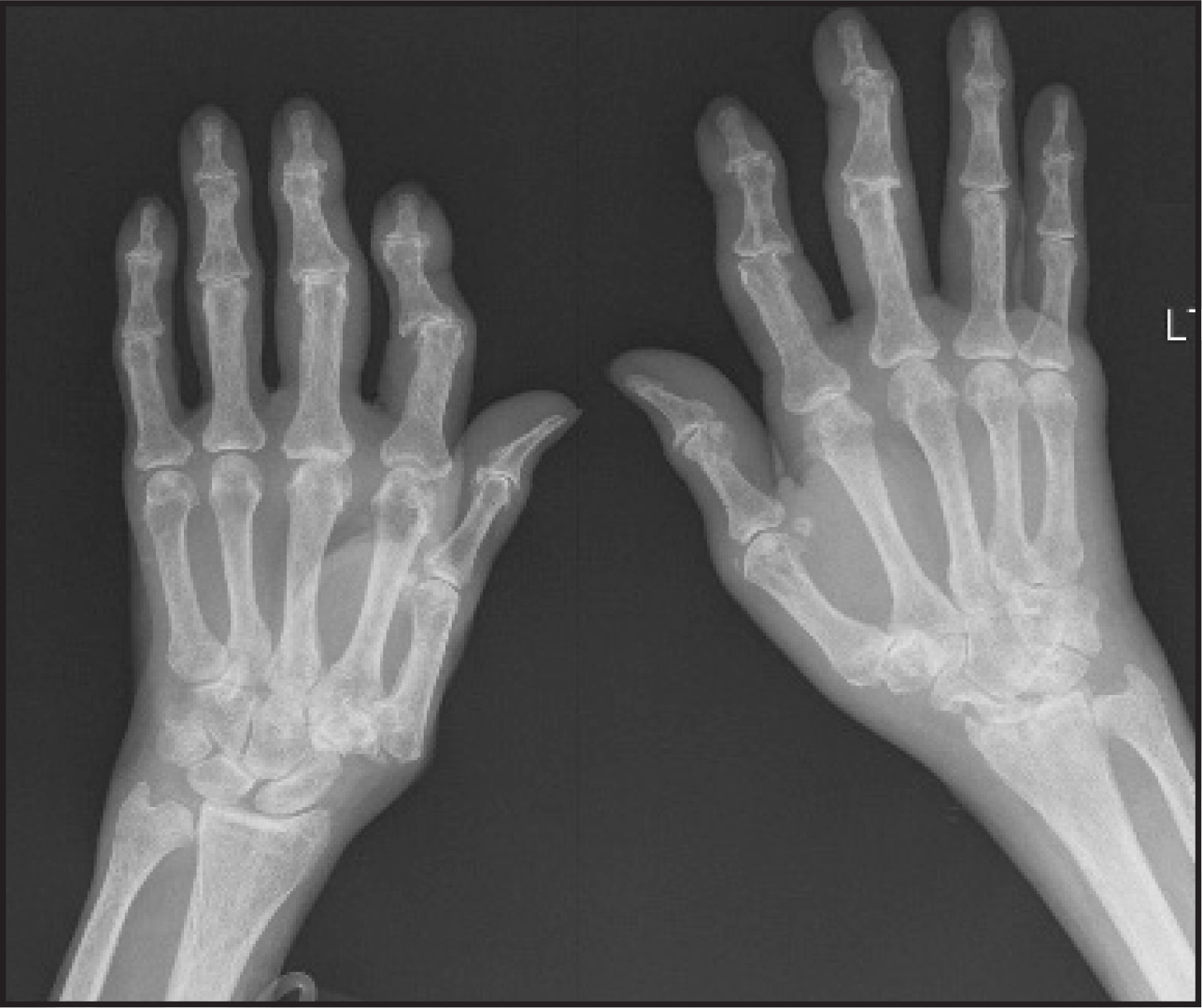
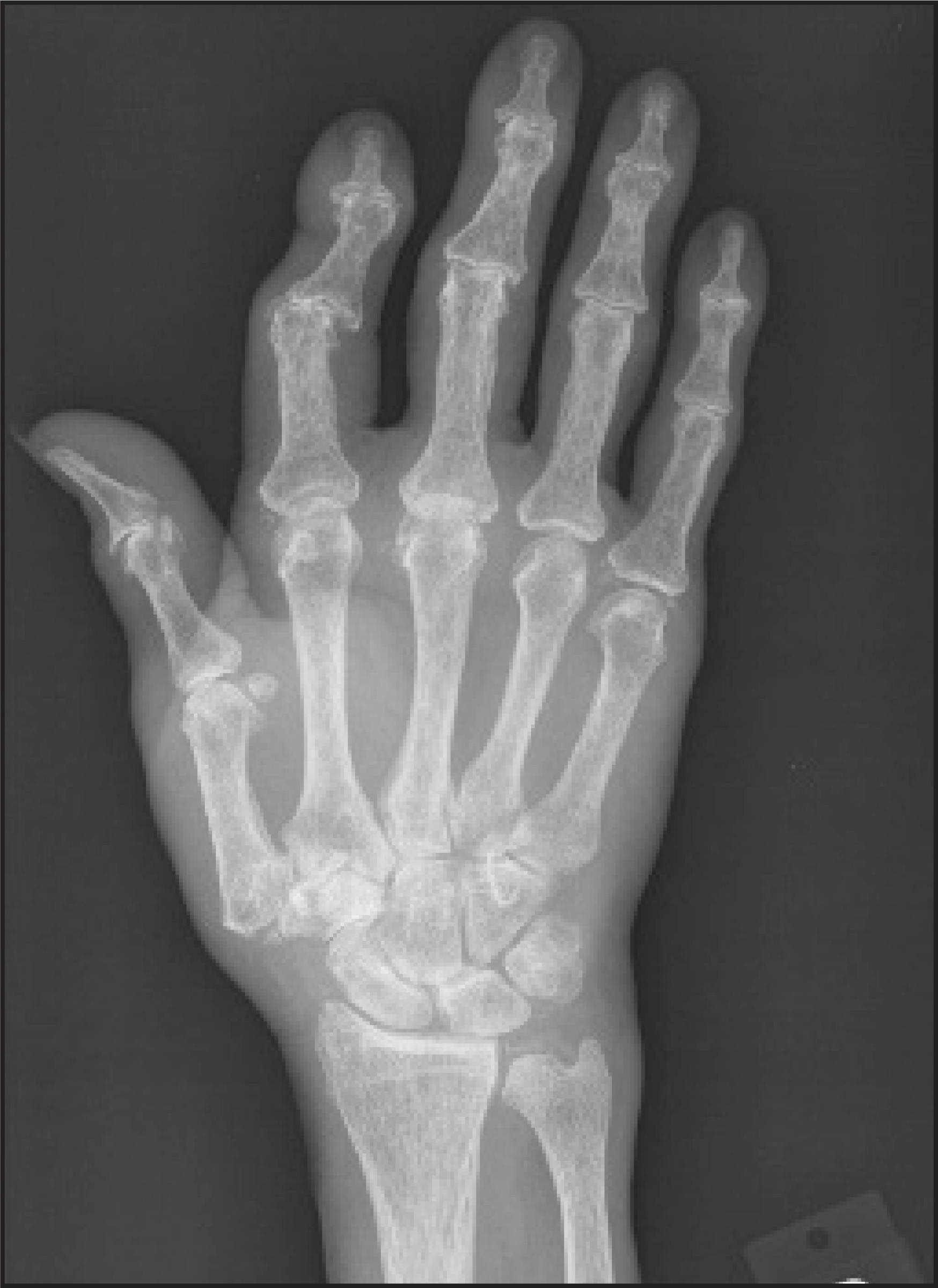
A physical examination revealed a thin, elderly white woman in no distress. Musculoskeletal examination was significant for grossly deformed digits on both hands. Tender, erythematous Heberden and Bouchard nodes were present over multiple distal interphalangeal (DIP) and proximal interphalangeal (PIP) joints. Joint swelling was noted in the left second, left third, right first, and right second PIP joints. Significant squaring of the bilateral carpometacarpal joints and malalignment of the index fingers were also present. The stability of the interphalangeal joints of both the thumbs was compromised. A clinical diagnosis of erosive osteoarthritis (EOA) was made, and plain radiographs were obtained to confirm the diagnosis (Figures 1 and 2).
IMAGING FINDINGS
Frontal and oblique radiographs of the left and right hand showed diffuse mild osteopenia (Figures 1 and 2). There were multiple areas of joint space narrowing with osteophyte formation; these were most notable at the left third DIP joint and the right second PIP interphalangeal joint. Several interphalangeal joints contained central erosions, which were especially prominent in the right second DIP. There was medial sub- luxation of the middle phalanx as compared with the proximal phalanx of the right second digit. Similar subluxation was also noted on the same level on the left hand. Bilateral osteoarthritic changes were noted on the first carpometacarpal joints bilaterally; chages on the right were greater than those on the left. Incidentally, chondrocalcinosis was seen in the triangular fibrocartilage.
DIAGNOSIS
Advanced erosive osteoarthritis (EOA)
DISCUSSION
Kellgren and Moore1 first described the condition that is now known as erosive osteoarthritis in 1952. More recently, Ehrlich2 coined the term “inflammatory osteoarthritis” to emphasize the clinical signs of inflammation that are routinely present: swelling, tenderness, erythema, and warmth. Erosive osteoarthritis usually begins abruptly with pain and morning stiffness in the DIP joints before advancing to the PIP joints, whereas OA has a more insidious, generalized onset. Rarely, large joints such as the hip and shoulder can become involved.2,3
The differential of EOA includes osteoarthritis of the hand, rheumatoid arthritis, gout, and psoriatic arthritis. Osteophytes, Heberden’s nodes, Bouchard’s nodes, and joint space narrowing can be seen in both EOA and osteoarthritis, but central erosions are characteristic of only EOA. Instability and ankylosis of interphalangeal joints are exclusively present in EOA when compared with osteoarthritis. Compared with the central erosions of EOA, rheumatoid arthritis erosions are typically marginal and do not result in the “gull-wing” appearance seen in EOA. “Gull-winging” results from a central erosion on the proximal plate with marginal proliferation in the distal plate at both the DIP and PIP joints. This can be contrasted to psoriatic arthritis, which exhibits marginal erosions in the proximal plate and marginal periostitis in the distal plate at the DIP joints. In gout, tophaceous deposits are present and the erosions appear as “overhanging edges,” neither of which is seen in EOA.3,4
Erosive OA has been associated with systemic diseases, including hypothyroidism, autoimmune thyroiditis, hyperparathyroidism, chronic renal disease, scleroderma, Sjögren’s syndrome, and calcium pyrophosphate dihydrate arthropathy (CPPD). Some authors believe that these associations are anecdotal. Our patient’s radiographs revealed chondrocalcinosis of the triangular cartilage, which is suggestive of CPPD, but synovial fluid crystal analysis was not pursued.4
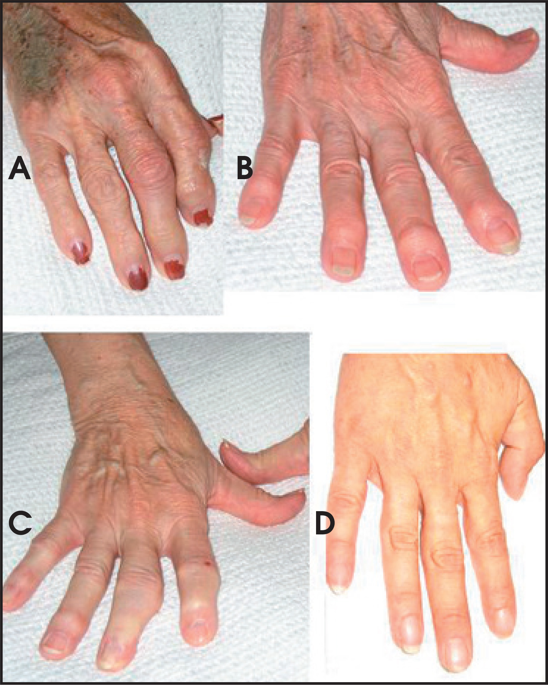
Although the true etiology is unknown, several investigators have suggested hormonal influences, metabolic disorders, and autoimmunity. Erosive OA exhibits a strong family history and an overwhelming female preponderance, with most women at or near menopause.2,4 Interestingly, our patient’s 2 daughters and 1 granddaughter were present during the interview and examination. The daughters were both in their sixties and the granddaughter was in her late thirties. All 3 women exhibited clinical findings that were suggestive of EOA (Figure 3). The eldest daughter carried the diagnosis of EOA, and her sibling had previously undergone surgery to correct a worsening deformity of the right third digit.
Standardized trials for the treatment of EOA are lacking and no definitive therapeutic approach has been reported. Acetaminophen and nonsteroidal anti-inflammatory drugs (NSAIDs) are the recommended first- and second-line therapeutic interventions, respectively.2-4 In small studies, hydroxychloroquine has been shown to be effective and well-tolerated in NSAID refractory EOA.5 Our patient did not receive NSAIDs because of the concern of exacerbating her renal insufficiency, and she was started on acetaminophen and hydroxychloroquine.
CONCLUSION
We have described a case of erosive osteoarthritis in 3 generations of women that supports a strong genetic component and female preponderance of the disease.
References
- Kellgren J, Moore R. Generalized osteoarthritis and Heberden’s nodes. Br Med J. 1952;1:181-187.
- Ehrlich G. Erosive osteoarthritis: Presentation, clinical pearls, and therapy. Curr Rheumatol Rep. 2001;3:484-488.
- Greenspan A. Erosive osteoarthritis. Semin Musculoskelet Radiol. 2003;7(2):155-159.
- Punzi L, Ramonda R, Sfriso P. Erosive osteoarthritis. Best Pract Res Clin Rheumatol. 2004;18:739-758.
- Bryant L, des Rosier K, Carpenter M. Hydroxychloroquine in the treatment of erosive osteoarthritis. J Rheumatol. 1995;22:1527-1531.
Citation
. RCOM RADIOLOGICAL CASE OF THE MONTH. Applied Radiology. 2007;36(12):48A-48C. doi:10.37549/AR1573.