RCOM RADIOLOGICAL CASE OF THE MONTH
Applied Radiology — Vol. 36 , Issue 12 , pp. 44 -46
DOI: 10.37549/AR1577
Published: December 1, 2007
Categories
CASE SUMMARY
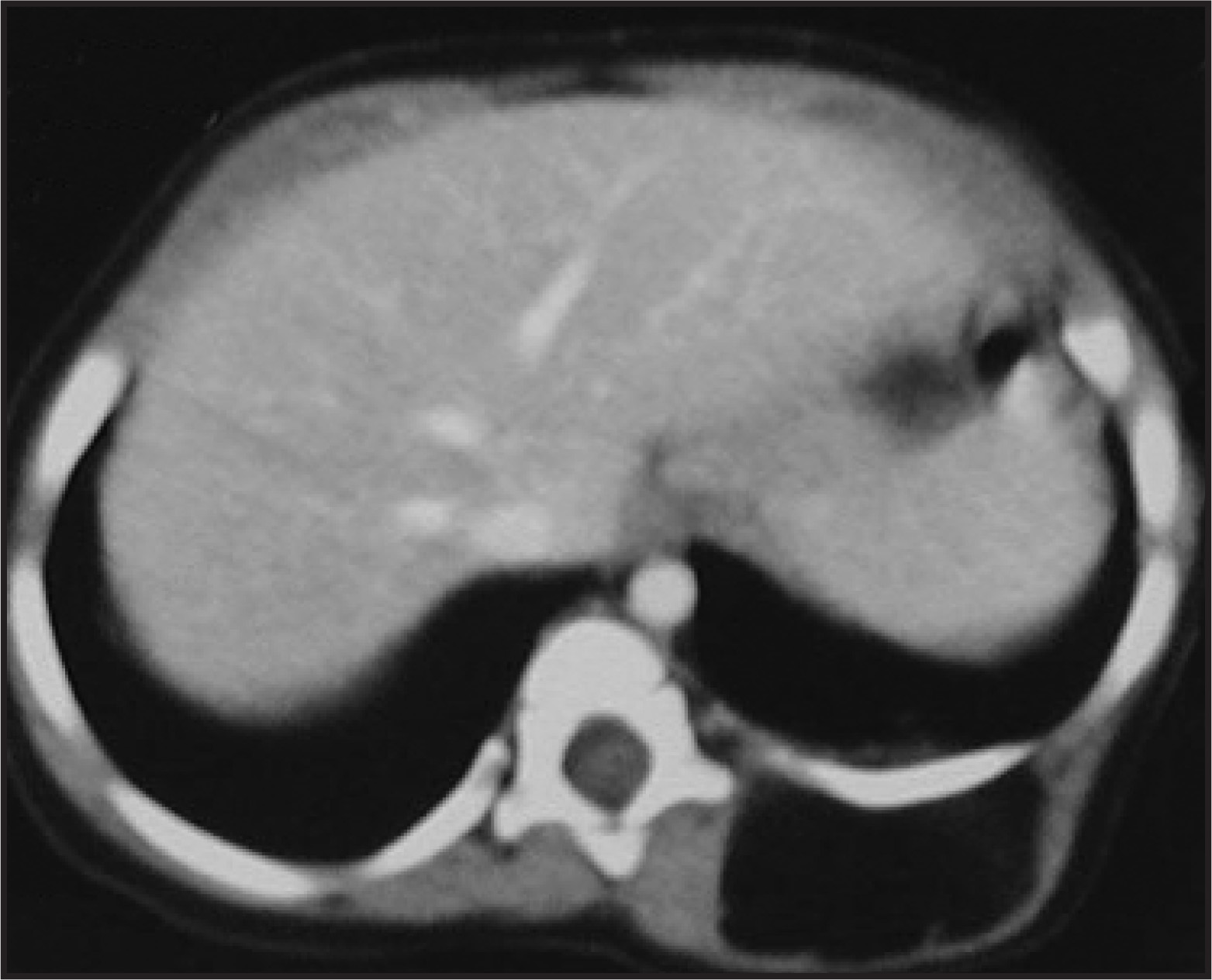
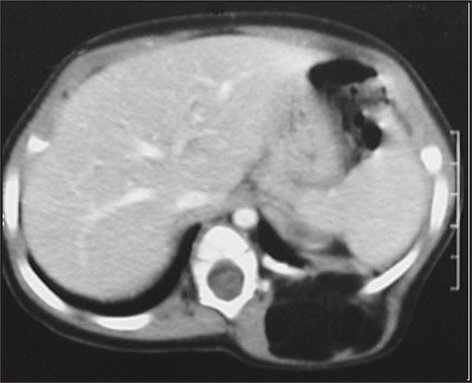
A 7-month-old girl presented with a 2-month history of a growing mass over her left posterior chest wall. The mass was soft and painless on palpation, beneath a normal skin. Physical examination was otherwise normal. A chest radiograph was unremarkable. Computed tomography (CT) of the chest was performed (Figures 1 and 2).
IMAGING FINDINGS
CT revealed an ovoid space-occupying lesion that was located into the paravertebral muscles (Figure 1). This lesion was of low attenuation, similar to that of subcutaneous fat, and contained soft tissue septations (Figures 1 and 2). The muscles adjacent to the lesion appeared to be displaced but were not invaded or atrophic. The mass bulged into the chest cavity through the intercostal spaces without rib involvement (Figure 2). The tumor was resected in its entirety. Histopathology was consistent with intramuscular lipoblastoma.
DIAGNOSIS
Thoracic wall lipoblastoma
DISCUSSION
Lipoblastoma is a rare tumor composed of fetal-embryonal fat that occurs exclusively in infants and children. Nearly 90% of these tumors occur in children <3 years of age. Males are affected more commonly than females by a ratio of 3:1.1 The tumor presents in 2 forms: a localized well-circumscribed lesion (lipoblastoma) and an unencapsulated diffuse type (lipoblastomatosis).2 Histologically, both forms are composed of lipoblasts, a plexiform capillary network, and myxoid stroma. Lipoblastoma most commonly involves the extremities.3 Other reported locations include the trunk, face, neck, mediastinum, and abdomen.3
Prognosis is excellent despite the tendency of the mass to invade locally and to recur if it is incompletely excised.1 When these tumors arise from the neck or mediastinum, they can grow rapidly, leading to recurrent respiratory infection or asphaxia. Otherwise, they can be detected as rapidly swelling, elastic, soft masses without any other symptoms. They can cause abdominal pain and vomiting if they arise in the peritoneal cavity.
Plain radiography in patients with lipoblastoma generally shows a nonspecific soft tissue mass. If superficial or large, the mass may show low density similar to fat. Ultrasound shows a homogeneous hyperechoic mass. On CT scans, lipoblastomas show fatty density and may contain soft tissue septa.
Although the tumor has a relatively rich capillary network, it does not enhance after injection of contrast media. This lack of enhancement suggests that the lesion exhibits relatively poor blood flow.4,5
Differential diagnosis of adipocytic tumors include mainly lipomas and liposarcomas. Rarely, other lesions such as myxoid tumors or hemangiomas that invade adipose tissue and/or engulf a portion of it may also simulate a fatty lesion. Lipomas tend to have well-defined borders, an exclusively fatty density, and a superficial location; they tend to displace rather than infiltrate adjacent structures.
Liposarcoma is the second most common soft tissue sarcoma encountered in adults, but it is rare in infants and young children.6 It usually appears as a nonspecific inhomogeneous mass with both soft tissue and fatty components, but it may also exhibit imaging findings similar to those of lipoblastoma. Liposarcomas are extremely rare in children <10 years of age; the myxoid variant represents the most frequent histologic subtype.6
Histologic differentiation of lipoblastoma from myxoid liposarcoma may be difficult, and cell cultures from fresh tissue in order to determine the tumor karyotype have been recommended.7
Age is an important parameter when confronted with growing, ill-defined fatty tumors that contain soft tissue components. In order to diagnose lesions preoperatively and to avoid large mutilating procedures, patient age must be considered in the differential diagnosis.
CONCLUSION
Lipoblastoma is a rare tumor of embryonal fat cells that occurs in early childhood. Differentiating it from other tumors, particularly liposarcoma, is very important. The definitive diagnosis is made only by the histopathologic examination.
References
- Coffin C. Lipoblastoma: An embryonal tumor of soft tissue related to organogenesis. Semin Diagn Pathol. 1994;11:98-103.
- Chung E, Enzinger F. Benign lipoblastomatosis. An analysis of 35 cases. Cancer. 1973;32:482-492.
- Chang P, Teng R, Tsou Yau K. Lipoblastomatosis in a newborn: Case report. Pediatr Surg Int. 1997;12:71-72.
- Black W, Burke J, Feldamn P. CT appearance of cervical lipoblastoma. J Comput Assist Tomogr. 1986;10:696-698.
- Kwak J, Ha D, Kim Y, Shim J. Lipoblastoma of the parietal pleura in a 7-month-old infant. J Comput Assist Tomogr. 1999;23:952-954.
- Mentzel T, Calonje E, Fletcher C. Lipoblastoma and lipoblastomatosis: A clinicopathological study of 14 cases. Histopathology. 1993;23:527-533.
- Miller G, Yanchar N, Magee J, Balir G. Tumor karyotype differentiates lipoblastoma from liposarcoma. J Pediatr Surg. 1997;32:1771-1772.
Citation
. RCOM RADIOLOGICAL CASE OF THE MONTH. Applied Radiology. 2007;36(12):44-46. doi:10.37549/AR1577.