RCOM RADIOLOGICAL CASE OF THE MONTH
Applied Radiology — Vol. 37 , Issue 1 , pp. 34 -36
DOI: 10.37549/AR1579
Published: January 1, 2008
Categories
CASE SUMMARY
A 10-year-old mentally disabled boy presented to the emergency room after a fall. Lateral X-rays of the skull were obtained (Figure 1). Upon questioning the parents, his history of long-standing epilepsy was revealed. The child also had a “port-wine stain” on the right side of his face along the distribution of the trigeminal nerve.
IMAGING FINDINGS
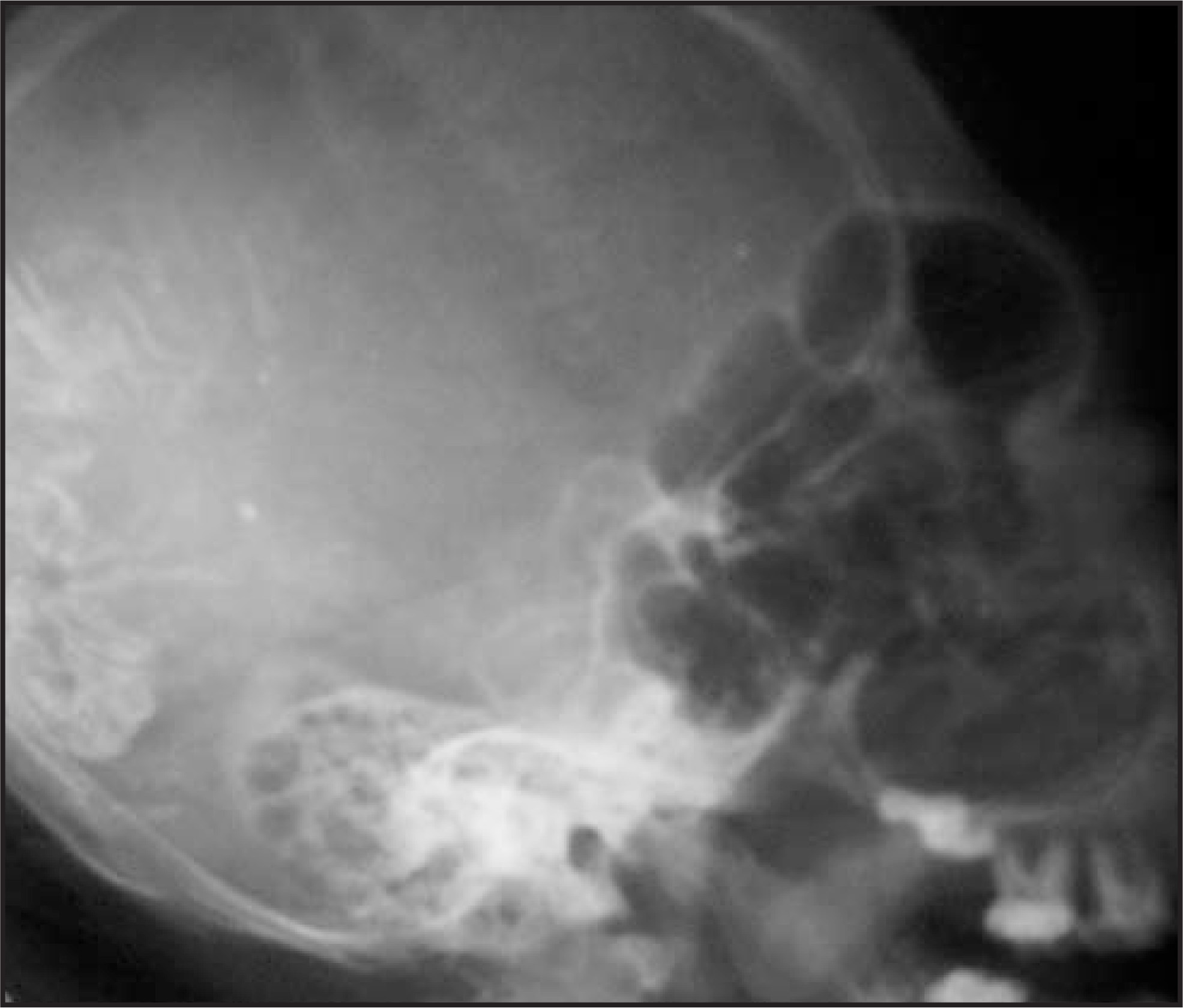
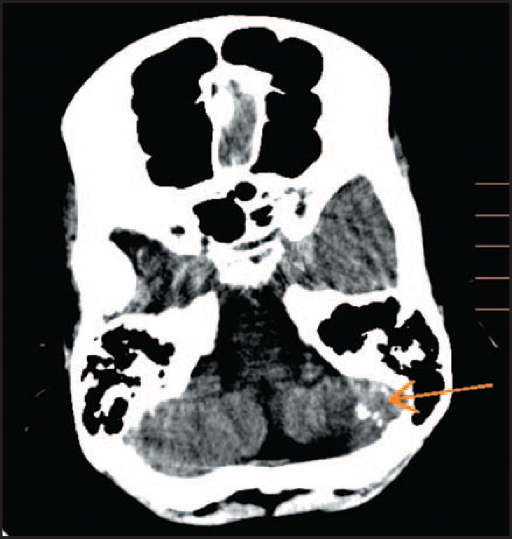
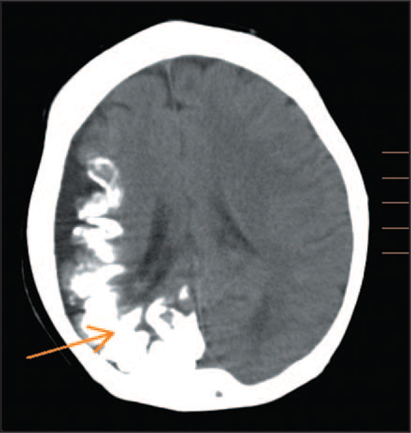
A lateral X-ray of the skull showed the typical gyriform calcification, “tram-track” calcification, and enlargement of the paranasal sinuses (Figure 1). A computed tomography (CT) scan subsequently revealed extensive gyriform calcification in the right parieto-occipital region with ipsilateral cerebral cortical atrophy. Calcific foci were also noted in the left cerebellar hemisphere (Figures 2 and 3).
DIAGNOSIS
Sturge-Weber syndrome
DISCUSSION
Sturge-Weber syndrome (encephalotrigeminal angiomatosis) is a sporadic phakomatosis of unknown etiology that may be characterized by1:
“Port wine” vascular nevus flammeus in the trigeminal nerve distribution;
Leptomeningeal venous angiomatosis;
Seizures, dementia, hemiplegia;
Hemianopsia, bupthalmos, and glaucoma;
Possible associated cleral and choroidal angiomata;
Possible associated angio-osteohypertrophy of Klippel-Trenaunay syndrome; and/or
Intracranial calcification, a common radiologic manifestation.
In patients with Sturge-Weber syndrome, X-ray findings in the skull usually show a tram-track pattern of calcification that is caused by calcification in opposing gyri on either side of an intervening dilated sulcus. On CT, curvilinear calcifications in a gyral pattern are often seen, primarily in the occipital and posterior parietal lobes ipsilateral to the facial angioma. Progressive cortical atrophy, ipsilateral calvarial thickening, and enlargement of the paranasal sinuses and mastoid may also be seen. On contrast-enhanced CT, very prominent medullary and subependymal veins may be seen in some cases. Magnetic resonance imaging and angiography can provide detailed information about vascular anomalies.2,3
CONCLUSION
Sturge-Weber syndrome is sporadic neurocutaneous syndrome. Its imaging findings in patients presenting with facial angioma and epilepsy are classic.
References
- Elster A, Chen M. MR imaging of Sturge-Weber syndrome: Role of gadopentetate dimeglumine and gradient-echo techniques. AJNR Am J Neuroradiol. 1990;11:685-689.
- Benedikt R, Brown D, Walker R. Sturge-Weber syndrome: Cranial MR imaging with Gd-DTPA. AJNR Am J Neuroradiol. 1993;14:409-415.
- Martí-Bonmatí L, Menor F, Poyatos C, Cortina H. Diagnosis of Sturge-Weber syndrome: Comparison of the efficacy of CT and MR imaging in 14 cases. AJR Am J Roentgenol. 1992;158:867-871.
Citation
. RCOM RADIOLOGICAL CASE OF THE MONTH. Applied Radiology. 2008;37(1):34-36. doi:10.37549/AR1579.