Renal Arteriovenous Fistula
Applied Radiology — Vol. 55 , Issue 5
Published: June 1, 2026
1 UCLA Department of Biology, University of California, Los Angeles, California
2 Community Regional Medical Center, Fresno, California
* Corresponding author: Luke Carson (lukecarson32@gmail.com)
Abstract
Arteriovenous (AV) fistulas are rare vascular anomalies that can cause high-output cardiac failure due to significant AV shunting. We report a case of a middle-aged patient presenting with progressive heart failure and abdominal pain. The patient was initially suspected of having hydronephrosis on grayscale US. However, color Doppler imaging subsequently revealed extensive vascular flow within the renal pelvis, excluding obstruction. CTA confirmed a large AV fistula between the right renal artery and right renal vein, with marked dilation of the renal vasculature and inferior vena cava. This case highlights the importance of Doppler imaging in avoiding misdiagnosis and demonstrates the role of CTA in characterizing renal vascular pathology. Early recognition of renal AV fistulas is critical given their potential for severe hemodynamic consequences.
Keywords
arteriovenous fistula, renal arteriovenous fistula, high-output heart failure
Categories
Case Summary
An adult with a several-month history of progressive congestive heart failure presented to the emergency department with shortness of breath and generalized abdominal pain. Initial grayscale-only abdominal US demonstrated findings interpreted as severe right hydronephrosis. The patient was discharged but presented several weeks later to his primary care provider with continued shortness of breath, prompting concern for renal pathology based on the prior imaging findings.
Imaging Findings
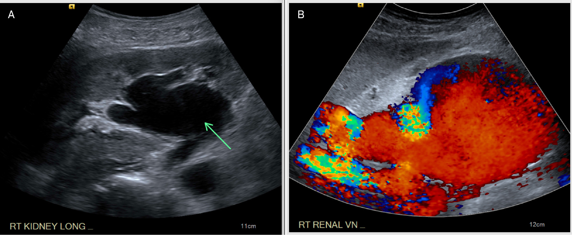
Grayscale sagittal US of the right kidney (Figure 1) demonstrated an anechoic region within the renal hilum concerning for renal collecting system dilatation. Color Doppler imaging, however, revealed extensive arterial and venous flow within the same region, excluding obstruction and raising concern for a high-flow vascular abnormality. Marked turbulent flow was identified within a markedly dilated renal vein, suggesting the presence of an AV shunt.
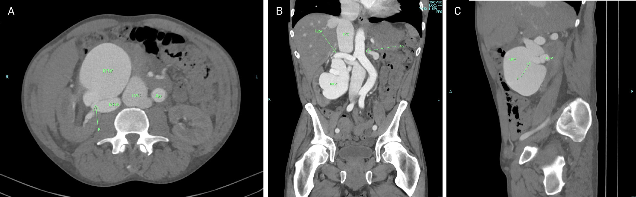
Axial CTA (Figure 2) confirmed a large renal AV fistula with marked dilation of the right renal artery, right renal vein, and inferior vena cava. The right renal artery measured up to 1.6 cm in diameter, with direct communication to the right renal vein at the renal hilum. The right renal vein measured up to 9.8 cm in diameter centrally, tapering at its junction with the inferior vena cava. The inferior vena cava was enlarged, with reflux into the hepatic veins reflecting elevated right-sided pressures. Coronal and sagittal images (Figure 2) further delineated the fistulous tract and extent of venous dilation, with associated mass effect on adjacent structures.
Diagnosis
Congenital right renal AV fistula resulting in high-output cardiac failure.
Differential diagnosis includes hydronephrosis, renal vein thrombosis, venous malformation, and arteriovenous malformation (AVM).
Discussion
Renal AV fistulas are abnormal direct communications between the renal arterial and venous systems. Most of these entities are acquired, accounting for approximately 70% of cases, but about 20% are congenital.1 Acquired fistulas most often result from iatrogenic injury following renal biopsy, surgery, or other percutaneous interventions.2 Less commonly, they may arise from trauma, inflammation, renal malignancy, or rupture of a renal artery aneurysm.2 Their reported incidence ranges from 0.3% to 19% in native kidneys and 6% to 8% in renal allografts.1 Renal AV fistulas significantly alter normal hemodynamics by allowing arterial blood to bypass the renal parenchyma and shunt directly into the venous system. This abnormal flow leads to progressive dilation of the renal vein and inferior vena cava and increases venous return to the heart. Over time, the chronic volume load may precipitate high-output cardiac failure, particularly in large, high-flow fistulas.3,4
Clinical manifestations include hematuria, flank pain, abdominal bruit, hypertension, and, in severe cases, high-output cardiac failure.2,5
Management depends on fistula size and symptomatology. Small, asymptomatic AV fistulas may resolve spontaneously, whereas large or symptomatic lesions generally require intervention. Endovascular embolization is the preferred treatment owing to its minimally invasive nature and high success rates.6 Surgical options, including arterial ligation or nephrectomy, are typically reserved for cases where embolization is unsuccessful or not feasible.1
The differential diagnosis in this case also includes congenital renal AVM. These lesions may present clinically in a similar fashion to renal AV fistulas and are treated using similar endovascular techniques. However, renal AVMs are characterized by abnormal communication between the renal arterial and venous systems through multiple tangled, dilated, and tortuous vessels forming a discrete vascular nidus. They may occur in isolation or in association with Osler-Weber-Rendu syndrome.7
In this case, there was no history of Osler-Weber-Rendu syndrome, no vascular nidus identified on CT angiography, and no history of hereditary hemorrhagic telangiectasia or prior renal intervention. Although an unrecognized remote traumatic etiology cannot be entirely excluded, the absence of identifiable acquired risk factors supports a presumptive congenital origin. This patient was successfully treated with endovascular embolization via arterial access.
Conclusion
Renal AV fistulas are rare but potentially life-threatening vascular anomalies. This case demonstrates how grayscale US findings may mimic hydronephrosis and emphasizes the essential role of color Doppler in detecting abnormal vascular flow. CTA allows definitive diagnosis and treatment planning. Early recognition is critical, as prompt intervention can prevent progressive cardiac decompensation and reduce morbidity.
References
- Serrado M, Kusel K, Jones J. Renal arteriovenous fistula.
- Tiwary N, Landis G, Siegel D, Etkin Y. Renal arteriovenous fistula associated high-output heart failure treated with embolization. J Vasc Surg Cases Innov Tech. 2023;9(1). doi:10.1016/j.jvscit.2022.07.012.
- Maldonado J, Sheps S, Bernatz P, Deweerd J, Harrison E. Renal arteriovenous fistula. a reversible cause of hypertension and heart failure. Am J Med. 1964;37(4):499-513. doi:10.1016/0002-9343(64)90064-6.
- Mehta P, Dubrey S. High output heart failure. QJM. 2009;102(4):235-241. doi:10.1093/qjmed/hcn147.
- Laberge J. Interventional management of renal transplant arteriovenous fistula. Semin Intervent Radiol. 2004;21(4):239-246. doi:10.1055/s-2004-861558.
- deSouza N, Reidy J, Koffman C. Arteriovenous fistulas complicating biopsy of renal allografts: treatment of bleeding with superselective embolization. AJR Am J Roentgenol. 1991;156(3):507-510. doi:10.2214/ajr.156.3.1899745.
- Wang J. Renal arteriovenous malformation. Urol Sci. 2012;23(2):52-54. doi:10.1016/j.urols.2012.04.001.
Disclosures
The authors have no conflicts of interest to disclose. None of the authors received outside funding for the production of this original manuscript and no part of this article has been previously published elsewhere.
Citation
. Renal Arteriovenous Fistula. Applied Radiology. 2026;55(5). doi:10.37549/AR-D-26-0009.