Report from the Annual Meeting of the Society of Nuclear Medicine
Applied Radiology — Vol. 33 , Issue 8 , pp. 41 -43
DOI: 10.37549/AR1273
Published: August 1, 2004
Categories
Hybrid imaging, particularly the combination of single-photon emission computed tomography (SPECT) and computed tomography (CT), was the highlight of the 51st Annual Meeting of the Society of Nuclear Medicine (SNM), held June 19–23, 2004 in Philadelphia, PA.
Siemens Medical Solutions (Hoffman Estates, IL) and Royal Philips Electronics (Best, The Netherlands) both introduced new SPECT/CT systems that feature multislice CT (MSCT) components.
Siemens introduces TruePoint
The Siemens’ system, TruePoint SPECT/CT, integrates the company’s e.cam SPECT imaging technology with their MSCT technology on the Siemens Symbia platform.
This system allows clinicians to perform three separate studies—SPECT, MSCT, and SPECT/CT—all on one unit, designed to fit in a room approximately 11′ × 21′. The system’s scalable architecture allows the company to offer a variety of Symbia models, ranging from systems offering attenuation correction to premium MSCT capabilities. Symbia offers various MSCT configurations with speeds of up to 0.6 seconds per rotation, allowing acquisition of a diagnostic quality CT scan in <30 seconds.
“The addition of diagnostic CT will improve all aspects of SPECT imaging, and minimize the ‘scientific guestimates’ we’ve relied upon previously,” said Homer A. Macapinlac, MD, Deputy Chairman, Department of Nuclear Medicine, M.D. Anderson Cancer Center, Houston, TX. “In fact, nuclear medicine has been nicknamed ‘unclear medicine’ in the past. Having the ability to fuse and register SPECT images with the anatomical images of diagnostic CT scans will instead make it the ‘clear medicine’ for the future.”
The integration of the company’s proprietary syngo software platform provides a common user interface and enables fully automated and integrated user interaction counting tools, so that SPECT and CT datasets can be acquired and processed on a single workstation.
“The introduction of new technology, like TruePoint SPECT·CT, offers the opportunity to re-examine how the diagnostic process works—the order in which studies are performed and the order in which the whole care pathway is constructed,” said Macapinlac. “It may well be that a hybrid imaging device that’s capable of high-quality scans in both SPECT and CT will offer all the diagnostic and therapeutic advantages you might expect. It is also closer to ‘one-stop shopping’ for the patient, who only has to make one appointment and undergo a seamless procedure.”
Philips unveils Precedence
The new Philips Precedence SPECT/ CT system incorporates the company’s SPECT system with the Brilliance MSCT system (Figure 1). It can also be used for either SPECT or MSCT imaging individually, or as a hybrid system. Based on the company’s Vequion information technology (IT) platform, the Precedence Workspace facilitates multimodality acqui- sition, processing, and review, as well as integration with other imaging modalities.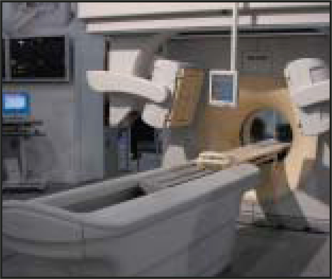
“This breakthrough hybrid technology offers unique benefits for all parties: better, quicker exams for patients, higher levels of confidence for physicians, and a competitive advantage, long-term investment, and reduced costs for hospitals and research facilities,” said Ian Farmer, Vice President, SPECT and PET, for Philips.
Philips also highlighted several other new products and works in progress:
The JETStream Workspace, a nuclear medicine workstation, is designed for enterprise-level implementation. It encompasses display, processing, review, reporting, and image archiving in a single environment.
GEMINI with Brilliance 16 Power CT system, multislice, open positron emission tomography (PET)/CT, is now available in a mobile setting.
GE Healthcare highlights cardiac applications
GE Healthcare (Chalfont St. Giles, UK), exhibiting for the first time under the new division name, focused much of their exhibit on the com-pany’s cardiac solutions, including advanced applications on the Discovery ST PET/CT system, the Infinia Hawkeye hybrid imaging technology, and Myoview, the company’s myocardial perfusion imaging agent.
“We are entering a new era in noninvasive cardiology, where hybrid imaging will play a major role in managing heart disease,” said Karim Benali MD, MSc, Global Manager, Cardiology PET/CT and Nuclear Medicine at GE Healthcare.
The latest enhancement to the Discovery PET/CT system is a new advanced application that aligns the vascular coronary tree created by CT images on the 3D functional data from PET images. Called HeartFusion, this application allows physicians to quantify and analyze the impact of the atherosclerotic lesions on the heart muscle.
“PET/CT with HeartFusion provides a tool to noninvasively evaluate fused coronary anatomy and myocardial perfusion information for a more comprehensive assessment of coronary artery disease,” said Ernest V. Garcia, PhD, Professor and Vice Chairman (Research), Department of Radiology, Emory University Hospital. “This exam has the potential to eventually replace diagnostic invasive coronary arteriography in specific patient populations.”
The company also featured the latest enhancements to the nuclear medicine system, Infinia, which can now be combined with their Hawkeye SPECT/CT technology (Figure 2). This system provides CT-based attenuation correction for cardi-ology SPECT imaging.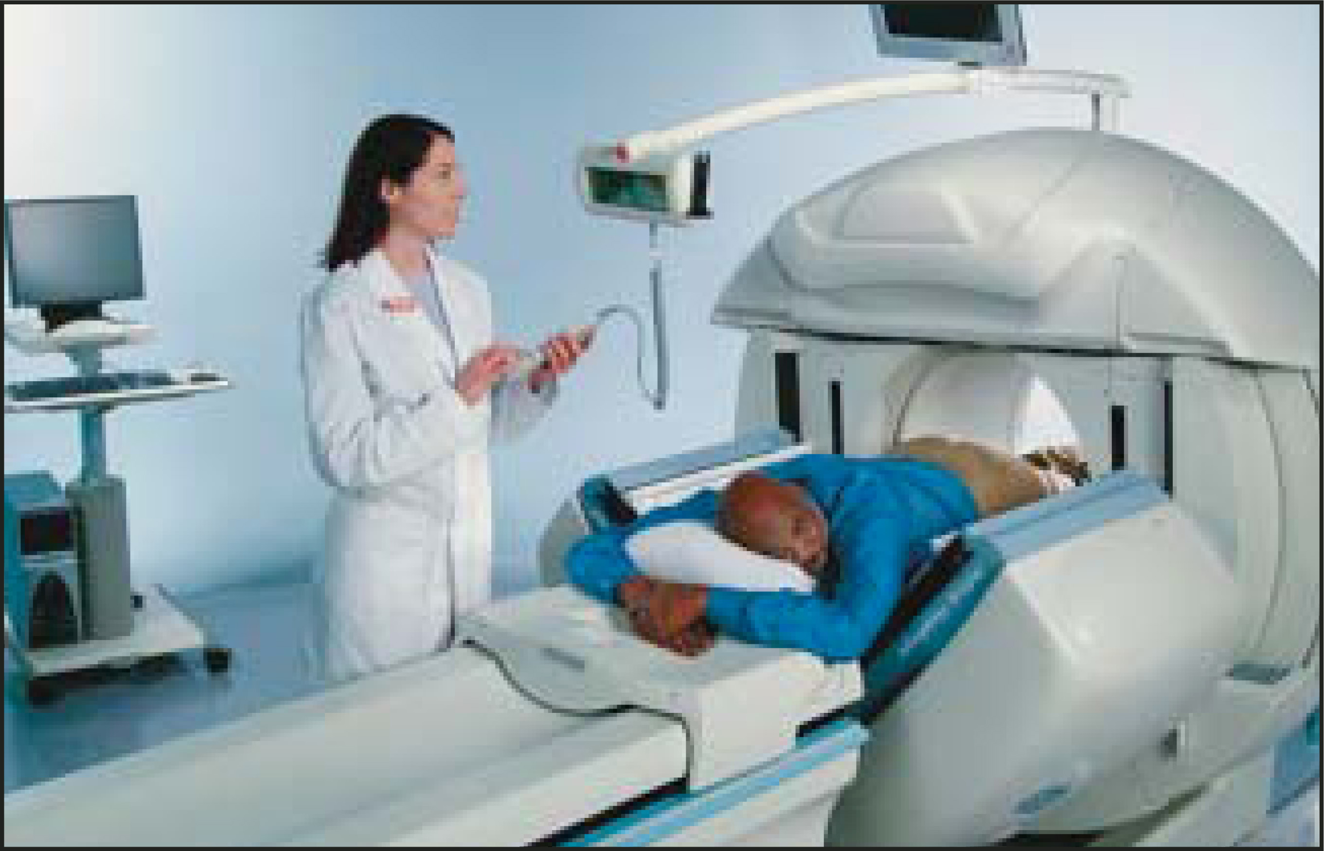
The system now includes a 5-mm CT axial resolution with Hawkeye SPECT for enhanced SPECT/CT lesion detection, fan-beam collimation for enhanced brain SPECT scanning, and a camera-based PET option for extended clinical utility. In addition, new detector motions were added to accommodate a greater range of patient positions.
“Flexibility is a major issue in medical imaging,” said Professor Ora Israel, Nuclear Medicine Physician, Rambam Hospital, Haifa, Israel. “With Infinia, we can examine patients quickly—whether they’re standing, seated, confined to a wheelchair, or on a stretcher.”
Infinia systems are integrated with the Xeleris workstations through Ignite, which enables the operator to complete a scan-and-review cycle in a single click.
The company also displayed Myoview (Kit for the Preparation of Technetium Tc-99m Tetrofosmin for Injection). Used to help diagnose heart disease by visualizing blood flow to the heart muscle, this agent was granted FDA approval for use in the assessment of left ventricular function in patients with coronary artery disease.
CTI and PETNET focus on education
PETNET Solutions, a subsidiary of CTI Molecular Imaging (Knoxville, TN) introduced two new educational products at this year’s SNM: PET/CT University and Taking it to the Streets: Marketing PET to Oncologists Training Seminar.
PET/CT University was designed as a training resource for interpreting physicians, referring physicians, and technologists. The educational goals include im- proving PET and PET/CT interpretation skills, increasing knowledge of PET utility, and establishment of PET practice guidelines for the referring physician.
The program consists of a range of online case studies, which include full datasets with DICOM viewer and pathology reports, as well as assigned readings in PET/CT protocols and algorithms, monthly quizzes, and a final exam. In addition, the program has a library of reference articles, an “Ask the Professor” section, and bulletin boards.
The Taking it to the Streets program was developed to help to educate the people responsible for promoting a PET imaging center to the medical community. “Knowledge on the appropriate utilization of PET in oncology combined with selling and presentation skills increases their confidence when interacting with referring physicians,” said Joan Washburn, Vice President of Sales and Marketing.
Course modules include a PET market overview and updates, lessons in the clinical utility of PET in oncology, basic selling and presentation skills, lessons on how to handle objections, and how to market PET to referring physicians.
The company notes that those who complete this seminar “will have increased knowledge about PET imaging and its applications. They will be able to educate referring physicians about the benefits of PET imaging for their patients, including how and when to order a PET scan.”
In addition, CTI Molecular Imaging also announced the formation of its new research and development entity, CTI Molecular Technologies, Inc. (MTI). The company reports that this unit will be “the focal point of biomarker research and development activities” within CTI.
“MTI serves as the hub within the company for the development of new molecular biomarkers for a wide array of disease targets, as well as for the development of enabling technologies designed to meet the needs of biologists and pharmaceutical scientists to help integrate and facilitate their research in developing more informative diagnostics and more effective therapeutics,” said Ronald Nutt, PhD, company President and Chief Executive Officer.
Toshiba highlights gamma camera enhancements
Toshiba America Medical Systems, Inc. (Tustin, CA) demonstrated the latest upgrades to its t.cam Signature Series gamma camera system. This variable angle, dual-detector system now features the e.soft version 3.5 nuclear medicine software platform with workflow environment enhancements. The company also highlighted the new e.soft express version 2.0 remote viewing software solution designed for cardiac, general nuclear medicine, and PET/CT data.
The latest enhancements to the e.soft version 3.5 software platform enable the use of the quality control activity to motion correct data acquired via Profile Attenuation Correction. This feature promotes greater diagnostic accuracy during cardiac examinations, as well as during the imaging of organs surrounding the heart.
The optional e.soft express version 2.0 software enables remote review of e.soft data. Versions are also available for cardiac, general nuclear medicine, and PET/CT data with complete workflow functionality.
According to Martha Bonino, Product Manager, Nuclear Medicine Business Unit, Toshiba America Medical Systems, “The t.cam delivers the most innovative technology to meet today’s clinical needs for superior image quality and efficiency in nuclear medicine.”
Other nuclear medicine news
Dilon Technologies uses nuclear imaging for breast cancer diagnosis
Dilon Technologies (Newport News, VA) is using nuclear imaging for diagnosis of breast cancer with its breast-specific gamma camera system, the Dilon 6800. Unlike anatomic breast imaging, scintimammography looks at the metabolic difference between breast tissue and breast cancer.
An early multicenter study found that scintimammography’s sensitivity for detect-ing breast cancer was similar to mammography, but its specificity was higher. “Unlike mammography, where the specificity is approximately 20% to 30%, the specificity was >80%,” noted Rachel Brem, MD, Director of Breast Imaging and Intervention at George Washington University Medical Center and Professor of Radiology and Biomedical Engineering, Vice Chairman for Faculty Development and Research. A second study then found that this method was not affected by breast density.
These early studies, however, used a traditional gamma camera, and the average cancer found was 2.2 cm. The system was much less effective with smaller tumors. “That’s because the intrinsic resolution of the general purpose gamma camera is limited,” explained Brem. “The other problem is that the traditional gamma camera is not designed for optimal breast imaging. That’s why the high-resolution, breast-specific Dilon 6800 gamma camera was developed.”
The camera incorporates more than 3,000 3-mm × 3-mm scintillation crystals and 48 position-sensitive photomultiplier tubes (PMT) arranged in a 6 × 8-inch array. The camera is mounted on a 40-inch gantry arm that can be positioned as needed. This flexibility allows for a variety of views not normally seen in breast imaging, including craniocaudal views performed with the detector against the patient’s chest wall. Standard mammographic views can also be obtained to allow for the direct comparison of the anatomic information provided by X-rays with metabolic information provided by the scintimammography.
“This is a really novel approach to breast cancer diagnosis, and it really asks questions that allow for improved diagnosis that were simply not asked before,” concluded Brem.
Coming next month:
Film Mammography in the Digital Age
Citation
. Report from the Annual Meeting of the Society of Nuclear Medicine. Applied Radiology. 2004;33(8):41-43. doi:10.37549/AR1273.