Respiratory Motion Management in Lung Cancer Radiation Therapy: A Practical Review and Decision Framework for Technique Selection and Treatment Delivery
Applied Radiation Oncology — Vol. 15 , Issue 3
Published: September 1, 2026
1 Department of Radiation Oncology, Taussig Cancer Institute, Cleveland Clinic, Cleveland, OH
2 Cleveland Clinic Lerner College of Medicine of Case Western Reserve University, Cleveland, OH
3 Department of Radiation and Cellular Oncology, The University of Chicago, Chicago, IL
4 Department of Radiation Oncology, Sidney Kimmel Medical College, Thomas Jefferson University, Philadelphia, PA
* Corresponding author: Nader Ghassemi (ghassen@ccf.org)
Abstract
Respiratory motion remains a defining geometric and dosimetric challenge in lung radiation therapy, but not every moving target requires advanced motion control. This narrative review synthesizes existing guidance into a consequence-based clinical checklist for selecting and verifying the simplest reproducible strategy for an individual patient. The clinically relevant question is whether the observed motion is consequential for target coverage, organ-at-risk sparing, image-guidance confidence, or treatment-delivery robustness. 4-dimensional CT (4DCT) anchors motion assessment and selection of free-breathing vs managed respiratory-state workflows. For many patients, internal target volume (ITV)-based planning with robust image guidance remains an appropriate baseline strategy when motion can be safely encompassed. Escalation to breath hold, respiratory gating, or abdominal compression is best reserved for situations in which the managed state provides a patient-specific geometric or dosimetric advantage, can be reproduced consistently, and can be verified throughout treatment. External surrogates and surface guidance can support monitoring, but they do not replace internal target verification. This review summarizes 4DCT-based motion assessment, ITV-based planning, gating, breath hold, abdominal compression, surrogate monitoring, image guidance, and quality assurance, emphasizing that the preferred strategy is usually the least complex approach that preserves coverage, protects organs at risk, and can be delivered reproducibly.
Keywords
lung cancer, respiratory motion management, stereotactic body radiation therapy, 4-dimensional CT, internal target volume, respiratory gating, breath hold, abdominal compression, surface-guided radiation therapy, quality assurance
Categories
Introduction
Respiratory motion is a defining geometric and dosimetric problem in lung radiation therapy, not because every moving tumor requires an advanced intervention, but because unrecognized or poorly managed motion can compromise target coverage, enlarge normal-tissue exposure, and weaken confidence in image guidance.1-3 The practical question is therefore not which motion-management device is most sophisticated; it is whether the observed motion is consequential for the intended plan, and whether a simpler or more selective strategy can be reproduced safely over the course of treatment.1,4-6
Current practice reflects that consequence-based approach. In the American Association of Physicists in Medicine (AAPM) Task Group 324 survey, most respondents reported using respiratory motion management, but internal target volume (ITV)-based approaches remained more common than gating or tracking workflows.1,4 That pattern is clinically reasonable: advanced methods can narrow the motion envelope in selected patients, but they also add coaching, treatment time, image-verification demands, quality assurance (QA) requirements, and additional opportunities for error.1,5,7,8 The remaining gap is not a lack of technique-specific guidance, but rather the absence of a concise, consequence-based pathway linking simulation findings to treatment-room verification and decisions about de-escalation.
Accordingly, this review is intended for clinical teams choosing among motion-encompassing planning, respiratory gating, breath hold, abdominal compression, and surrogate-based monitoring. The central thesis is deliberately conservative: the best technique is usually the least complex approach that preserves target coverage, protects organs at risk (OARs), and can be verified and reproduced safely.1,4,5,8 The emphasis is photon-based lung radiation therapy, including stereotactic body radiation therapy (SBRT) and conventionally fractionated treatment, with technology-specific discussion limited to workflows represented in the cited clinical and technical literature.
Respiratory Motion as a Clinical Problem
Respiratory motion becomes clinically important when it changes a decision. In practice, this may mean changing the ITV/planning target volume (PTV) approach, increasing margins, shifting from free breathing to a managed respiratory state, approaching lung V20/MLD/V5 or serial-organ constraints, or reducing confidence in image registration. For a peripheral upper-lobe tumor with limited excursion, free-breathing ITV-based planning may be robust and efficient. For a lower-lobe or peridiaphragmatic lesion, tumor motion may be larger and more phase-dependent, and a motion-encompassing plan may enlarge the treated volume enough to affect lung, heart, esophagus, or proximal bronchial tree constraints.1,2,9,10 The same measured motion also has different consequences in a 3-fraction or 5-fraction SBRT plan than in a conventional 30-fraction plan because SBRT uses high fractional dose, tight margins, and steep dose gradients.5-7,11
Motion uncertainty is also not confined to the planning CT. Respiratory baseline can shift, the tumor may not follow an external marker perfectly, and intrafraction variation can erode the apparent advantage of a narrow treatment window.12-15 This is why motion management should be evaluated as an end-to-end process: simulation, contouring dataset, dose calculation, image guidance, treatment delivery, and stop rules all have to describe the same respiratory state.1,16-18
The clinical stakes are particularly apparent in SBRT. SBRT established high local control for medically inoperable early-stage non-small cell lung cancer, but central anatomy introduced a different risk profile, with toxicity concerns that made image guidance, dose constraints, and geometric reliability non-negotiable.6,11,19,20 For conventionally fractionated thoracic radiation therapy, respiratory management is usually less about millimeter-scale gradient preservation and more about maintaining coverage while avoiding unnecessary lung and cardiac dose, a concern reinforced by modern data in stage III non-small cell lung cancer linking thoracic dose distributions to outcomes and toxicity.1,10,21
4DCT Simulation and Motion Assessment
Four-dimensional CT (4DCT) simulation and motion assessment remains the practical foundation for lung motion assessment because it provides a phase-resolved view of the target and thoracic anatomy during breathing.3,22-24 Its value is not the production of a single motion number, but the ability to assess tumor trajectory, phase dependence, proximity to OARs, and whether respiratory motion changes the contouring or dose-calculation problem.1,2,24
A useful simulation is both an imaging study and a feasibility test. If breath hold, gating, or abdominal compression is being considered, the simulation should show that the selected maneuver improves geometry or dosimetry for the patient.8,25-27 If a respiratory belt, marker block, spirometric device, or surface system will be used for sorting or delivery, the respiratory-monitoring chain should be as consistent as possible between simulation and treatment; differences between respiratory monitoring systems have been shown to matter in combined 4DCT and gated workflows.1,17,18
Maximum-intensity projection (MIP) can support ITV generation in selected lung lesions with adequate contrast, while average-intensity projection (AIP) images are often useful for free-breathing dose calculation. However, direct review of phase datasets remains important for tumors near the diaphragm, mediastinum, chest wall, or atelectatic lung.24,28,29 Irregular breathing and sorting artifacts can misrepresent target excursion or shape, so a poor-quality 4DCT should not be allowed to define a narrow margin or justify a complex motion strategy without additional verification.1,23,30 Table 1 summarizes the practical roles, common pitfalls, and verification implications of the main 4DCT-derived datasets used in lung radiation therapy planning.
| Dataset or Image Type | Practical Role | Main Advantage | Common Limitation | Verification Implication |
|---|---|---|---|---|
| Phase-resolved 4-dimensional CT images | Direct review of tumor motion, trajectory, deformation, and relationship to adjacent anatomy | Best representation of respiratory motion pattern across the breathing cycle | Can be affected by irregular breathing, sorting artifacts, or poor surrogate signal | Phase images should be reviewed before relying on MIP, AIP, or reduced-margin strategies |
| Maximum-intensity projection | ITV generation for selected lung lesions with good tumor-to-lung contrast | Efficient visualization of the full motion envelope | Can overestimate or underestimate target extent near mediastinum, diaphragm, vessels, atelectasis, or chest wall | Should not be used blindly; confirm target boundary on phase images |
| Average-intensity projection | Dose calculation for free-breathing treatment in selected cases | Represents time-averaged anatomy for free-breathing dose calculation | Does not fully show motion extremes or transient tumor positions | Should be paired with appropriate target definition from phase images or ITV dataset |
| Mid-ventilation or mid-position dataset | Planning around a representative respiratory position when residual motion is handled by margins or image guidance | May reduce treated volume compared with full ITV approaches | Requires careful motion modeling, image guidance, and residual uncertainty assessment | Margin reduction should be justified by institutional verification and reproducibility |
| Breath-hold CT | Planning in a managed respiratory state, commonly DIBH | Can reduce motion and improve thoracic geometry in selected patients | Depends on reproducible breath-hold depth and patient tolerance | Treatment imaging must confirm the target in the same breath-hold state |
| Gated-phase or gated-window dataset | Planning around the respiratory phase or amplitude range intended for gated delivery | Narrows the treated motion envelope | Sensitive to duty cycle, baseline drift, and internal-external correlation changes | Internal target verification in the gated state is essential |
Clinical Decision Framework
A practical decision framework starts with 4 questions. (1) Is the motion real and representative? (2) Does it affect target coverage, OAR sparing, registration confidence, or dose-gradient robustness? (3) Does a managed respiratory state provide a meaningful gain over robust free breathing? (4) Can the patient and institution reproduce and verify that state at every fraction?1,4,5,7 Figure 1 translates these questions into a consequence-based pathway for selecting the least complex technique that still preserves target coverage, OAR protection, and treatment-room verifiability. If the answer to the 2nd or 3rd question is “no,” escalation beyond motion-encompassing planning is often unnecessary. If the answer to the 4th question is “no,” escalation may be unsafe even when the planning dosimetry looks attractive.1,8,16,31
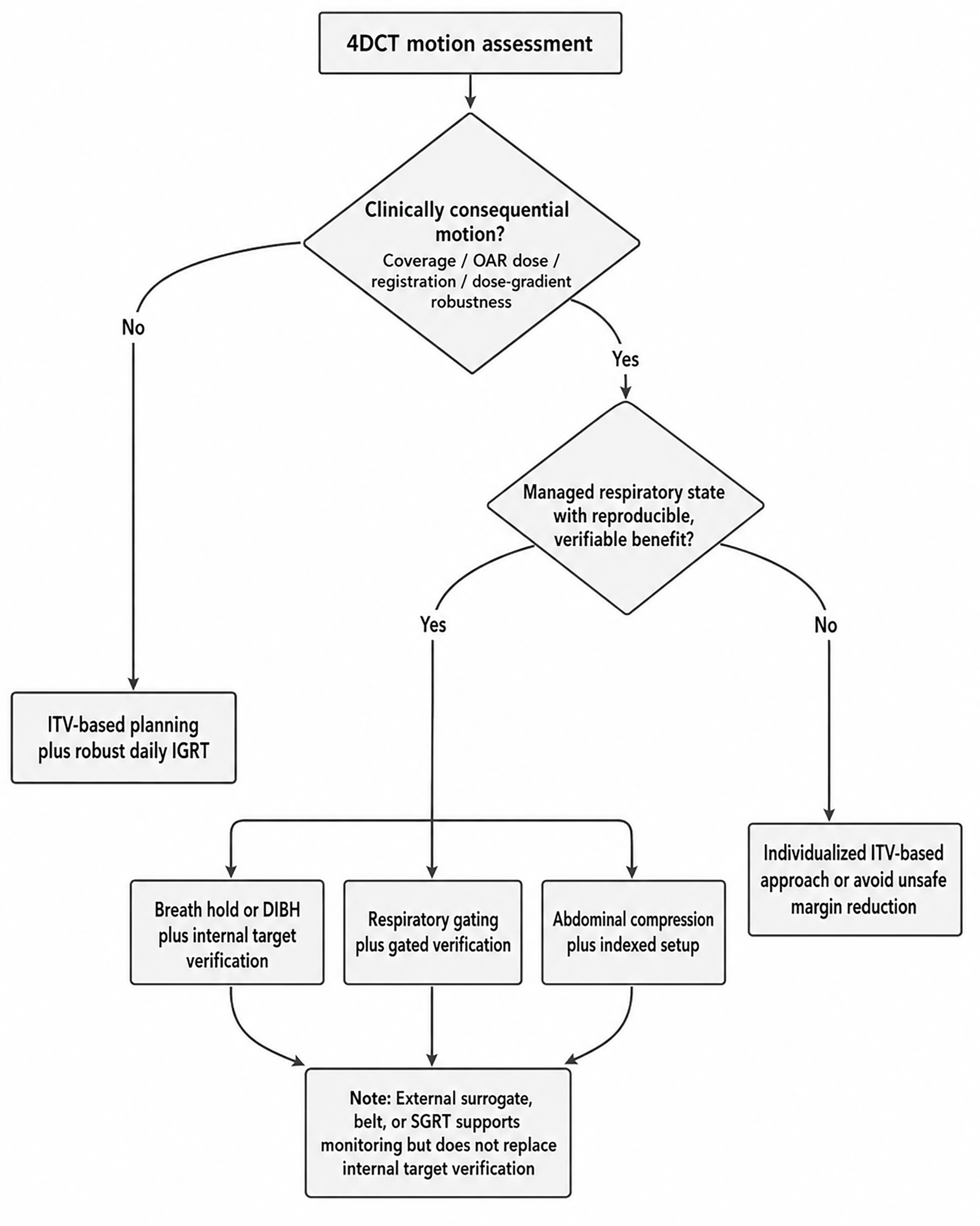
The decision should not be driven by motion amplitude alone. Tumor location, fractionation, OAR proximity, pulmonary reserve, patient comfort, breath-hold capacity, breathing regularity, treatment time, staffing, and image-guidance strength all influence the safest choice.1,4,7,21 In SBRT, the tolerance for residual geometric uncertainty is generally lower because high-dose gradients and hypofractionation magnify the consequences of a miss, especially for central tumors or targets close to serial thoracic structures.6,7,19,20
The resulting hierarchy is pragmatic rather than prescriptive and should be adapted to institutional equipment, team expertise, tumor visibility, patient tolerance, and treatment-room verification capability. ITV-based planning is the default when encompassing motion is dosimetrically acceptable and image guidance is robust.1,4,5,31 Breath hold can be favored when the patient can reproduce the hold state and the plan gains meaningful lung, heart, or target-stability benefit.8,32-34 Gating can be useful when motion is substantial, breath hold is not preferred or not feasible, and breathing is regular enough to support a clinically efficient window.1,25,35,36 Abdominal compression is best reserved for patients in whom simulation confirms a benefit and tolerance is acceptable.9,26,27 Table 2 summarizes how this hierarchy plays out clinically by comparing the best-fit scenario, expected gain, verification burden, and fallback pathway for each motion-management option.
| Clinical Scenario | Usually Favored Approach | Why It Fits | Main Caution | Fallback If Not Reproducible |
|---|---|---|---|---|
| Limited tumor motion and acceptable free-breathing dosimetry | ITV-based or motion-encompassing planning | Robust, efficient, and less dependent on patient coaching | Do not assume motion is limited if 4DCT quality is poor | Repeat or supplement motion assessment if uncertainty is high |
| Irregular breathing, cough, anxiety, or poor coaching tolerance | ITV-based planning with robust IGRT | More reliable than complex techniques that require reproducibility | Larger treated volume may increase OAR dose | Use conservative margins and daily image guidance |
| Large motion with reproducible breath hold and meaningful dosimetric gain | Breath hold or DIBH | Can reduce motion and may improve lung or cardiac sparing | Benefit must persist beyond simulation and coaching | Revert to ITV or gating if breath hold becomes inconsistent |
| Large motion, no reliable breath hold, but regular coached breathing | Respiratory gating | Narrows beam delivery to a selected respiratory window | Duty cycle, latency, and baseline drift may reduce benefit | Use ITV-based approach if gated delivery becomes inefficient or unstable |
| Lower-lobe or peridiaphragmatic lesion with compression benefit at simulation | Abdominal compression as adjunct | May reduce motion envelope or improve planning geometry while preserving free breathing | Benefit is patient specific, and tolerance may be limited | Remove compression and use ITV or another verified strategy |
| Reliable external monitoring, but uncertain internal-external correlation | Surrogate-assisted workflow with internal image verification | Useful for coaching, sorting, gating, or breath-hold monitoring | External signal does not equal tumor position | Avoid margin reduction based on surrogate trace alone |
| Central or ultracentral SBRT with steep gradients near critical structures | Individualized approach with strong IGRT and conservative uncertainty management | Small geometric errors may have high clinical consequence | Overly aggressive margin reduction can be unsafe | Prefer reproducible, verifiable technique over theoretically optimal technique |
| Conventional fractionation with acceptable dosimetry | ITV-based planning or simple motion-encompassing strategy | Efficiency and reproducibility may outweigh marginal geometric gains | Daily setup and anatomy changes still matter | Escalate only if motion clearly compromises coverage or OAR sparing |
ITV-Based and Motion-Encompassing Planning
ITV-based planning remains the baseline strategy for many lung radiation therapy patients because it is robust to irregular breathing and does not require repeated breath holds or a stable duty cycle.1,4 In peripheral lung SBRT, ITV-based planning remains compatible with implementation guidance when combined with appropriate immobilization, contouring discipline, and image guidance.5,7,31 In this context, robust image-guided radiation therapy (IGRT) generally means daily cone-beam CT (CBCT) or equivalent volumetric imaging, soft-tissue or tumor matching when visible, review of adjacent bronchovascular and bony anatomy when needed, physician review for high-risk or ambiguous matches, and predefined action thresholds. Its strength is not that it is crude, but rather that it can be highly reliable when the dosimetric price of encompassing motion is acceptable.1,4,5
The limitation of ITV-based planning is the treated volume. If excursion is substantial, encompassing the full respiratory envelope may increase uninvolved lung dose or worsen overlap with the heart, esophagus, chest wall, proximal bronchial tree, or great vessels.1,10,24 This tradeoff is most important when the free-breathing plan approaches OAR constraints or when the target lies near serial structures, as in central SBRT.6,7,19,20 A motion-encompassing approach is therefore a decision to accept a larger but robust envelope, not a reason to relax image guidance or QA.1,16,31
MIP-based ITV construction and related derived datasets should be used with attention to lesion visibility and anatomy. Underberg et al support MIP use for selected lung targets, but Borm et al highlight that MIP and AIPs can differ from the actual moving target representation depending on size, contrast, and motion.24,28,29 Mid-ventilation and other motion-representative strategies may reduce margins in selected workflows, but they require a validated residual-uncertainty model and strong treatment-room verification. Clinical validation and institutional experience remain important when margin reduction is used; Bellec et al specifically evaluated ITV vs mid-ventilation in lung SBRT using in-treatment 4D-CBCT and framed margin adequacy as an image-guided question.37-40
Respiratory Gating
Respiratory gating restricts beam delivery to a selected respiratory phase or amplitude window, so treatment occurs within a narrower motion envelope than unrestricted free breathing.1,35,36 The technique is most defensible when the observed motion is large enough to matter, the gating window produces a meaningful geometric or dosimetric advantage, and the patient can maintain a reproducible respiratory pattern that supports an acceptable duty cycle.1,25,36,41 In practice, the selected gate should balance residual-motion reduction with treatment efficiency and should be verified with fluoroscopy, respiratory-correlated CBCT, or another institutional imaging strategy when feasible. Saito et al found that 3-dimensional and craniocaudal clinical target volume (CTV) motion and craniocaudal CTV position were predictive of lung-dose reduction from respiratory gating, supporting selective rather than routine use.25
The main weaknesses of gating are operational and related to verification. Beam-on efficiency can decrease, treatment time can increase, and the workflow is vulnerable to latency, baseline drift, irregular breathing, and changes in internal-external correlation.14,15,36,41 A smooth external trace is not enough; gated treatment still leaves residual internal motion and requires confirmation that the internal target is in the intended state.13-15,42 This helps explain why gating is less common than ITV-based planning in practice, despite clear value in selected patients.1,4
For conventional fractionation, gating may be considered when a narrower motion envelope materially improves lung or heart dosimetry without creating an impractical workflow.1,10,25 For SBRT, gating can be attractive for larger excursions or lower-lobe/peridiaphragmatic targets near abdominal OARs when breath hold is not preferred or not feasible, but the team should explicitly account for residual motion in the gate, image-registration uncertainty, and the consequences of a missed fraction-level setup.5,7,40,43 Gating should be abandoned or de-escalated when the patient cannot maintain a stable pattern or when verification shows that the internal target does not match the external signal.1,14,15
Breath-Hold Techniques
Breath-hold techniques replace cyclic respiratory motion with repeated stable respiratory states during which imaging and treatment are performed. Active breathing control and deep inspiration breath hold (DIBH) have long been studied as methods to reduce breathing motion and, in some patients, alter thoracic geometry favorably.32,33,44,45 Modern implementation guidance emphasizes that breath hold is a workflow, not just a patient instruction: coaching, reproducibility testing, imaging in the hold state, and fallback criteria are required.8,44 End-exhale breath hold and adjuncts such as supplemental oxygen may be used in selected institutional workflows, but patient comfort, achievable hold time, reproducibility, and system compatibility should be confirmed before treatment.
DIBH may be considered when it improves the relationship among the tumor, functional lung volume, heart, subcardiac structures, and, in selected lower-lobe or peridiaphragmatic cases, nearby abdominal OARs. Early lung tumor studies showed potential value for immobilization and dose distribution, and contemporary lung cancer dosimetric data suggest that selected patients may have improved pulmonary, cardiac, and subcardiac metrics compared with free breathing.32-34 These are dosimetric and workflow advantages, not proof that DIBH is universally superior to ITV or gating; the benefit has to be demonstrated for the individual patient and reproduced over the full fraction and across fractions, with internal target confirmation in the intended hold state.8,21,34
Patient selection is the limiting factor. Dyspnea, cough, pain, anxiety, cognitive barriers, or inability to reproduce depth can make a nominally attractive DIBH plan less safe than a robust free-breathing plan.1,8,44 For SBRT, repeated short holds may be feasible for some patients, but treatment time, image acquisition, and intrafraction verification must be planned around the actual hold length and reproducibility rather than around an idealized simulation performance.7,8,31,32 For conventionally fractionated therapy, DIBH may be especially worth testing when cardiac or subcardiac sparing is a major planning objective.10,34
Abdominal Compression
Abdominal compression is intended to limit diaphragmatic excursion and thereby reduce respiratory motion or improve planning geometry while preserving a free-breathing workflow.1,9,26 It can be attractive for selected lower-lobe or peridiaphragmatic lesions when breath hold is not feasible and gating would add disproportionate complexity. However, compression should not be assumed to reduce motion; when feasible, benefit can be assessed with fluoroscopy or by comparison with an uncompressed condition, recognizing that a second 4DCT may be reserved for cases with excessive residual motion.9,26,27
The evidence supports a patient-specific view. Negoro et al described motion reduction and setup considerations with an immobilization approach, while Heinzerling et al showed that tumor and organ motion can vary at different compression levels.9,26 Qi et al reported that abdominal compression did not uniformly reduce target motion amplitude in peripheral lung stereotactic radiation therapy, although geometric volumes such as internal gross tumor volume and PTV could decrease.27 Clinically, that means compression may still be useful if it improves the plan, but the endpoint should be reproducible benefit, not simply the presence of a compression device.1,26,27
Tolerance is part of the treatment technique. Compression that causes pain, reflux, nausea, dyspnea, respiratory distress, or increases discomfort, worsens breathing regularity, or changes patient position can create more uncertainty than it removes.1,9,26 If compression is used, the setup should be indexed, the pressure or device position documented, and daily imaging reviewed with the same caution used for any other motion-managed state.7,16,26,31
External Surrogates, Respiratory Belts, and Surface-Guided Monitoring
External respiratory signals are enabling tools. Marker blocks, belts, spirometry, and surface-guided platforms can support 4DCT sorting, coached breathing, breath-hold monitoring, gating, and intrafraction observation, but they do not directly localize the lung tumor.1,12,13,17 Studies of external surrogates and internal-external correlation show that diaphragm or surface motion may correlate imperfectly with tumor position and may vary by patient, anatomy, respiratory phase, and time.12-15 When a surrogate is used for gating, breath hold, or margin reduction, internal-external correlation should be established at simulation using available internal imaging and monitored during treatment for baseline drift, hysteresis, trace changes, or mismatch on repeated imaging.13-15,40
This limitation is most important when the surrogate is used to justify a smaller treatment envelope. Residual tumor motion can persist within a gated window, and internal-external correlation can degrade through baseline drift, hysteresis, fatigue, or cough.14,15,42 Respiratory-monitoring systems also should not be interchanged casually between simulation and treatment, because system differences can affect the signal used for sorting or gating.1,18 The safest operational rule is simple: the more indirect the signal, the stronger the internal-image verification should be.16,17,40,43
Surface-guided radiation therapy (SGRT) adds useful nonionizing information about patient surface position and intrafraction change, and AAPM Task Group (TG)-302 provides a framework for SGRT commissioning, QA, and clinical use.17 In lung and abdominal SBRT workflows, SGRT has been used with IGRT for setup support and intrafraction monitoring, and frameless surface-guided positioning has been evaluated for lung SBRT.46,47 The key word is with: surface guidance can strengthen the workflow, but it should not replace internal target verification when respiratory motion or tumor position is the clinical question.17,40,46,47
Image Guidance, Verification, and QA
Image guidance is the clinical test of whether the planned respiratory strategy is being delivered. CT-based IGRT QA recommendations and lung SBRT implementation guidance emphasize localization accuracy, consistent imaging protocols, and documented action thresholds.5,7,16,31 For motion-managed lung treatment, the team should define in advance which dataset is the reference, which anatomy is registered, how residual motion is handled, and what degree of mismatch triggers re-coaching, re-imaging, adaptation, or technique de-escalation.1,8,16,31 Figure 2 maps this end-to-end alignment from simulation through delivery and highlights the stages at which residual uncertainty can enter the workflow.
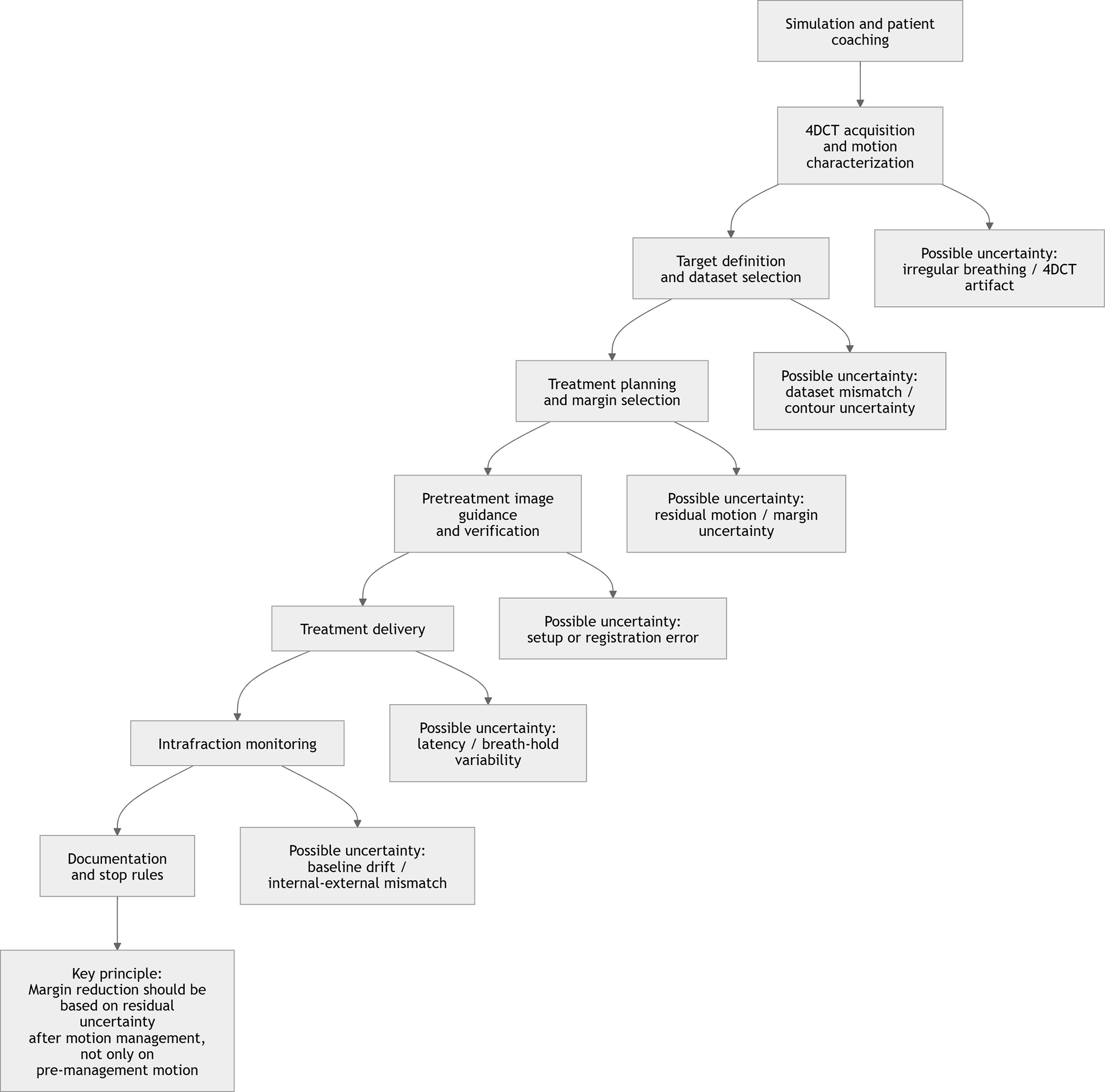
CBCT, respiratory-correlated CBCT, and 4D-CBCT can provide treatment-room information that a planning 4DCT cannot. Sonke et al and Li et al established technical approaches for respiratory-correlated or 4-dimensional CBCT, while Purdie et al and Bissonnette et al showed the value of CBCT-based localization and intrafraction or interfraction motion assessment in lung SBRT.40,43,48,49 These tools are especially important when the plan uses tight margins, central anatomy, gating, breath hold, compression, or mid-ventilation assumptions.7,39,40,43
QA should match technique complexity. An ITV-based plan requires accurate 4DCT acquisition, contour review, dose-calculation dataset selection, and daily image guidance.1,5,16 Gating requires end-to-end tests of the gating interface, latency, audiovisual coaching or monitoring, and image verification in the gate.35,36,41,50 Breath hold requires reproducibility checks and imaging in the hold state, with triggered or repeated imaging considered in compatible workflows when the target is visible and clinically warranted, while SGRT requires commissioning and routine QA of cameras, calibration, thresholds, and workflow integration.8,17,46,47 Linac and image-guidance QA frameworks remain the floor on which all of these motion-specific tests are built.16,50 Table 3 summarizes these technique-specific verification expectations, intrafraction monitoring priorities, and stop-rule triggers so that escalation remains reversible if the selected workflow is not reproducible in the treatment room.
| Workflow Stage | Key Question | Technique-Specific Checks | Stop-Rule Trigger |
|---|---|---|---|
| Simulation | Can the patient tolerate the intended position and respiratory maneuver? | Assess comfort, coaching response, breathing regularity, breath-hold ability, and compression tolerance | Patient cannot reproduce the intended state or tolerate setup |
| 4DCT acquisition | Is the 4DCT representative of treatment breathing? | Review breathing trace, sorting quality, artifacts, tumor visibility, and phase consistency | Severe artifact, irregular breathing, or unreliable surrogate signal |
| Target definition | Is the target dataset appropriate for the selected strategy? | Confirm phase images, MIP/AIP use, ITV construction, breath-hold CT, or gated-window dataset | Target boundary uncertain or dataset does not match intended treatment condition |
| Treatment planning | Does the motion strategy meaningfully improve coverage or OAR sparing? | Compare free-breathing and managed-state dosimetry when relevant | Added complexity produces no meaningful geometric or dosimetric benefit |
| Pretreatment imaging | Is the internal target confirmed in the intended respiratory state? | Verify target position with CBCT, 4D-CBCT, fluoroscopy, or other available imaging | Target mismatch exceeds institutional action threshold |
| Respiratory gating | Is the gating window stable and efficient? | Check duty cycle, latency, baseline drift, audiovisual coaching, and gated-state imaging | Unstable trace, poor duty cycle, or loss of reproducible gating window |
| Breath hold/DIBH | Is the breath hold reproducible during treatment? | Verify breath-hold depth, duration, surface or spirometric signal, and image guidance in hold state | Inconsistent hold depth, short hold duration, or target mismatch |
| Abdominal compression | Is the compression setup reproducible and beneficial? | Confirm indexed setup, patient comfort, respiratory pattern, and target position | Discomfort, altered breathing, or no reproducible geometric benefit |
| Surrogate or SGRT monitoring | Does the external signal remain reliable? | Check camera/belt/marker calibration, thresholds, baseline drift, and correlation with internal imaging | External trace changes without confidence in internal target position |
| Documentation | Can another team member reproduce the workflow safely? | Record setup, respiratory instructions, imaging match criteria, thresholds, and fallback plan | Missing or ambiguous instructions in the treatment chart |
A practical QA culture also includes stop rules. Therapists should know when to pause for unstable breathing, failed breath hold, loss of surface signal, unexpected CBCT position, or patient discomfort. Physicians and physicists should know when to accept residual uncertainty and when to resimulate or simplify the plan.1,8,16,17 The safest escalation strategy is reversible: if the selected technique is not reproducible at treatment, the team should be able to return to a validated, usually less complex alternative.1,4,31
Future Directions
The evidence base would be more clinically useful if future studies reported not only motion reduction, but also residual uncertainty, treatment time, image-guidance workload, failure rates, patient tolerance, staff burden, and dosimetric consequences after realistic verification.4,25,27,34 Head-to-head clinical outcome data comparing ITV, gating, breath hold, compression, and SGRT-assisted workflows remain limited, so broad claims of superiority should remain restrained.4,8,25,34
MR-guided, adaptive and motion-resolved workflows will likely continue to improve, but implementation should remain consequence-based. More imaging does not automatically produce safer treatment unless the information changes a decision and is incorporated into margins, delivery, and QA.16,17,40,48 This review intentionally leaves 4DCT ventilation-based functional avoidance outside the main discussion; it is a separate planning objective and should not be conflated with respiratory-state selection unless a protocol explicitly integrates both questions.
The most practical future direction may be standardization rather than novelty: clearer reporting of respiratory state, surrogate type, gating window, breath-hold reproducibility, compression setting, imaging dataset, registration method, and residual uncertainty.1,8,16,17 Such reporting would make it easier for radiation oncologists, physicists, dosimetrists, and therapists to judge whether a technique worked because it was intrinsically better or because it was selected carefully, verified rigorously, and executed reproducibly.4,5,7,21
Conclusion
A practical rule for lung motion management is to measure the motion, judge its consequence, choose the simplest reproducible strategy, verify the internal target, and define when to stop or de-escalate. Motion should be measured, usually with interpretable 4DCT, and then judged by its consequences for coverage, OAR sparing, image guidance, and delivery robustness.1-3,23 ITV-based planning remains the usual baseline when motion can be encompassed safely. Breath hold, gating, and abdominal compression are valuable when they provide a patient-specific gain that can be reproduced and verified.4,5,8,25
External surrogates, respiratory belts, and surface monitoring can improve workflow awareness, but they are not substitutes for internal target verification.13,14,17,46 Across SBRT and conventionally fractionated lung treatment, the most defensible technique is usually the least complex approach that preserves target coverage, protects OARs, and can be delivered safely by the team every day.1,4,7,16
References
- Keall P. The management of respiratory motion in radiation oncology report of AAPM task group 76. Medical Physics. 2006;33(10):3874-3900. doi:10.1118/1.2349696.
- Mageras G, Pevsner A, Yorke E. Measurement of lung tumor motion using respiration-correlated CT. International Journal of Radiation Oncology*Biology*Physics. 2004;60(3):933-941. doi:10.1016/j.ijrobp.2004.06.021.
- Vedam S, Keall P, Kini V. Acquiring a four-dimensional computed tomography dataset using an external respiratory signal. Phys Med Biol. 2003;48(1):45-62. doi:10.1088/0031-9155/48/1/304.
- Ball H, Santanam L, Senan S. Results from the AAPM Task Group 324 respiratory motion management in radiation oncology survey. J Appl Clin Med Phys. 2022;23(11). doi:10.1002/acm2.13810.
- Guckenberger M, Andratschke N, Dieckmann K. ESTRO ACROP consensus guideline on implementation and practice of stereotactic body radiotherapy for peripherally located early stage non-small cell lung cancer. Radiother Oncol. 2017;124(1):11-17. doi:10.1016/j.radonc.2017.05.012.
- Videtic G, Donington J, Giuliani M. Stereotactic body radiation therapy for early-stage non-small cell lung cancer: executive summary of an ASTRO evidence-based guideline. Pract Radiat Oncol. 2017;7(5):295-301. doi:10.1016/j.prro.2017.04.014.
- Benedict S. Stereotactic body radiation therapy: the report of AAPM task group 101. Medical Physics. 2010;37(8):4078-4101. doi:10.1118/1.3438081.
- Aznar M, Carrasco de Fez P, Corradini S. ESTRO-ACROP guideline: recommendations on implementation of breath-hold techniques in radiotherapy. Radiother Oncol. 2023;185:109734. doi:10.1016/j.radonc.2023.109734.
- Negoro Y, Nagata Y, Aoki T. The effectiveness of an immobilization device in conformal radiotherapy for lung tumor: reduction of respiratory tumor movement and evaluation of the daily setup accuracy. International Journal of Radiation Oncology*Biology*Physics. 2001;50(4):889-898. doi:10.1016/S0360-3016(01)01516-4.
- Bradley J, Paulus R, Komaki R. Standard-dose versus high-dose conformal radiotherapy with concurrent and consolidation carboplatin plus paclitaxel with or without cetuximab for patients with stage IIIA or IIIB non-small-cell lung cancer (RTOG 0617): a randomised, two-by-two factorial phase 3 study. Lancet Oncol. 2015;16(2):187-199. doi:10.1016/S1470-2045(14)71207-0.
- Timmerman R, Paulus R, Galvin J. Stereotactic body radiation therapy for inoperable early stage lung cancer. JAMA. 2010;303(11):1070-1076. doi:10.1001/jama.2010.261.
- Vedam S, Kini V, Keall P. Quantifying the predictability of diaphragm motion during respiration with a noninvasive external marker. Medical Physics. 2003;30(4):505-513. doi:10.1118/1.1558675.
- Hoisak J, Sixel K, Tirona R, Cheung P, Pignol J. Correlation of lung tumor motion with external surrogate indicators of respiration. International Journal of Radiation Oncology*Biology*Physics. 2004;60(4):1298-1306. doi:10.1016/j.ijrobp.2004.07.681.
- Berbeco R, Nishioka S, Shirato H, Chen G, Jiang S. Residual motion of lung tumours in gated radiotherapy with external respiratory surrogates. Phys Med Biol. 2005;50(16):3655-3667. doi:10.1088/0031-9155/50/16/001.
- Ionascu D, Jiang S, Nishioka S, Shirato H, Berbeco R. Internal‐external correlation investigations of respiratory induced motion of lung tumors. Medical Physics. 2007;34(10):3893-3903. doi:10.1118/1.2779941.
- Bissonnette J, Balter P, Dong L. Quality assurance for image‐guided radiation therapy utilizing CT‐based technologies: a report of the AAPM TG‐179. Medical Physics. 2012;39(4):1946-1963. doi:10.1118/1.3690466.
- Al‐Hallaq H, Cerviño L, Gutierrez A. AAPM task group report 302: surface‐guided radiotherapy. Medical Physics. 2022;49(4). doi:10.1002/mp.15532.
- Liu J, Lin T, Fan J. Evaluation of the combined use of two different respiratory monitoring systems for 4D CT simulation and gated treatment. J Appl Clin Med Phys. 2018;19(5):666-675. doi:10.1002/acm2.12434.
- Timmerman R, McGarry R, Yiannoutsos C. Excessive toxicity when treating central tumors in a phase II study of stereotactic body radiation therapy for medically inoperable early-stage lung cancer. J Clin Oncol. 2006;24(30):4833-4839. doi:10.1200/JCO.2006.07.5937.
- Bezjak A, Paulus R, Gaspar L. Safety and efficacy of a five-fraction stereotactic body radiotherapy schedule for centrally located non-small-cell lung cancer: NRG oncology/RTOG 0813 trial. J Clin Oncol. 2019;37(15):1316-1325. doi:10.1200/JCO.18.00622.
- Cole A, Hanna G, Jain S, O’Sullivan J. Motion management for radical radiotherapy in non-small cell lung cancer. Clinical Oncology. 2014;26(2):67-80. doi:10.1016/j.clon.2013.11.001.
- Pan T, Lee T, Rietzel E, Chen G. 4D-CT imaging of a volume influenced by respiratory motion on multi-slice CT. Med Phys. 2004;31(2):333-340. doi:10.1118/1.1639993.
- Rietzel E, Pan T, Chen G. Four‐dimensional computed tomography: image formation and clinical protocol. Medical Physics. 2005;32(4):874-889. doi:10.1118/1.1869852.
- Rietzel E, Chen G, Choi N, Willet C. Four-dimensional image-based treatment planning: target volume segmentation and dose calculation in the presence of respiratory motion. International Journal of Radiation Oncology*Biology*Physics. 2005;61(5):1535-1550. doi:10.1016/j.ijrobp.2004.11.037.
- Saito T, Sakamoto T, Oya N. Predictive factors for lung dose reduction by respiratory gating at radiotherapy for lung cancer. J Radiat Res. 2010;51(6):691-698. doi:10.1269/jrr.10084.
- Heinzerling J, Anderson J, Papiez L. Four-dimensional computed tomography scan analysis of tumor and organ motion at varying levels of abdominal compression during stereotactic treatment of lung and liver. International Journal of Radiation Oncology*Biology*Physics. 2008;70(5):1571-1578. doi:10.1016/j.ijrobp.2007.12.023.
- Qi Y, Li J, Zhang Y. Effect of abdominal compression on target movement and extension of the external boundary of peripheral lung tumours treated with stereotactic radiotherapy based on four-dimensional computed tomography. Radiat Oncol. 2021;16(1). doi:10.1186/s13014-021-01889-0.
- Underberg R, Lagerwaard F, Slotman B, Cuijpers J, Senan S. Use of maximum intensity projections (MIP) for target volume generation in 4DCT scans for lung cancer. International Journal of Radiation Oncology*Biology*Physics. 2005;63(1):253-260. doi:10.1016/j.ijrobp.2005.05.045.
- Borm K, Oechsner M, Wiegandt M. Moving targets in 4D-CTs versus MIP and AIP: comparison of patients data to phantom data. BMC Cancer. 2018;18(1). doi:10.1186/s12885-018-4647-4.
- Yamamoto T, Langner U, Loo B, Shen J, Keall P. Retrospective analysis of artifacts in four-dimensional CT images of 50 abdominal and thoracic radiotherapy patients. International Journal of Radiation Oncology*Biology*Physics. 2008;72(4):1250-1258. doi:10.1016/j.ijrobp.2008.06.1937.
- Hurkmans C, Cuijpers J, Lagerwaard F. Recommendations for implementing stereotactic radiotherapy in peripheral stage IA non-small cell lung cancer: report from the Quality Assurance Working Party of the randomised phase III ROSEL study. Radiat Oncol. 2009;4(1):1. doi:10.1186/1748-717X-4-1.
- Hanley J, Debois M, Mah D. Deep inspiration breath-hold technique for lung tumors: the potential value of target immobilization and reduced lung density in dose escalation. International Journal of Radiation Oncology*Biology*Physics. 1999;45(3):603-611. doi:10.1016/S0360-3016(99)00154-6.
- Barnes E, Murray B, Robinson D. Dosimetric evaluation of lung tumor immobilization using breath hold at deep inspiration. International Journal of Radiation Oncology*Biology*Physics. 2001;50(4):1091-1098. doi:10.1016/S0360-3016(01)01592-9.
- Singh B, Chughtai A, Stock J. Comparative dosimetric analysis of deep inspiration breath-hold versus free breathing radiotherapy in lung cancer: special emphasis on cardiac and subcardiac structure protection. Technical Innovations & Patient Support in Radiation Oncology. 2026;37:100371. doi:10.1016/j.tipsro.2025.100371.
- Kubo H, Hill B. Respiration gated radiotherapy treatment: a technical study. Phys Med Biol. 1996;41(1):83-91. doi:10.1088/0031-9155/41/1/007.
- Jiang S. Technical aspects of image-guided respiration-gated radiation therapy. Med Dosim. 2006;31(2):141-151. doi:10.1016/j.meddos.2005.12.005.
- Wolthaus J, Schneider C, Sonke J. Mid-ventilation CT scan construction from four-dimensional respiration-correlated CT scans for radiotherapy planning of lung cancer patients. International Journal of Radiation Oncology*Biology*Physics. 2006;65(5):1560-1571. doi:10.1016/j.ijrobp.2006.04.031.
- Wolthaus J, Sonke J, van Herk M. Comparison of different strategies to use four-dimensional computed tomography in treatment planning for lung cancer patients. International Journal of Radiation Oncology*Biology*Physics. 2008;70(4):1229-1238. doi:10.1016/j.ijrobp.2007.11.042.
- Bellec J, Arab-Ceschia F, Castelli J, Lafond C, Chajon E. ITV versus mid-ventilation for treatment planning in lung SBRT: a comparison of target coverage and PTV adequacy by using in-treatment 4D cone beam CT. Radiat Oncol. 2020;15(1). doi:10.1186/s13014-020-01496-5.
- Bissonnette J, Franks K, Purdie T. Quantifying interfraction and intrafraction tumor motion in lung stereotactic body radiotherapy using respiration-correlated cone beam computed tomography. International Journal of Radiation Oncology*Biology*Physics. 2009;75(3):688-695. doi:10.1016/j.ijrobp.2008.11.066.
- Ford E, Mageras G, Yorke E. Evaluation of respiratory movement during gated radiotherapy using film and electronic portal imaging. International Journal of Radiation Oncology*Biology*Physics. 2002;52(2):522-531. doi:10.1016/S0360-3016(01)02681-5.
- Korreman S, Mostafavi H, Le Q, Boyer A. Comparison of respiratory surrogates for gated lung radiotherapy without internal fiducials. Acta Oncologica. 2006;45(7):935-942. doi:10.1080/02841860600917161.
- Purdie T, Bissonnette J, Franks K. Cone-beam computed tomography for on-line image guidance of lung stereotactic radiotherapy: localization, verification, and intrafraction tumor position. International Journal of Radiation Oncology*Biology*Physics. 2007;68(1):243-252. doi:10.1016/j.ijrobp.2006.12.022.
- Mah D, Hanley J, Rosenzweig K. Technical aspects of the deep inspiration breath-hold technique in the treatment of thoracic cancer. International Journal of Radiation Oncology*Biology*Physics. 2000;48(4):1175-1185. doi:10.1016/S0360-3016(00)00747-1.
- Wong J, Sharpe M, Jaffray D. The use of active breathing control (ABC) to reduce margin for breathing motion. International Journal of Radiation Oncology*Biology*Physics. 1999;44(4):911-919. doi:10.1016/S0360-3016(99)00056-5.
- Heinzerling J, Hampton C, Robinson M. Use of surface-guided radiation therapy in combination with IGRT for setup and intrafraction motion monitoring during stereotactic body radiation therapy treatments of the lung and abdomen. J Appl Clin Med Phys. 2020;21(5):48-55. doi:10.1002/acm2.12852.
- Sarudis S, Karlsson A, Bäck A. Surface guided frameless positioning for lung stereotactic body radiation therapy. J Appl Clin Med Phys. 2021;22(9):215-226. doi:10.1002/acm2.13370.
- Sonke J, Zijp L, Remeijer P, van Herk M. Respiratory correlated cone beam CT. Medical Physics. 2005;32(4):1176-1186. doi:10.1118/1.1869074.
- Li T, Xing L, Munro P. Four‐dimensional cone‐beam computed tomography using an on‐board imager. Medical Physics. 2006;33(10):3825-3833. doi:10.1118/1.2349692.
- Klein E, Hanley J, Bayouth J. Task group 142 report: quality assurance of medical accelerators. Medical Physics. 2009;36(9part1):4197-4212. doi:10.1118/1.3190392.
Disclosures
The authors have no conflicts of interest to disclose. None of the authors received outside funding for the production of this original manuscript and no part of this article has been previously published elsewhere.
Citation
. Respiratory Motion Management in Lung Cancer Radiation Therapy: A Practical Review and Decision Framework for Technique Selection and Treatment Delivery. Applied Radiation Oncology. 2026;15(3). doi:10.37549/ARO-D-26-0012.