Small cell carcinoma with cystic metastasis to the brain
Applied Radiology — Vol. 37 , Issue 9 , pp. 44 -47
DOI: 10.37549/AR1646
Published: September 1, 2008
Categories
CASE SUMMARY
The patient is a 59-year-old African American man with a medical history significant for splenectomy pursuant to remote trauma, significant coronary artery disease, and hypertension with consequential chronic heart failure, and bradycardia requiring a pacemaker/cardioverter-defibrillator. He presented with complaints of over-the-counter nonsteroidal anti-inflammatory drug (NSAID) recalcitrant headache, recurrent nausea and vomiting, and subacute weight loss. The patient’s symptoms began approximately 8 weeks prior to admission with a progressive 18-pound weight loss. This was followed by a bitemporal nonradiating and piercing headache approximately 1 week prior to admission that failed to recede with ibuprofen. Three days prior to this presentation, approximately 5 to 12 episodes of bilious nonbloody vomiting ensued, which finally led him to seek medical care.
In the emergency department, the patient was found to be afebrile, with stable vital signs. In general, the patient was an overweight African-American man who was slightly somnolent but easily arousable and was oriented to time, place, and person. Physical examination disclosed focal rales at the left upper lobe area. He reported quitting smoking cigarettes 6 years prior to admission with a previous tobacco burden of 75 pack-years. The patient had not traveled outside of the United States.
IMAGING FINDINGS
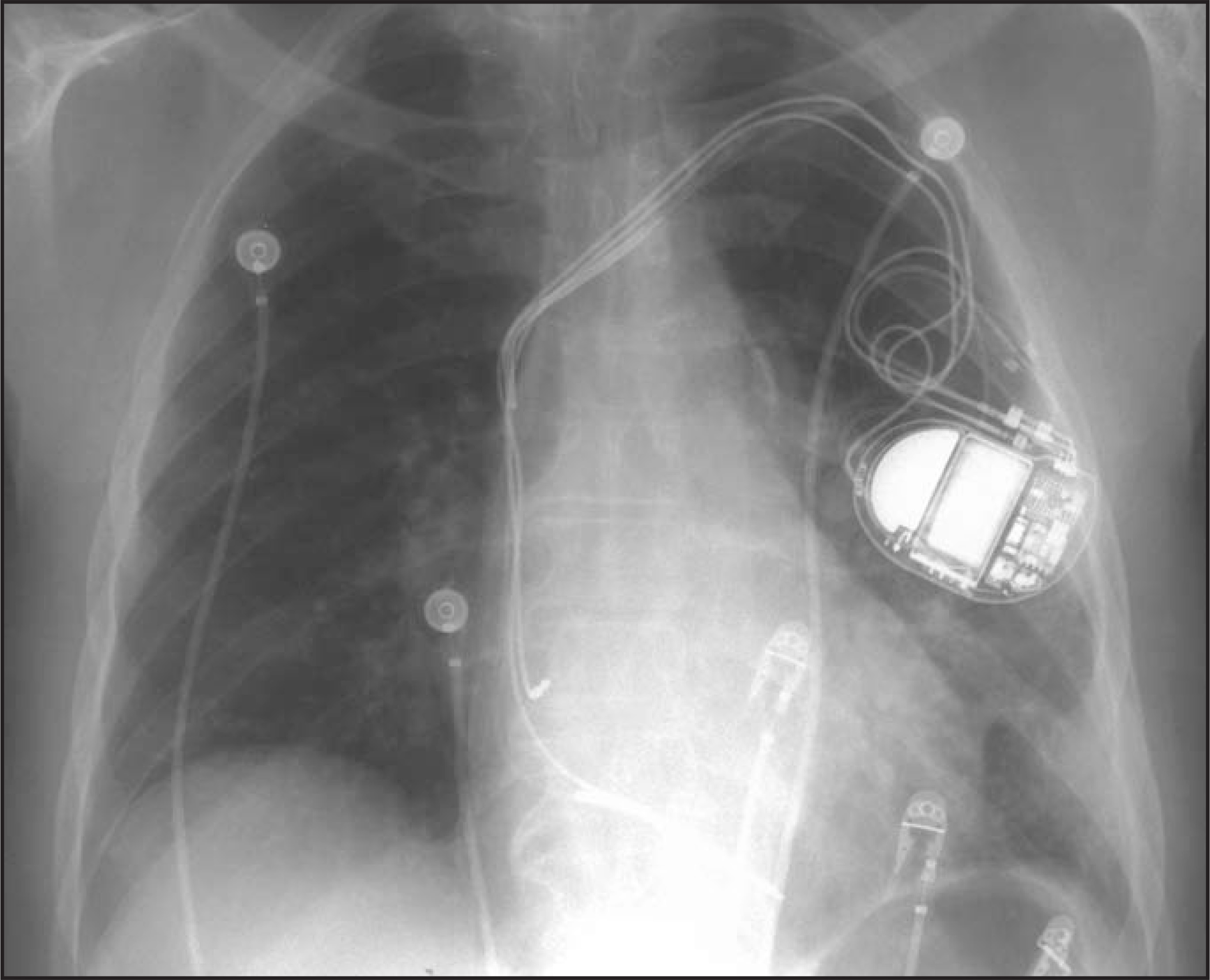
On admission, a plain chest radiograph revealed poorly defined patchy opacities within the left lower lobe that are consistent with air-space disease or multiple lung masses (Figure 1). Head CT with and without intravenous contrast was performed and showed that the cortical sulci, ventricles, and basilar cisterns showed mass effect upon the quadrigeminal plate and some encroachment on the suprasellar cistern. There were multiple cysts and masses throughout the brain. The largest mass was a hypodense, noncystic mass that was located at the left temporal parietal lobe and measured 4 × 4.7 cm. There was a 2.6 × 2.1-cm cystic lesion along the midline of the cerebellum, containing an internal fluid level;these findings likely represented necrotic debris, abscess, or hemorrhage. There were several smaller cystic lesions within the cerebellar hemispheres. Some of the cystic lesions were ring-enhancing. A relatively higher-density mass with surrounding edema was identified within the left frontal. A large 4.1 × 1.4 × 2.1-cm high-density mass suggesting hemorrhagic transformation was seen within the region of the right caudate, encroaching on the right lateral ventricle. There were no osseous abnormalities (Figure 2).
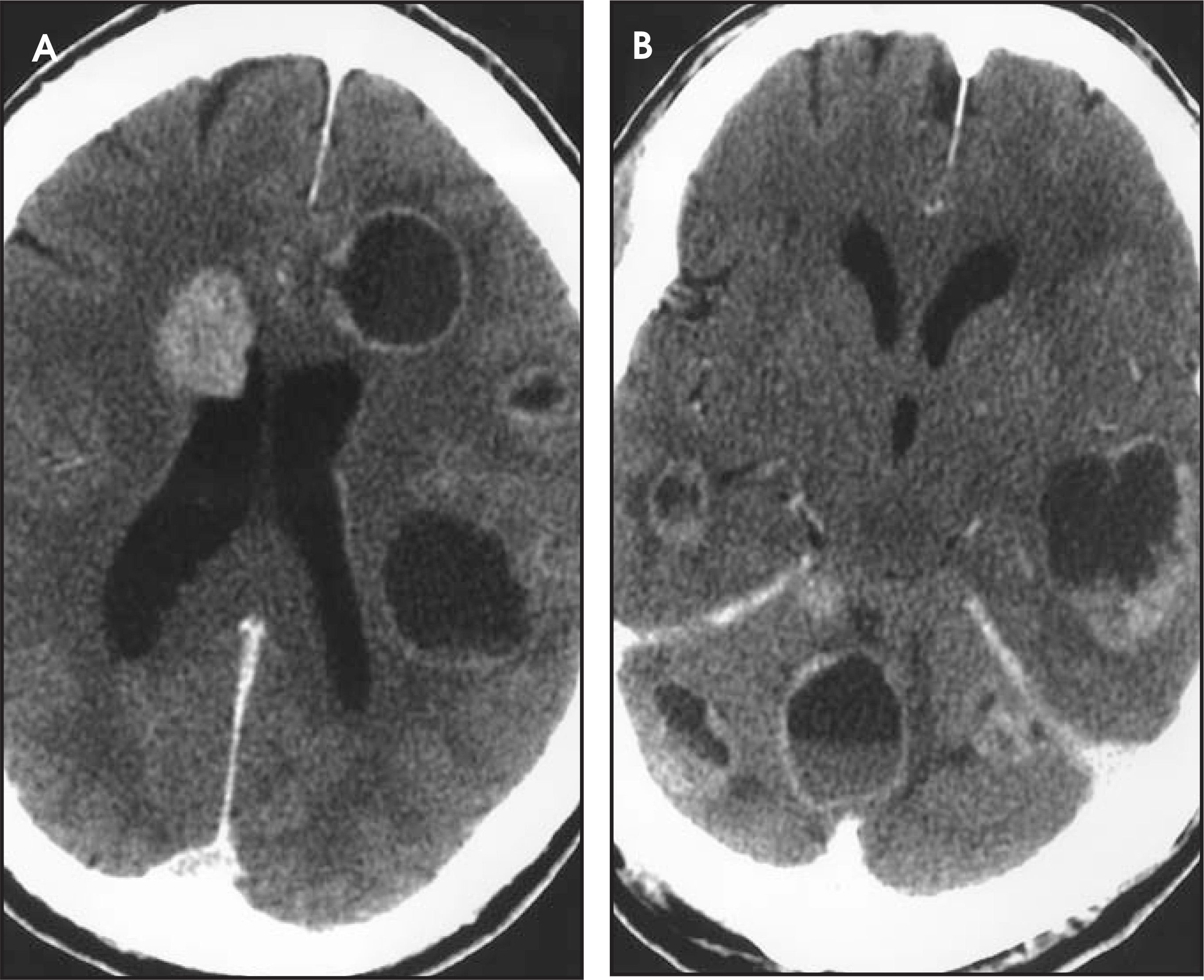
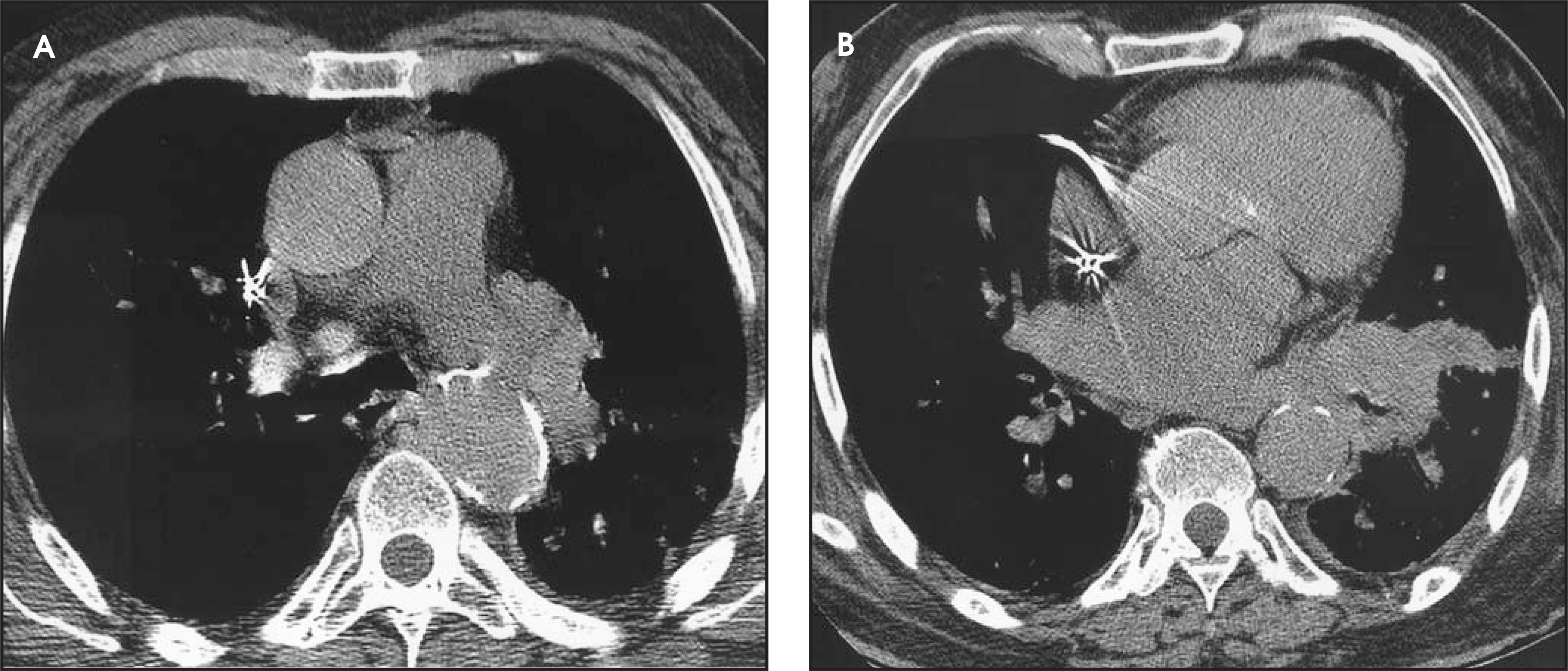
The patient was transferred to the medical intensive care unit and was started on intravenous dexamethasone and empiric antibiotics in addition to an antifungal agent. Laboratory values, sputum, and culture results were negative. To evaluate a potential source for the metastatic,infectious, or neoplastic process and to further clarify the extent of the left perihilar disease, a chest/abdomen/pelvis CT was performed.The examination showed a mass in the left infrahilar region that measured approximately 6× 4 cm. There were also scattered multiple 1- to 3-cm lymph nodes present in the left perihilar region (Figure 3). The lobar bronchi to the lingula and the left lower lobe were narrowed. There wa salso scarring present in the left major fissure. Mild cardiomegaly was noted, and there was no evidence of hepatic, splenic, or adrenal involvement.
CLINICAL COURSE
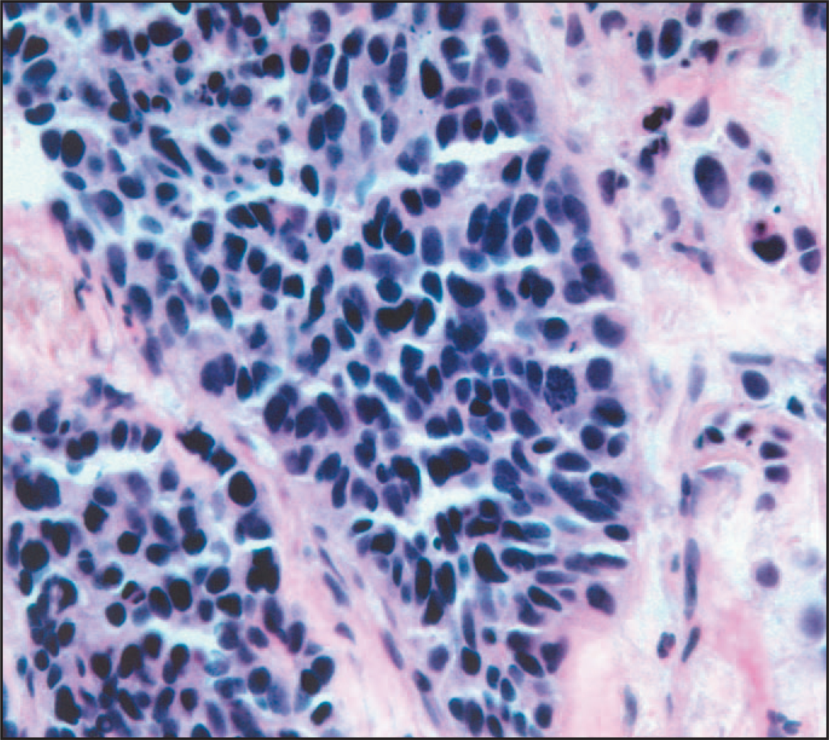
Following 3 days of steady improvement in the patient’s somnolence, he underwent a CT-guided transthoracic needle biopsy. The biopsy disclosed islands of cells infiltrating a desmoplastic stroma. The cells were characterized by hyperchromatic, irregular nuclei, indistinct nucleoli, and scant cytoplasm. In some areas, there appeared to be “molding.” The cell block preparation showed islands of malignant cells infiltrating a desmoplastic stroma (Figure 4). Immunoperoxidase studies were performed on core biopsies from the case and showed positive staining with chromogranin, synaptophysin, keratin, and thyroid transcription factor. These findings supported the initial cytological diagnosis of small cell carcinoma. After extensive discussion with his family, the patient opted for lone palliative cranial radiation therapy. Two weeks after the diagnosis, the patient was discharged on dexamethasone and continued to function without appreciable limitation.
DIAGNOSIS
Small cell carcinoma with cystic metastasis to the brain
DISCUSSION
The differential diagnosis for a patient whose findings include the presence of multiple cerebral cystic lesions involving both hemispheres includes disseminating microorganisms such toxoplasma and cystercercosis. These infectious diseases are more prevalent in the immunocompromised host. We have presented a case report on a metastatic process that presented with similar radiographic findings as the latter infectious agents, but in an immunocompetent patient.
Small cell lung carcinoma (SCLC) is an aggressive neoplasm that can be associated with cerebral metastases in 10% of affected patients, at the time of their initial presentation; this may be present in 65% of patients at the time of autopsy.1,2 Neuroradiological findings of metastaticlesions include leptomeningeal involvement and carcinomatous meningitis,3miliary cerebral metastases,4and larger single or multiple densities,some with central necrosis, with or without hemorrhagic transformation. However, to our knowledge, the morphological description of multiple cystic-appearing lesions, including some lesions with internal fluid levels, has not previously been reported for a patient in their initial SCLC presentation. The cerebral pathology, as evaluated by head CT, indeed argued for infectious microorganisms such as neurocystercercosis ortoxoplasma. Our case report would suggest that with evidence of multiple cystic changes within the cerebrum, one should also entertain the possibility of metastatic SCLC.
Extensive stage disease (as defined by pericardial and/or pleural effusions, or bilateral pulmonary parenchymal involvement, or any evidence of disease outside the thorax) carries a median life expectancy of 35 weeks from the date of first treatment, which is further attenuated to 22 weeks in those with brain involvement.5Therapy for SCLC, in general, is dependent upon the extent of the disease at the time of diagnosis and is detailed in a recent comprehensive literature review by Simon et al.6
References
- Stahel R. Diagnosis, staging, and prognostic factors of small cell lung cancer. Curr Opin Oncol. 1991;3:306-311.
- Richardson G, Venzon D, Edison M. Application of an algorithm for staging small-cell lung cancer can save one third of the initial evaluation costs. Arch Intern Med. 1993;153:329-337.
- Rosen S, Aisner J, Makuch R. Carcinomatous leptomeningitis in small cell lung cancer: A clinicopathologic review of the National Cancer Institute experience. Medicine (Baltimore). 1982;61:45-53.
- Bhushan C. “Miliary” metastatic tumors in the brain. Case report. J Neurosurg J Neurosurg.
- Sagman U, Maki E, Evans W. Small-cell carcinoma of the lung: derivation of a prognostic staging system. J Clin Oncol. 1991;9:1639-1649.
- Simon G, Wagner H. Small cell lung cancer. Chest. 2003;123(1 suppl):259S-271S.
Citation
. Small cell carcinoma with cystic metastasis to the brain. Applied Radiology. 2008;37(9):44-47. doi:10.37549/AR1646.