Sonographic findings of Down’s syndrome
Applied Radiology — Vol. 31 , Issue 9 , pp. 9 -14
DOI: 10.37549/AR1123
Published: September 1, 2002
Categories
The prevalence of Down’s syndrome during the second trimester has risen in the last three decades due to an increasing number of births in women older than the age of 35. In 1997, the prevalence had increased to 1 in 504 births,1 or approximately 0.2%; it continues to be the most common chromosomal abnormality among newborns. Screening protocols for fetal Down’s syndrome have advanced simultaneously, improving detection in both high-risk and low-risk populations. Most recent advances relate to the first trimester examination, with an emphasis on nuchal translucency (NT). Second trimester sonographic screening has also improved with recognition of structural and nonstructural abnormalities and incorporation of the presence or absence of such findings to modify the risk of fetal Down’s syndrome.
First trimester sonographic findings
With recent advances in technology, first trimester sonography can now detect, or at least suggest, many structural abnormalities. However, structural abnormalities in the first trimester are identified much more commonly in trisomies 18 and 13 than in trisomy 211 The focus of first trimester screening for Down’s syndrome is rather on fetal NT.
Nicolaides et al2 introduced the term “nuchal translucency” to describe the collection of fluid under the skin behind the neck of fetuses between 11 and 14 weeks of gestation (Figure 1). An increase in this fluid is seen in chromosomally abnormal fetuses, most commonly trisomy 21, and has since been recognized to be a useful sonographic marker for the detection of fetal Down’s syndrome in the first trimester.1,3 Its relatively low false-positive rate further enhances its screening potential. The origin of the fluid is unknown, and maybe multifactorial. It is thought to be possibly linked to cardiac dysfunction, venous congestion, lymphatic abnormality, altered extracellular matrix, or other unknown causes.4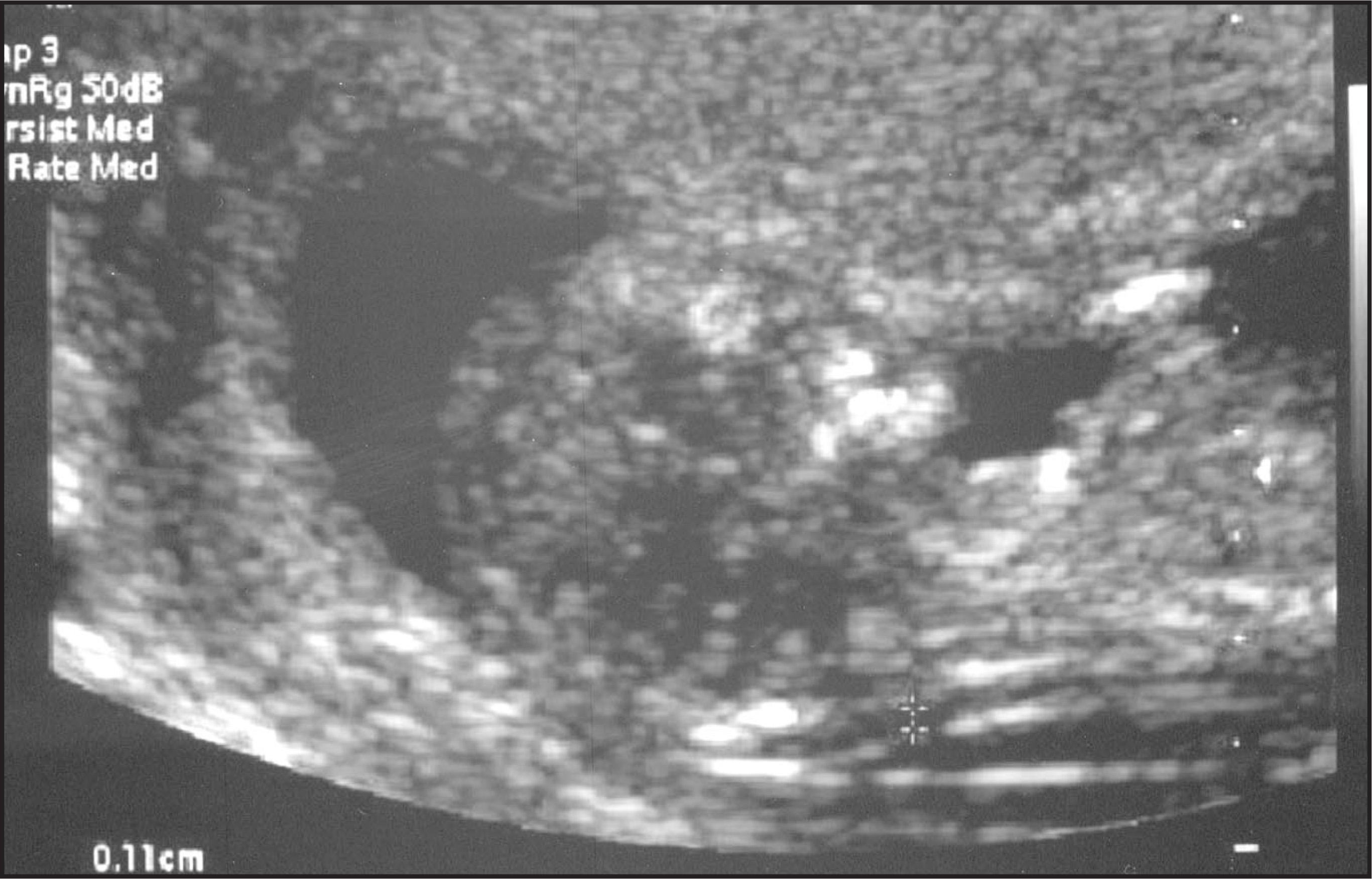
A reproducible specific method for measurement of NT is very important, as the variation of measurement between abnormal and normal is so small. The Fetal Medicine Foundation (FMF) has established standardized technical guidelines for the correct measurement of NT (also available online at: http://www.fetalmedicine.com).4 It has also developed a training and accreditation program. Using this protocol, NT can be measured reproducibly in approximately 95% of cases transabdominally, with transvaginal scanning required in the remaining 5%. The criteria used to obtain uniform results are as follows: 1) good sagittal section of the fetus with a crown-rump length between 45 and 84 mm; 2) gestational age of 11 to 13 6/7 weeks; 3) magnification such that the fetus occupies at least three-quarters of the image and measurements are accurate to 0.1 to 0.2 mm; 4) measurement performed in neutral position, as flexion may decrease measurements and hyperextension may increase measurements; 5) visualization of the amnion separate from the fetal skin, and 6) correct placement of calipers from the inner to inner line.4
Multiple studies of NT have been performed. Souter and Nyberg1 cite 16 studies, with most early studies using a cut-off of 3.0 mm and most recent studies using gestational-age–specific thresholds for a positive result.1 The sensitivity rate for the detection of trisomy 21 varied from 28.6% to 90.3%. However, many of these studies did not implement strict criteria and quality control. A large study of approximately 100,000 cases from the FMF published in 1998 by Snijders and collegues3 reported a sensitivity rate for trisomy 21 of 82.2% with a false-positive rate of 8.3% using their established criteria. Subsequent studies have obtained similar results.3
The normal range for NT measurements is gestational age dependent. The 95th percentile increases from 2.2 mm at a crown-rump length of 38 mm to 2.8 mm at 84 mm.1,5 As such, NT measurements are usually expressed relative to gestational age or crown-rump length and positive results reported as >95th percentile, multiple of the median, or a delta value. These values can then be combined with maternal age to calculate a risk for trisomy 21.
During the second trimester, NT in both karyotypically normal and abnormal fetuses usually resolves or, in a few cases, evolves into either nuchal edema or cystic hygromas with or without generalized hydrops.6,7 Fetuses with abnormally increased NT measurements and normal chromosomes are still at risk for structural abnormalities, especially major cardiac defects, and a variety of genetic conditions. The risk increases with increasing NT and subsequent fetal echocardiography has been advised.8-10
Second trimester sonographic findings
Structural abnormalities
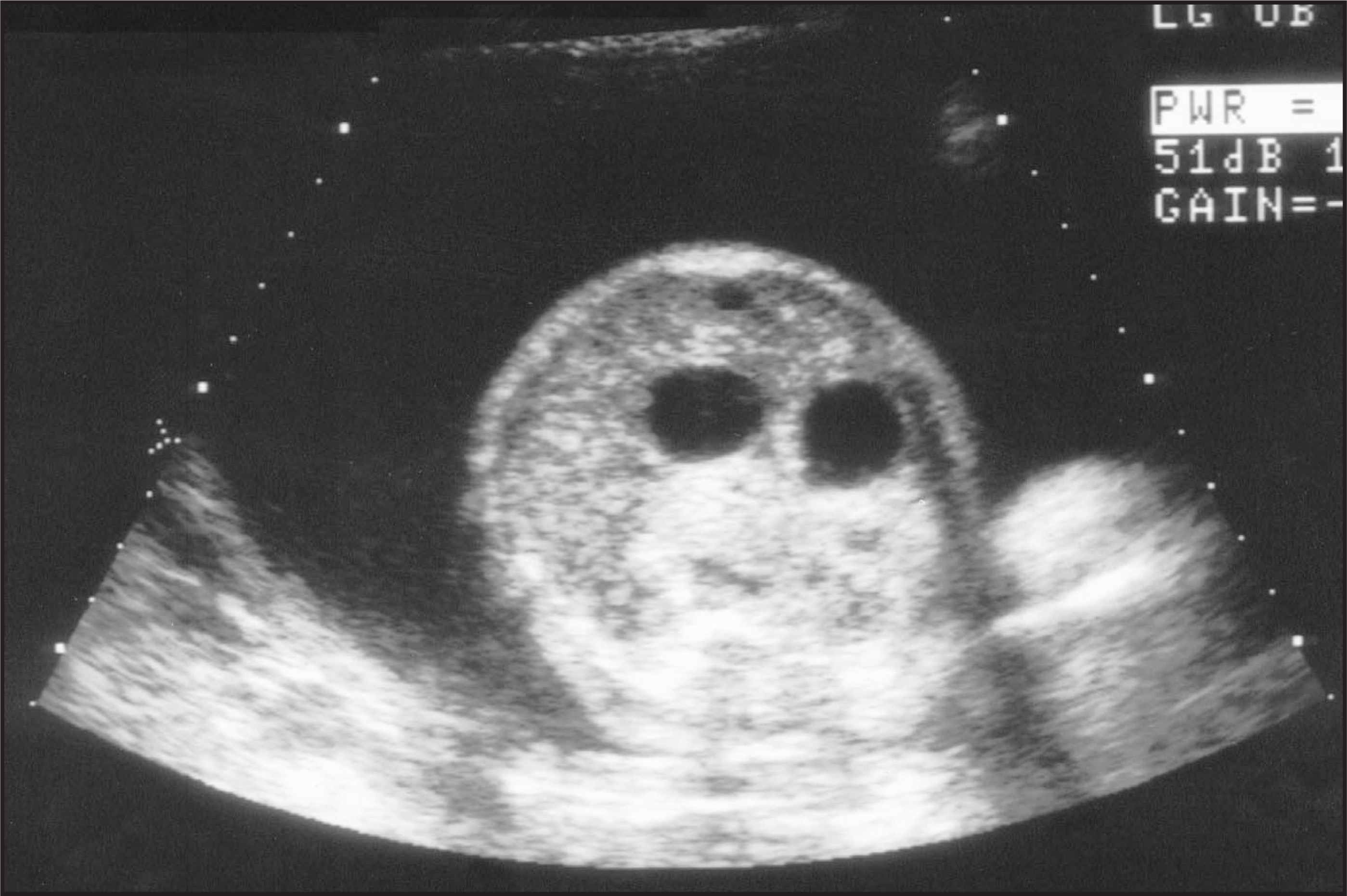
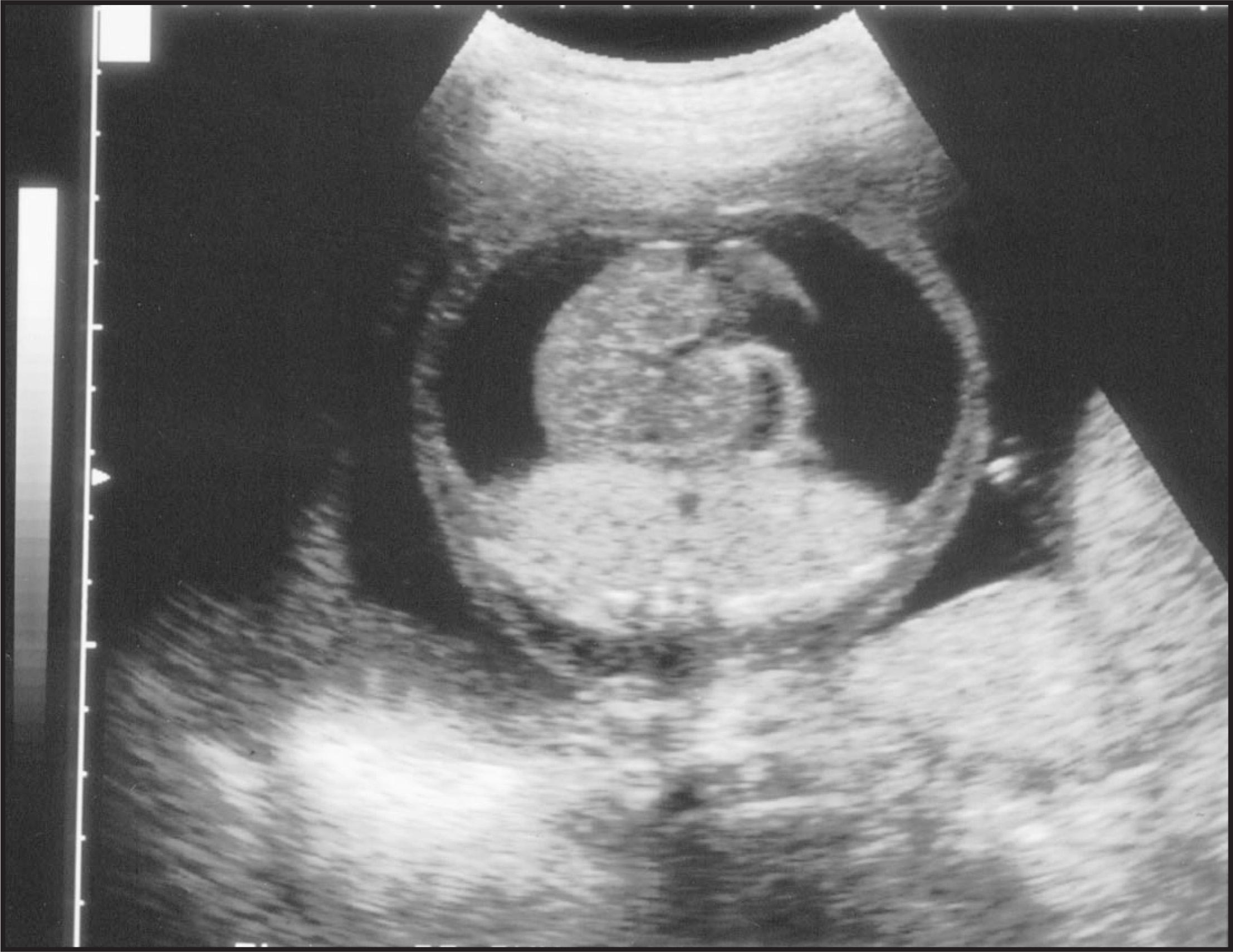
Although structural abnormalities are common with trisomies, they are less often seen sonographically in trisomy 21, and are usually identified in <25% of fetuses with trisomy 21.11,12 Major structural abnormalities associated with second trimester Down’s syndrome include cardiac defects, cystic hygroma (Figure 2), duodenal atresia (Figure 3), and hydrops (Figure 4).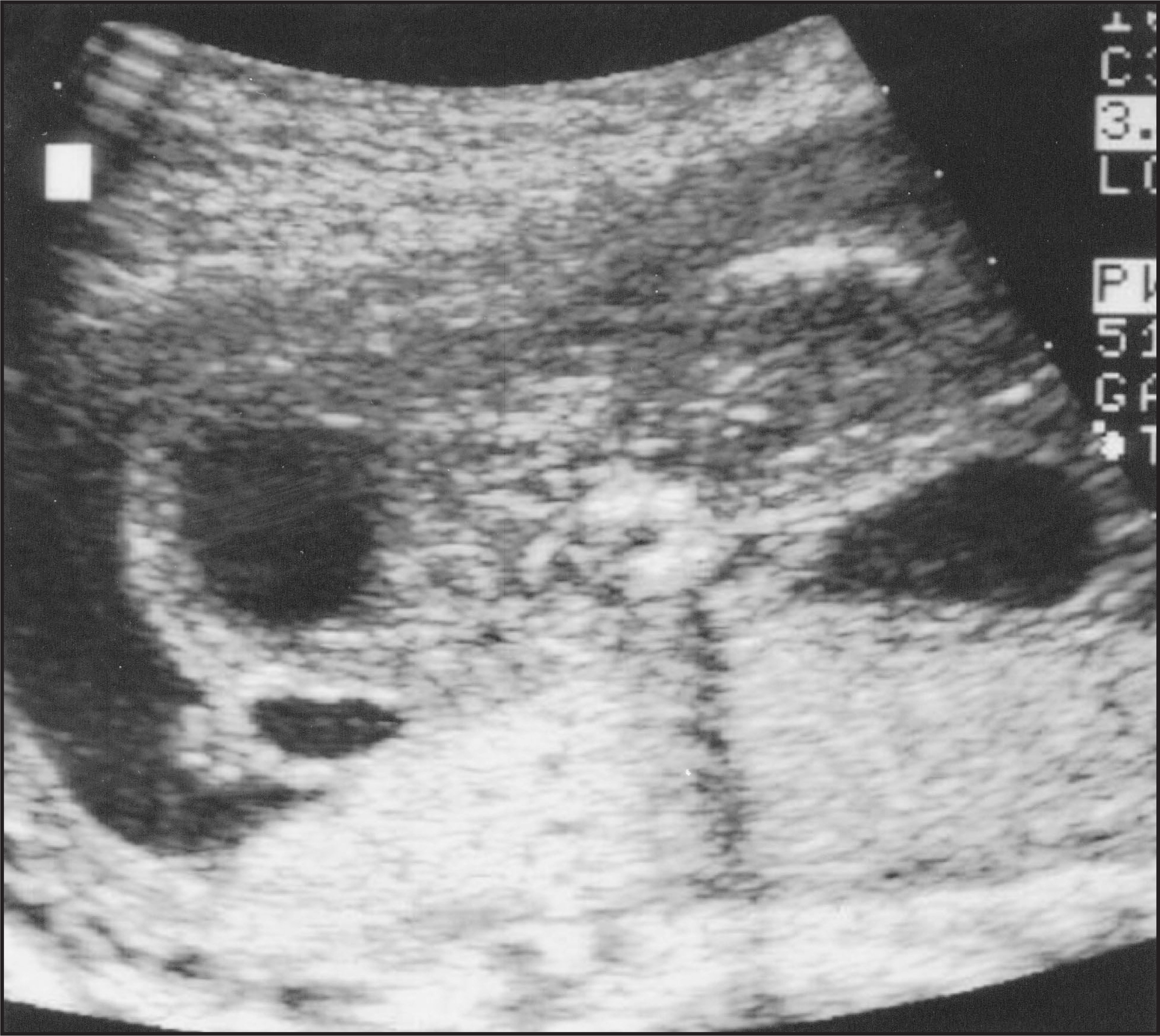
Cardiac defects such as ventricular septal defect (Figure 5) and endocardial cushion defect are commonly associated with Down’s syndrome clinically and pathologically. However, these are particularly difficult to detect sonographically, especially at the routine screening age of 16 to 20 weeks. Even at 24 weeks, an optimal gestational age for sonography, with prior knowledge of karyotype, Paladini et al13 detected only approximately one half of cardiac defects. Nyberg14 also reports low detection rates of cardiac defects of <10% in fetuses with Down’s syndrome. Using less specific cardiac findings—such as right-to-left chamber disproportion, pericardial effusion and tricuspid regurgitation—Devore15 identified cardiac abnormalities in 76% of affected fetuses. However, in practice, without prior knowledge of karyotype, there appears to be a low sensitivity for detection of cardiac defects in trisomy 21 with sonography.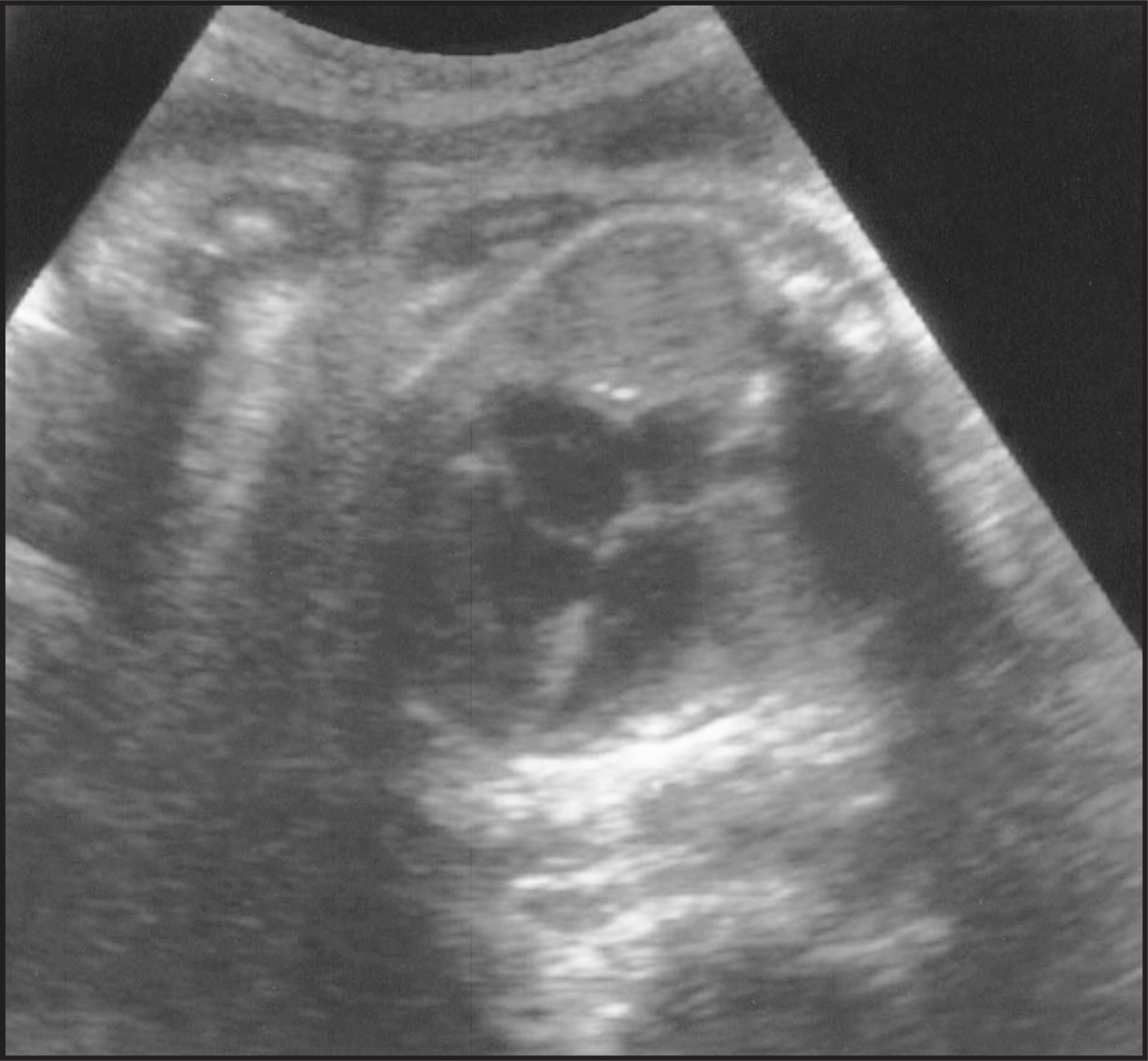
Sonographic markers
In addition to structural abnormalities, nonstructural findings, or sonographic markers, are important findings in trisomy 21. These sonographic markers are seen most often in normal fetuses, are nonspecific, are often transient, and by themselves do not alter fetal outcome. Their significance varies with the specific marker, increases with multiplicity of markers identified, and is interpreted most accurately with maternal age and other clinical variables. The most commonly recognized sonographic markers associated with trisomy 21 are nuchal thickening, hyperechoic bowel, echogenic intracardiac focus, short femur or humerus, and renal pyelectasis. Other proposed markers include widened pelvic angle, small ears, clinodactyly, shortened frontal lobes, small cerebellum, pericardial effusion, and others.14
Of these, nuchal thickening has the highest association with Down’s syndrome. Benacerraf et al16 first described this important second trimester marker as abnormal thickening of the soft tissues in the back of the fetal occiput. This is measured in the transverse plane at the level of the cerebellum with the view including the cerebellum, posterior fossa, and the occipital bone (Figure 6). It is measured from the outer edge of the occipital bone to the outer edge of the fetal skin.17 A measurement >5 mm is considered abnormal.18,19 This measurement appears to vary with gestational age, however, and age-specific criteria may be used to establish relative risk in the future.14 This finding generally resolves with time, but its transient nature does not deter from its initial significance.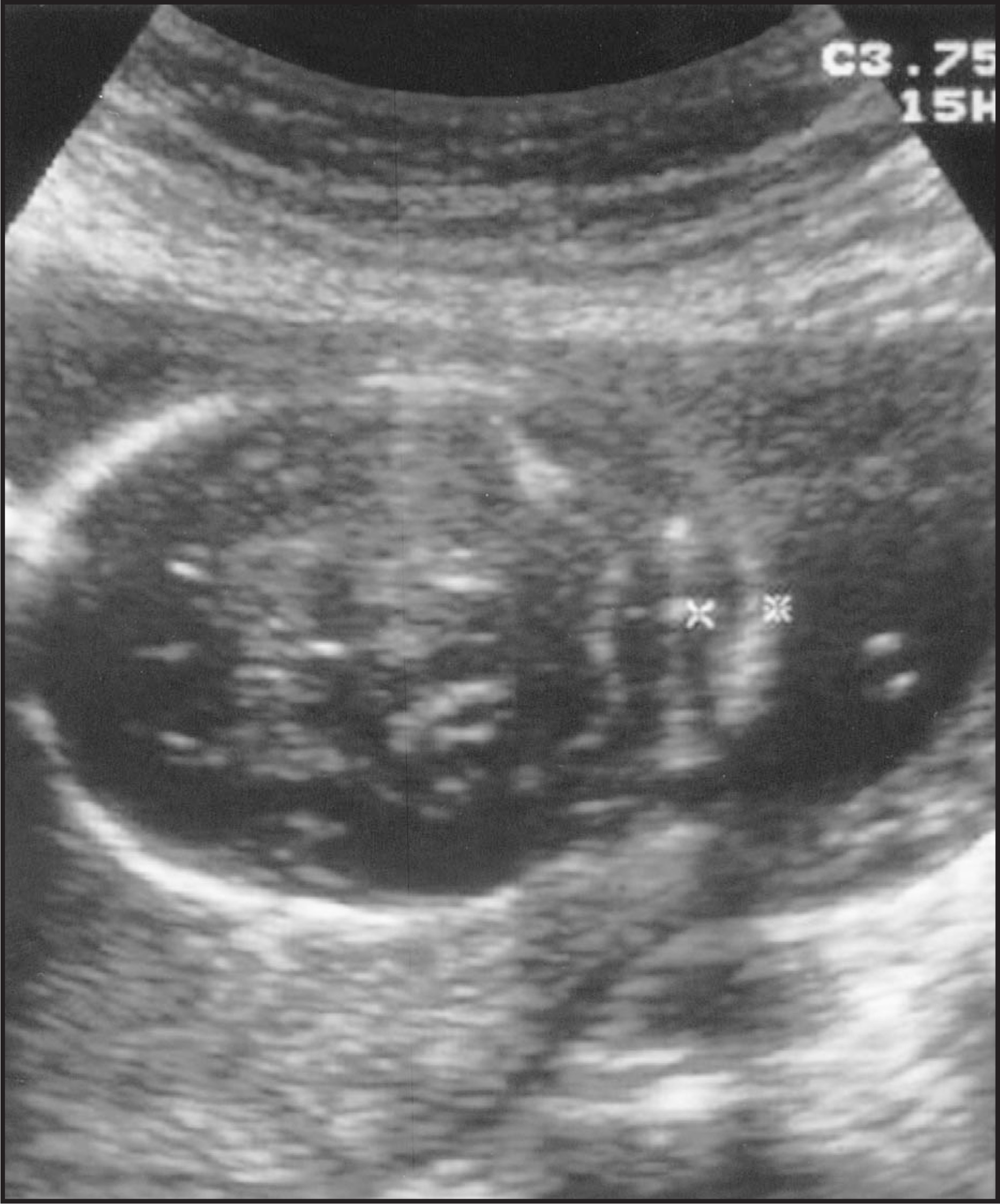
Echogenic bowel is another nonspecific marker that has been found in fetuses with aneuploidy (Figure 7). However, it is also seen in multiple other conditions, including bowel atresia, meconium ileus, thalassemia, intra-amniotic bleed, congenital infection, and intrauterine growth retardation.20-22 A grading classification has been established for echogenic bowel: grade 1—mildly echogenic and often diffuse; grade 2—moderately echogenic and often focal; and grade 3—very echogenic, equivalent to bone. Both grade 2 and grade 3 have been associated with aneuploidy, with a higher risk associated with grade 3.22 Care must be taken in grading the echogenicity of bowel, as the echogenicity of normal bowel increases with increasing transducer frequency.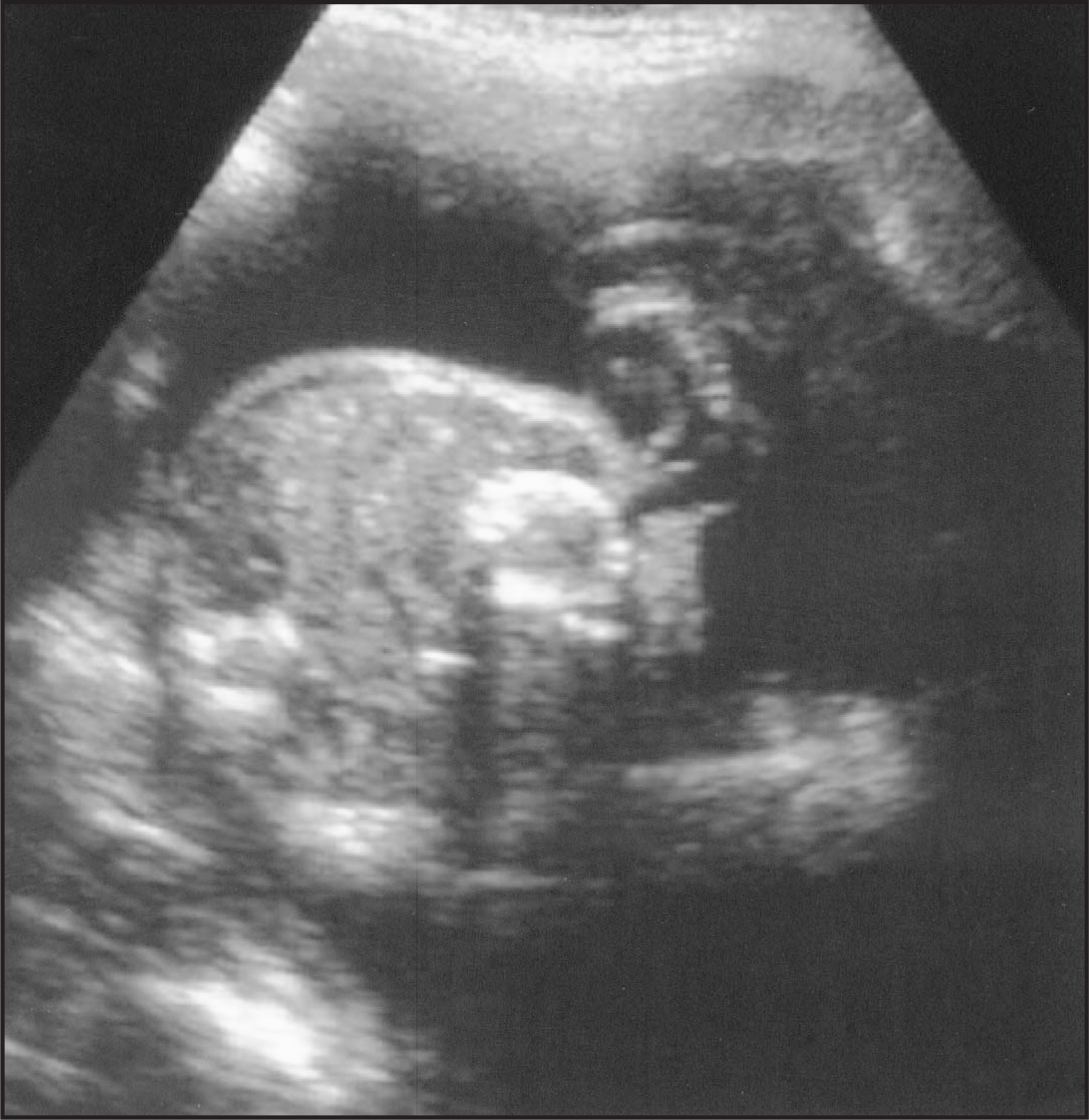
Echogenic intracardiac foci are a much-debated soft sonographic marker. They represent calcification of the papillary muscle and are seen best when the cardiac apex is pointed toward the transducer.23 They are observed in 3% to 4% of normal fetuses.24 Multiple studies yield conflicting conclusions as to the association of echogenic intracardiac foci with trisomy 21. However, multiple, large, or unusually conspicuous foci appear to carry a greater risk.14
Skeletal abnormalities associated with trisomy 21 include short proximal long bones. However, there is a large overlap between trisomy 21 and normal fetuses, and variations may be related to gestational age and ethnicity. A shortened humerus appears to be a more specific indicator for trisomy 21 than a shortened femur. Abnormalities are recognized from expected measurement based on the biparietal diameter at the time of the examination rather than the gestational age. The ratio of the measured femur/humerus length to the expected length is calculated. A measure:expected ratio of <0.90 for humerus length was found in 50% of afflicted fetuses by Benacerraf.25 Commonly utilized cutoffs included ratios of <0.90 for a short humerus and <0.91 for a short femur. These numbers may also change as investigators further establish likelihood ratios.
Renal pyelectasis is a measure of the fluid-filled fetal renal pelvis in the anteroposterior dimension on a transverse image of the kidneys (Figure 8). The commonly used cutoffs are 3 or 4 mm. Pyelectasis is also seen commonly in normal fetuses and may be related to the degree of maternal hydration, other genitourinary abnormalities, or fetal bladder distention.26,27 Nyberg et al14 observed pyelectasis in 3% of normal fetuses. Corteville et al28 reported an incidence of pyelectasis in 17.4% of fetuses with Down’s syndrome and 2% in the normal population.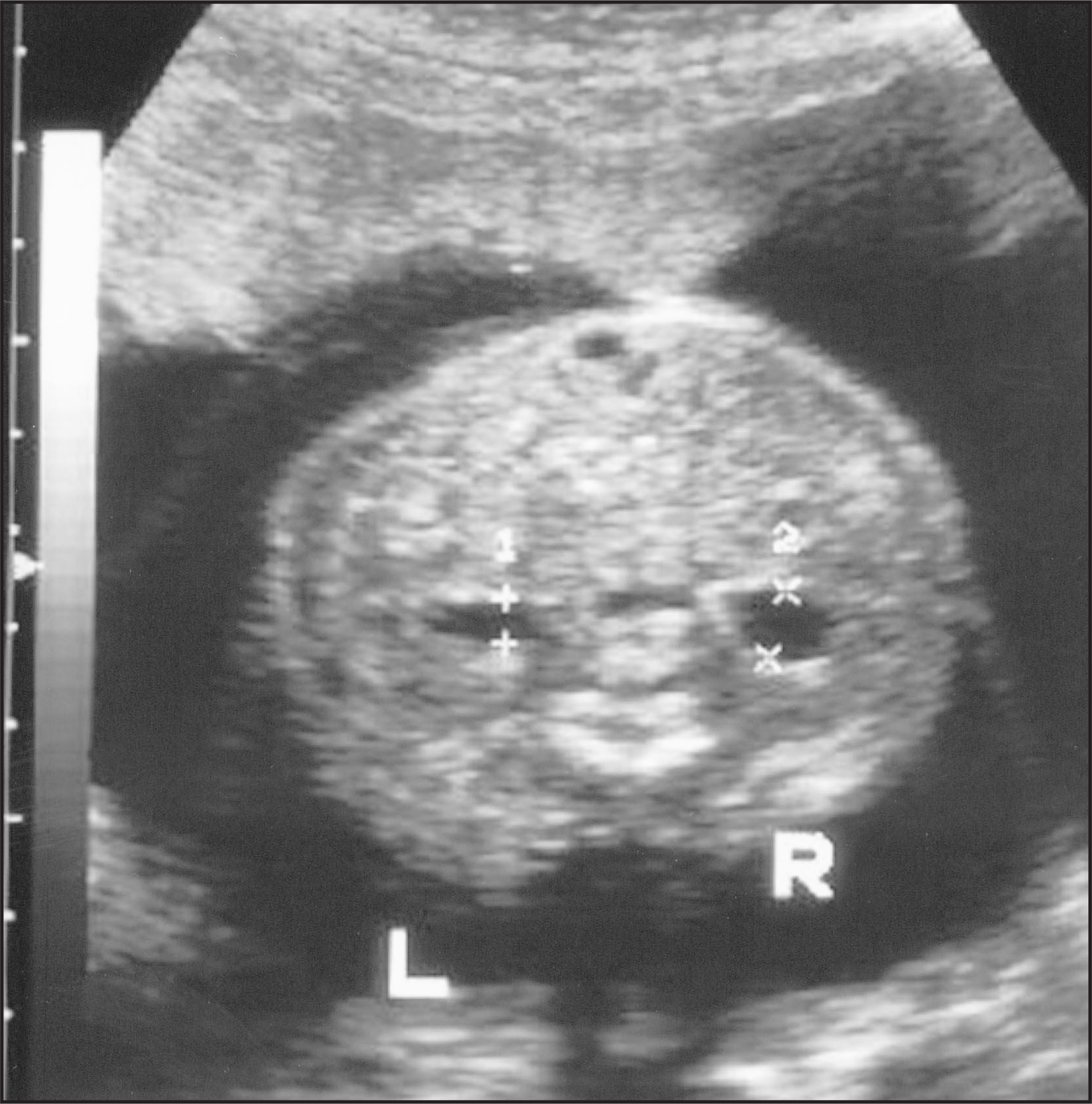
The importance of each of these findings lies not in isolation, but in interpretation with all the sonographic findings, the maternal age, and biochemical markers, to yield a more accurate risk assessment for trisomy 21. The genetic sonogram is such an assessment of sonographic abnormalities used to modify the risk of Down’s syndrome, either up or down for each fetus. Multiple scoring systems for sonographic findings have been proposed. Benacerraf and colleagues29,30 derived an excellent scoring index assigning 1 or 2 points for certain ultrasound findings, with a structural anomaly or nuchal thickening assigned 2 points; and with short humerus, short femur, hyperechoic bowel (grade 3), echogenic intracardiac focus, and pyelectasis each assigned 1 point. Maternal age ≥ 35 years also can be assigned 1 point. A score of 2 or more can result in a sensitivity of 75.5% and a false-positive rate of 5.7% for the detection of Down’s syndrome.29,30 Amniocentesis is usually offered with a score of 2 or more. Nyberg and colleagues31,32 have also derived an excellent method of integrating sonographic findings into prior maternal risk by an age-adjusted ultrasound risk assessment. Sonographic markers are expressed as likelihood ratios weighted by their individual importance, and the mother’s prior risk based on age is modified by these likelihood ratios to obtain a post-ultrasound risk, or post prior risk. This system helps minimize false-positive rates while providing a patient-specific risk estimate.
Conclusion
Both first and second trimester ultrasound screening for Down’s syndrome has made dramatic advances in recent years. It remains to be seen if one will prove more effective than the other, or a combination of the two will become the future standard in trisomy 21 screening. AR
References
- Souter V, Nyberg D. Sonographic screening for fetal aneuploidy.. J Ultrasound Med.. 2001;20:775-790.
- Nicolaides K, Azar G, Byrne D. Fetal nuchal translucency: Ultrasound screening for chromosomal defects in first trimester of pregnancy.. Br Med J.. 1992;304:867-869.
- Snijders R, Noble P, Seibre N. UK multicentre project on assessment of risk of trisomy 21 by maternal age and fetal nuchal-translucency thickness at 10-14 weeks of gestation.. Lancet.. 1998;351:343-346.
- Nicolaides K, Heath V, Liao A. The 11-14 week scan.. Clin Obstet Gynaecol. 2000;14,4:581-594.
- Pandya P, Snijders R, Johnson S. Screening for fetal trisomies by maternal age and fetal nuchal translucency thickness at 10 to 14 weeks of gestation.. Br J Obstet Gynaecol.. 1995;102:957-962.
- Pandya P, Snijders R, Johnson S. Natural history of trisomy 21 fetuses with increased nuchal translucency thickness.. Ultrasound Obstet Gynecol.. 1995;5:381-383.
- Salomon L, Bernard J, Taupin P. Relationship between nuchal translucency at 11-14 weeks and nuchal fold at 20-24 weeks of gestation.. Ultrasound Obstet Gynecol.. 2001;18:636-637.
- Souka A, Snijders R, Novakov A. Defects and syndromes in chromosomally normal fetuses with increased nuchal translucency thickness at 10-14 weeks of gestation.. Ultrasound Obstet Gynecol.. 1998;11:391-400.
- Hiippala A, Eronen M, Taipale P. Fetal nuchal translucency and normal chromosomes: A long-term follow-up study.. Ultrasound Obstet Gynecol. 2001;18:18-22.
- Hyett J, Perdu M, Sharland G. Using nuchal fetal translucency to screen for major congenital cardiac defects at 10-14 weeks of gestation: Population based cohort study.. Br Med J.. 1999;318:81-85.
- Nyberg D, Resta R, Luthy D. Prenatal sonographic findings of Down syndrome: Review of 94 cases.. Obstet Gynecol.. 1990;76((3 Pt 1)):370-377.
- Nyberg D, Souter V, El-Bastawissi A. Isolated sonographic markers for detection of fetal Down syndrome in the second trimester of pregnancy.. J Ultrasound Med.. 2001;20:1053-1063.
- Paladini D, Tartaglione A, Agangi A. The association between congenital heart disease and Down syndrome in prenatal life.. Ultrasound Obstet Gynecol.. 2000;15:104-108.
- Nyberg D, Souter V. Sonographic markers of fetal trisomies: Second trimester.. J Ultrasound Med.. 2001;20:655-674.
- DeVore G. Trisomy 21: 91% detection rate using second-trimester ultrasound markers.. Ultrasound Obstet Gynecol.. 2000;16:133-141.
- Benacerraf B, Frigoletto F, Laboda L. Sonographic diagnosis of Down syndrome in the second trimester.. Am J Obstet Gynecol.. 1985;153:49-52.
- Benacerraf B, Frigoletto F. Soft tissue nuchal fold in the second-trimester fetus: Standards for normal measurements compared with those in Down syndrome.. Am J Obstet Gynecol.. 1987;157:1146-1149.
- Gray D, Crane J. Optimal nuchal skin fold thresholds based on gestational age for prenatal detection of Down syndrome.. Am J Obstet Gynecol.. 1994;171:1282-1286.
- Borrell A, Costa D, Martinez J. Early midtrimester fetal nuchal thickness: Effectiveness as a marker of Down syndrome.. Am J Obstet Gynecol.. 1996;175:45-49.
- Sepulvada W, Sebire N. Fetal echogenic bowel: A complex scenario.. Ultrasound Obstet Gynecol.. 2000;16:510-514.
- Strocker A, Snijders R, Carlson D. Fetal echogenic bowel: Parameters to be considered in differential diagnosis.. Ultrasound Obstet Gynecol.. 2000;16:519-523.
- Nyberg D, Dubinsky T, Resta R. Echogenic fetal bowel during the second trimester: Clinical importance.. Radiology. 1993;188:527-531.
- Ranzini A, McLean D, Sharma S. Fetal intracardiac echogenic foci: Does visualization depend on the orientation of the four-chamber view? [abstract]. J Ultrasound Med. 1999;18((suppl 3)):S108.
- Levy D, Mintz M. The left ventricular echogenic focus: A normal finding.. AJR Am J Roentgenol.. 1998;150:85-86.
- Benacerraf B. The second-trimester fetus with Down syndrome: Detection using sonographic features.. Ultrasound Obstet Gynecol.. 1996;7:147-155.
- Petrikovsky B, Cuomo M, Schneider E. Isolated fetal hydronephosis: Beware the effect of bladder filling.. Prenat Diagn.. 1995;15:827-829.
- Robinson J, Tice K, Kolm P. Effect of maternal hydration on fetal renal pyelectasis.. Obstet Gynecol.. 1998;92:137-141.
- Corteville J, Dicke J, Crane J. Fetal pyelectasis and Down syndrome: Is genetic amniocentesis warranted?. Obstet Gynecol.. 1992;79((5 Pt 1)):770-772.
- Benacerraf B, Neuberg D, Bromley B. Sonographic scoring index for prenatal detection of chromosomal abnormalities.. J Ultrasound Med.. 1992;11:449-458.
- Benacerraf B, Nadel A, Bromley B. Identification of second-trimester fetuses with autosomal trisomy by use of a sonographic scoring index.. Radiology.. 1994;193:135-140.
- Winter T, Uhrich M, Souter V. The “Genetic Sonogram”: Comparison of the index scoring system with the age-adjusted US risk assessment.. Radiology.. 2000;215:775-782.
- Nyberg D, Luthy D, Resta R. Age-adjusted ultrasound risk assessment for fetal Down’s syndrome during the second trimester: Description of the method and analysis of 142 cases.. Ultrasound Obstet Gynecol.. 1998;12:8-14.
Citation
. Sonographic findings of Down’s syndrome. Applied Radiology. 2002;31(9):9-14. doi:10.37549/AR1123.