Spectrum of Cystic Mediastinal Lesions: An Imaging Review
Applied Radiology — Vol. 55 , Issue 5
Published: September 1, 2026
1 Department of Radiology, Division of Thoracic imaging, University of North Carolina, Chapel Hill, North Carolina
2 Division of Endocrinology, Diabetes, and Metabolism, Johns Hopkins University School of Medicine, Baltimore, Maryland
* Corresponding author: Behrooz Masuodi (dr.b.masuodi@gmail.com)
Abstract
Cystic and cyst-like lesions of the mediastinum encompass a broad spectrum of entities ranging from benign congenital abnormalities to inflammatory or malignant processes with cystic components. These lesions are frequently encountered with cross-sectional imaging and may present a diagnostic challenge due to overlapping imaging appearances. Accurate characterization is critical, as misclassification can lead to unnecessary surgical intervention or delayed diagnosis of clinically significant disease.
These lesions can be incidentally identified on routine chest radiographs as paramediastinal opacities. CT is typically the initial imaging modality for further evaluation of mediastinal lesions and provides essential information regarding lesion location, morphology, and relationship to adjacent structures. MRI offers superior soft-tissue characterization and plays a key role in problem-solving, particularly when CT findings are indeterminate.
The use of T1- and T2-weighted sequences, along with diffusion-weighted imaging, facilitates differentiation of simple cysts from lesions containing solid, proteinaceous, or malignant components. Awareness of characteristic imaging features of common congenital cysts, acquired cystic lesions, and important mimics further improves diagnostic confidence.
In conclusion, mediastinal cystic lesions require a structured imaging-based approach for accurate diagnosis. Integration of clinical context, anatomic location, and multimodality imaging findings, with particular emphasis on MRI, supports appropriate risk stratification and individualized patient management.
Keywords
mediastinal cysts, bronchogenic cyst, thymic cyst, pericardial cyst, esophageal duplication cyst, lymphangioma, mediastinal lesions
Categories
Introduction
Cystic and cyst-like lesions of the mediastinum comprise a heterogeneous group of entities that frequently pose diagnostic challenges, particularly when imaging findings are indeterminate.1-3 These lesions span a broad spectrum, ranging from benign congenital lesions to malignant cystic neoplasms or tumors undergoing cystic degeneration.1-3 Accurate differentiation is essential, as misinterpretation may lead to unnecessary imaging follow-up, surgical intervention, or delayed treatment of clinically significant disease.1,4
Congenital mediastinal cysts include bronchogenic cysts, esophageal duplication cysts, thymic cysts, pericardial cysts, neuroenteric cysts, and lymphangiomas.2,3,5 The classification, embryologic basis, and typical locations of mediastinal cystic lesions are summarized in Table 1. These lesions arise from distinct embryologic abnormalities such as aberrant foregut budding or incomplete recanalization and typically demonstrate characteristic imaging features, including well-defined margins, thin walls, unilocular configuration, and homogeneous fluid attenuation on CT or fluid signal intensity on MRI.2,3 Recognition of these features is highly important for confident diagnosis and appropriate clinical management.
| Type | Subtype | Embryologic/Pathologic Basis | Typical Location | Reference |
|---|---|---|---|---|
| Congenital | Bronchogenic cyst | Abnormal budding of tracheobronchial tree | Visceral mediastinum, near carina | 5,6 |
| Esophageal duplication cyst | Incomplete foregut recanalization, shares muscular wall | Adjacent to esophagus | 2,7 | |
| Pericardial cyst | Incomplete fusion of mesenchymal lacunae | Right cardiophrenic angle | 8 | |
| Thymic cyst | Persistent thymopharyngeal duct remnants | Prevascular mediastinum | 9 | |
| Lymphangioma | Sequestration of lymphatic tissue isolated from lymphatic system | Prevascular mediastinum | ||
| Acquired | Cystic degeneration of tumors | Degeneration/necrosis in thymomas, germ cell tumors, and so forth | Variable | 1,2 |
| Non-neoplastic processes | Infection, hemorrhage, abscess formation | Variable | 1,7 |
In contrast, acquired cyst-like mediastinal lesions may result from cystic degeneration or necrosis within malignant tumors such as thymomas, germ cell tumors, lymphomas, or metastatic disease, or from non-neoplastic processes including infection, hemorrhage, or abscess formation.1-3,8 Although these lesions may mimic benign cysts, the presence of thickened or irregular walls, enhancing or nodular internal septations, solid enhancing components, or restricted diffusion on MRI should raise concern for malignancy.1,3,9 We will provide a practical, imaging-based approach to cystic and cyst-like mediastinal lesions, with emphasis on classification, embryologic origin, characteristic imaging features, important mimics, and implications for diagnosis and management.
Imaging Modalities and Technical Considerations
Larger mediastinal cysts can be identified as circumscribed opacities silhouetting the cardio-mediastinal border on routine chest radiograph. CT is typically the first-line imaging modality for evaluation of mediastinal lesions and allows accurate assessment of lesion location, attenuation, wall characteristics, calcification, and relationships to adjacent mediastinal structures.1,3,10 CT plays a central role in initial lesion detection and anatomical localization; however, attenuation overlaps between proteinaceous fluid, hemorrhage, and necrotic tumors may limit diagnostic confidence in certain cases.1,3 Additionally, beam-hardening and streak artifacts may falsely elevate the measured attenuation of simple cysts.
MRI provides superior soft-tissue characterization and is particularly valuable when CT findings are equivocal.3,6,11 T1- and T2-weighted sequences facilitate differentiation of simple fluid from hemorrhagic or protein-rich content, while contrast-enhanced imaging assists in identifying solid enhancing components suggestive of malignancy.3,11 Diffusion-weighted imaging further improves lesion characterization by assessing diffusion properties and helps distinguish non-neoplastic cysts from solid or malignant lesions, demonstrating restricted diffusion.6,9
PET combined with CT may be useful in selected cases when malignancy is suspected. Increased fluorodeoxyglucose uptake suggests metabolically active components and supports the diagnosis of malignant or infectious processes rather than simple cysts.1,7 Key imaging characteristics of mediastinal cystic lesions across different imaging modalities are summarized in Table 2.
| Modality | Key Diagnostic Features | Reference |
|---|---|---|
| CT | Defines attenuation, wall calcification, relation to adjacent structures | 1,12 |
| MRI | Superior soft-tissue contrast; high T2 signal; absence of enhancement consistent with simple cyst | 3,13 |
| DWI | Differentiates cystic lesions from solid tumors; useful for indeterminate CT findings | 3,14 |
| PET/CT | Identifies metabolic activity in malignant or solid components | 4 |
Pathology-Based Review of Mediastinal Cystic Lesions
Bronchogenic Cyst
Bronchogenic cysts arise from abnormal budding of the tracheobronchial tree during embryogenesis and represent one of the most common congenital cystic lesions of the mediastinum.12,13 They are typically located in the visceral mediastinum near the carina or mainstem bronchi, although ectopic locations have also been described.2,3,12 Most lesions are asymptomatic and incidentally detected, while larger cysts may produce cough, chest pain, or compressive symptoms due to mass effect.12 Approximately 20% of these cysts are intraparenchymal and can cause recurrent infection and, rarely, pneumothorax.14
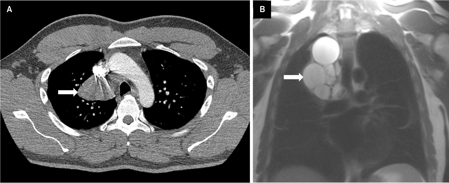
On imaging, bronchogenic cysts appear as well-defined, thin-walled cysts with simple fluid (water) attenuation or higher density fluid due to proteinaceous material on CT. These can be occasionally multilocular with thin internal septations. Air or hemorrhage is less common but can be seen within these cysts. On MRI, these lesions are characterized by high T2 signal intensity with variable T1 signal based on proteinaceous content, without enhancement or diffusion restriction (Figure 1).1,3,14 While most asymptomatic cases may be managed conservatively, symptomatic lesions or when diagnostic uncertainty exists, surgical resection is recommended. Malignant transformation is rare but has been reported.4,12,14,15
Esophageal Duplication Cyst
Esophageal duplication cysts result from incomplete recanalization of the foregut and are most commonly located adjacent to the esophagus in the visceral mediastinum.2,3 These lesions are frequently asymptomatic but may present with dysphagia or chest discomfort depending on size and location.2,5 Hemorrhage is a common complication of these cysts that can cause rapid enlargement of the cyst and chest pain.16 Malignant transformation is exceedingly rare.2
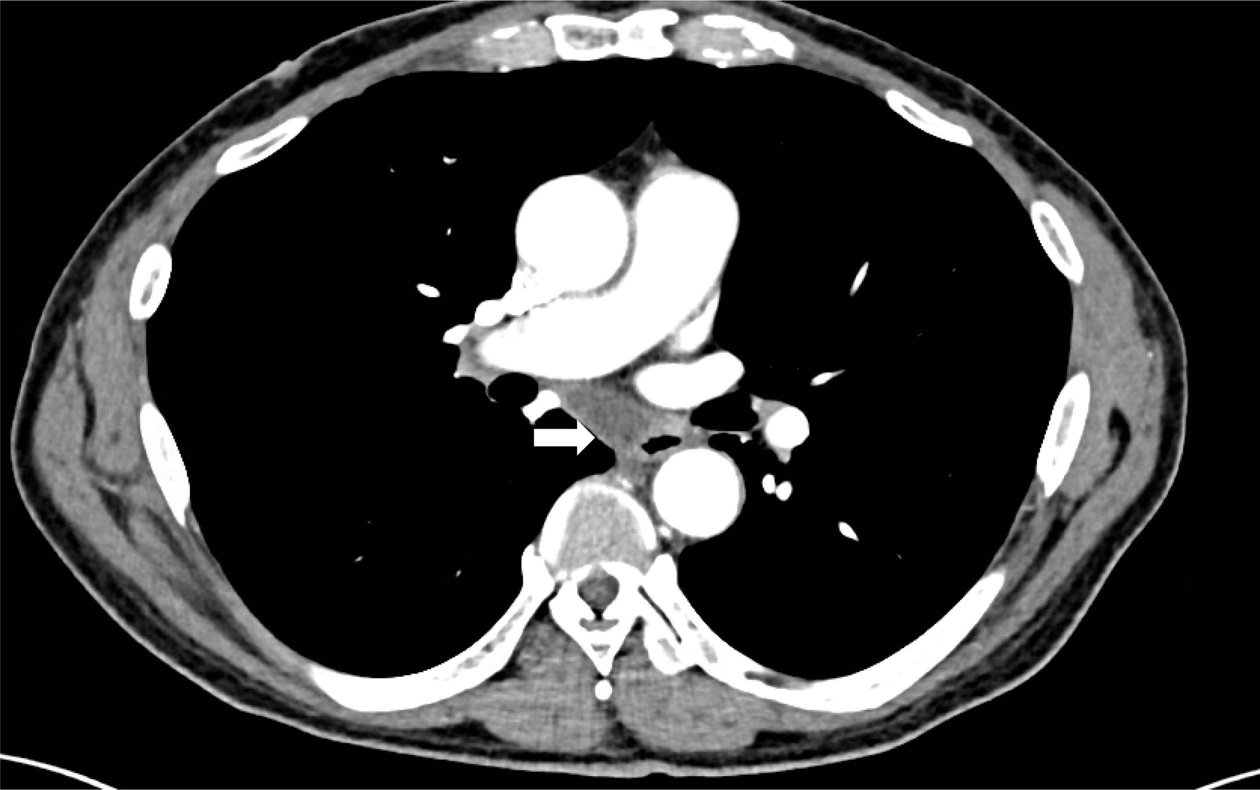
Imaging typically demonstrates a unilocular cyst closely associated with the esophageal wall, with homogeneous fluid characteristics on both CT and MRI (Figure 2).2,3 Surgical excision is generally recommended in symptomatic patients or when the diagnosis is uncertain.4
Pericardial Cysts
Pericardial cysts arise from incomplete fusion of mesenchymal lacunae during pericardial development and are most commonly located at the right cardiophrenic angle.13,17 These lesions are usually asymptomatic and incidentally detected, although symptoms may occur due to compression of adjacent structures.17
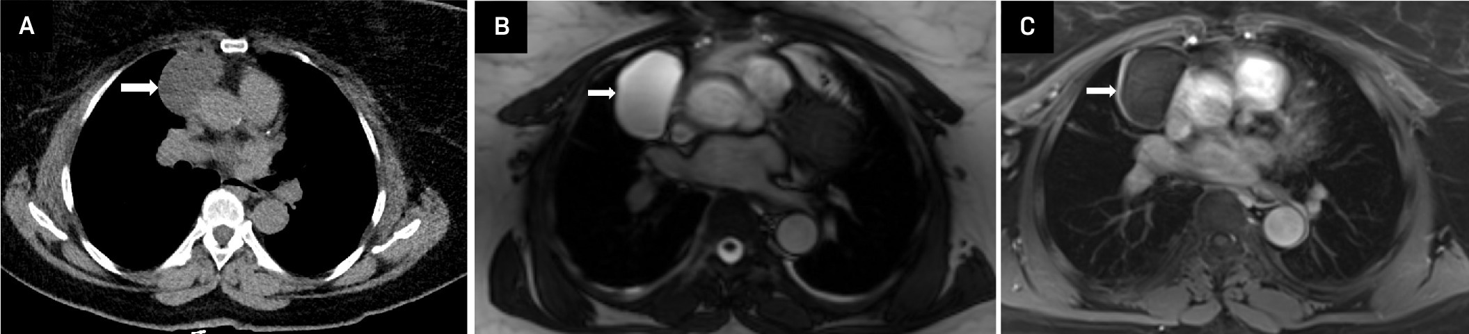
On imaging, pericardial cysts appear as well-circumscribed, thin-walled lesions with homogeneous fluid attenuation on CT and high T2 signal intensity on MRI, without enhancement (Figure 3).3,17 Larger pericardial cysts are detectable on frontal chest radiograph as well-defined basilar opacities silhouetting cardiac borders. The wall of the cyst can sometimes become calcified. There is no established malignant potential, and asymptomatic lesions may be managed conservatively, with intervention reserved for symptomatic or enlarging cysts.13
Thymic Cyst
Thymic cysts arise from persistent thymopharyngeal duct remnants and are typically located in the prevascular anterior mediastinum.10,18 Most lesions are asymptomatic and incidentally detected. While generally benign, multilocular thymic cysts may be associated with underlying thymic neoplasms.10,18
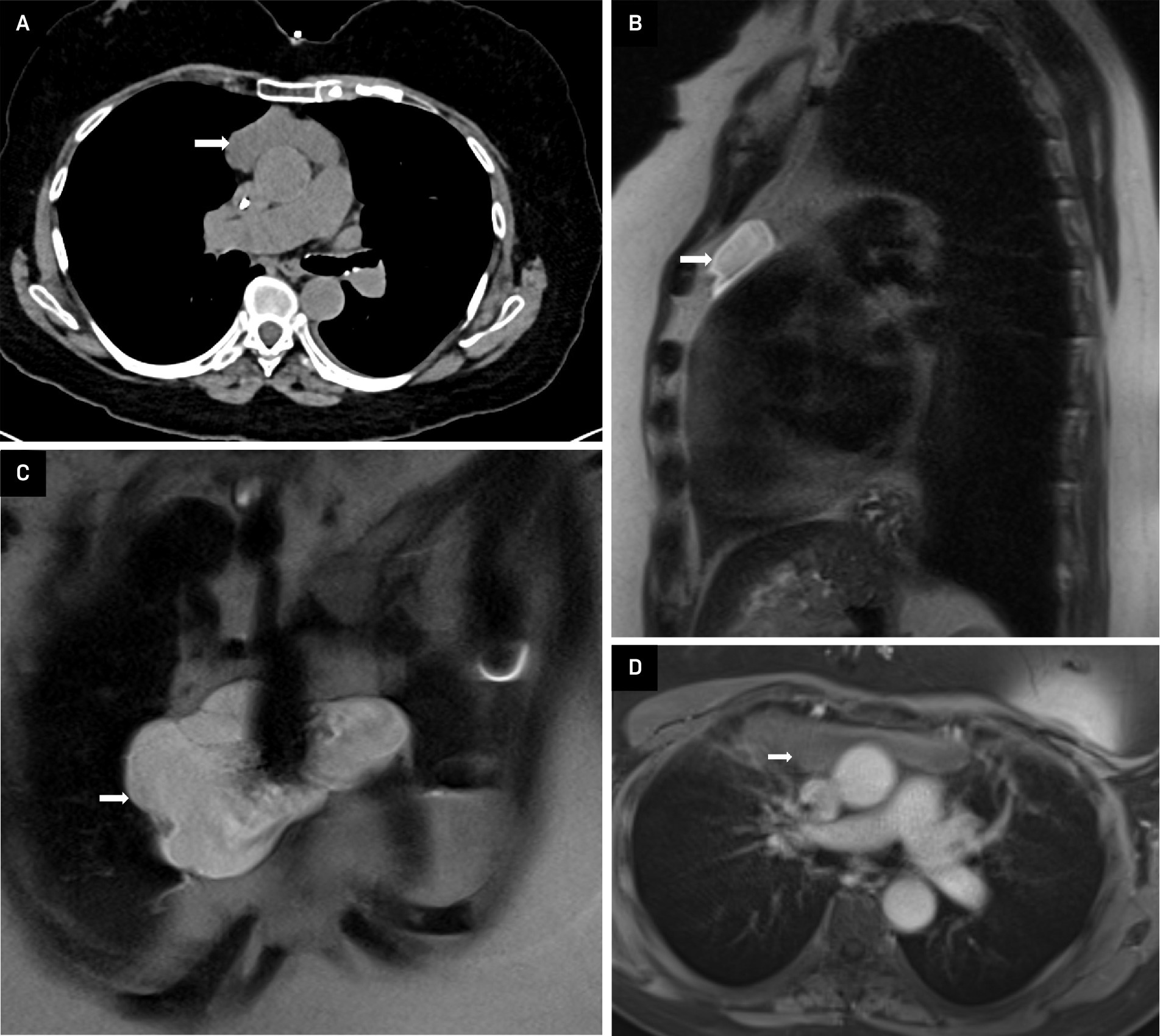
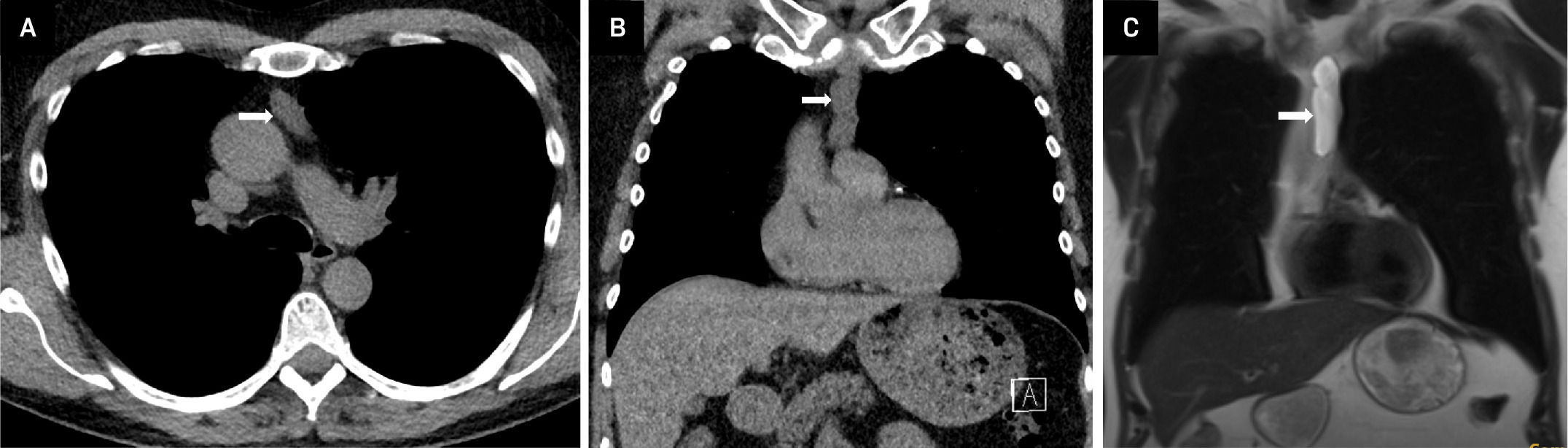
On imaging, thymic cysts appear as well-defined unilocular or multilocular cystic lesions with homogeneous fluid attenuation on CT and high T2 signal intensity on MRI (Figures 4, 5). MRI is particularly helpful in excluding internal solid or enhancing components that may raise concern for malignancy.3,10 Surgical resection is considered when imaging findings are atypical or malignancy cannot be confidently excluded.4,18
Lymphangioma
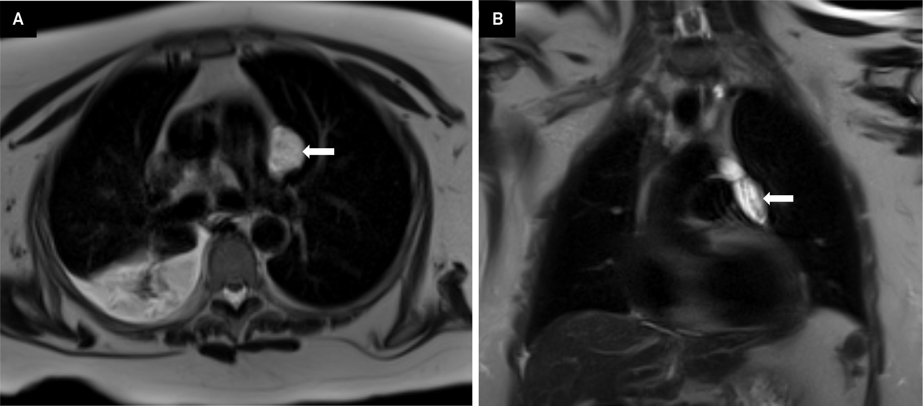
Mediastinal lymphangiomas are uncommon benign cystic structures that arise from focal proliferation of lymphatic vessels and are most commonly found in the superior aspect of prevascular mediastinum.19 They are usually asymptomatic and become apparent when there is mass effect on adjacent structures.20 Complications associated with lymphangiomas include infection, cystic hemorrhage, chylothorax, and chylopericardium. Standard treatment includes surgical excision if symptomatic. Recurrence is highly unlikely after excision. On CT, lymphangiomas appear as low-attenuation cysts with smooth contours and attenuation could be of simple to complex fluid and fat. On MR, they are isointense to muscle on T1-weighted images and hyperintense on T2; however, signal could vary based on cystic and proteinaceous content (Figure 6).
Differentials include necrotic tumors, teratomas, thymic cysts, pericardial or bronchogenic cysts, hematomas, goiters, and infectious fluid collections.21
Cystic Degeneration of Malignant Tumors
Cystic degeneration may occur within malignant mediastinal tumors such as thymomas, germ cell tumors, lymphomas, or metastatic disease.2,3,8 These lesions may present incidentally or with systemic or compressive symptoms depending on tumor burden.8 Imaging features suggestive of malignancy include irregular or thickened walls, internal septations, heterogeneous or nodular enhancement, and restricted diffusion on MRI.1,3,9 Management requires tissue diagnosis and appropriate oncologic treatment.4,22
Infectious and Inflammatory Cyst-Like Lesions
Cyst-like lesions may develop secondary to infection, hemorrhage, or abscess formation.1,8 Pseudocysts resulting from pancreatitis can also extend into the visceral mediastinum.23 Patients may present with fever, chest pain, or systemic signs of infection.1,3 Imaging typically demonstrates complex cystic lesions with surrounding inflammatory changes and possible enhancement.3 Management includes antibiotic therapy and drainage, with surgical intervention reserved for complicated cases.4,22
Important Mimics of Mediastinal Cystic Lesions
Several noncystic entities may closely mimic mediastinal cystic lesions on imaging and should be carefully considered in the differential diagnosis.1-3 Pericardial recesses represent normal anatomic variants that may appear as well-defined fluid collections along the pericardium, particularly adjacent to the great vessels.1,2 Recognition of their characteristic locations, continuity with the pericardial space, and lack of mass effect is essential to avoid misdiagnosis.3
Necrotic or cystic lymph nodes may also simulate mediastinal cysts, particularly in the setting of malignancy, infection, or inflammatory disease.1,2,22 Unlike simple cysts, necrotic lymph nodes often demonstrate irregular or thickened walls, peripheral or nodular enhancement, and restricted diffusion on MRI, which can aid in differentiation (Figure 7).9,22
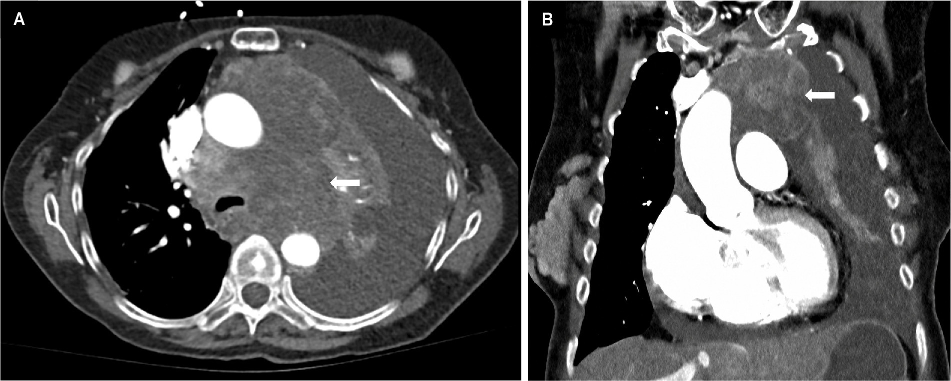
Other important mimics include cystic teratomas and chronic infectious or granulomatous processes.2,8,10 These entities may demonstrate heterogeneous internal components, fat, calcifications, or variable enhancement patterns, features that are atypical for simple cysts.2,10 Careful assessment of imaging findings in conjunction with clinical context and multimodality imaging is the key for distinguishing these conditions from true mediastinal cysts.1,5,22
Treatment of Cystic and Cyst-Like Mediastinal Lesions
Management is individualized based on symptoms, lesion growth, imaging characteristics, and diagnostic certainty.4,12,22,24 Surgical resection is recommended for symptomatic lesions, enlarging cysts, or lesions with suspicious imaging features, whereas conservative surveillance may be appropriate for asymptomatic congenital cysts with classic imaging appearances in patients with reliable follow-up.4,22
Conclusion
Cystic mediastinal lesions and their mimics have overlapping imaging features. It is very important to adopt an organized approach, including lesion localization, imaging features, and clinical information, which helps to narrow down the differentials. Even though CT is the first-line imaging technique, MRI plays a critical role in differentiating simple and benign lesions from complex and malignant lesions. Together, these complementary modalities enable confident diagnosis and guide appropriate treatment.
References
- Shah A, Rojas C. Imaging modalities (MRI, CT, PET/CT), indications, differential diagnosis and imaging characteristics of cystic mediastinal masses: a review. Mediastinum. 2023;7. doi:10.21037/med-22-31.
- Jeung M, Gasser B, Gangi A. Imaging of cystic masses of the mediastinum. Radiographics. Oct 2002;22 Spec No:S79-93. doi:10.1148/radiographics.22.suppl_1.g02oc09s79.
- Odev K, Arıbaş B, Nayman A. Imaging of cystic and cyst-like lesions of the mediastinum with pathologic correlation. J Clin Imaging Sci. 2012;2. doi:10.4103/2156-7514.97750.
- Barrios P, Avella Patino D. Surgical indications for mediastinal cysts-a narrative review. Mediastinum. 2022;6. doi:10.21037/med-22-27.
- Le Pimpec-Barthes F, Cazes A, Bagan P. Mediastinal cysts: clinical approach and treatment. Rev Pneumol Clin. Feb 2010;66(1):52-62. doi:10.1016/j.pneumo.2009.12.009.
- Hwang E, Paek M, Yoon S. Quantitative thoracic magnetic resonance criteria for the differentiation of cysts from solid masses in the anterior mediastinum. Korean J Radiol. May 2019;20(5):854-861. doi:10.3348/kjr.2018.0699.
- Kajiura K, Takao S, Kawano N. Evaluation of the components of mediastinal cystic lesions using imaging techniques. J Med Invest. 2019;66(1.2):106-111. doi:10.2152/jmi.66.106.
- Takahashi K, Al-Janabi N. Computed tomography and magnetic resonance imaging of mediastinal tumors. J Magn Reson Imaging. Dec 2010;32(6):1325-1339. doi:10.1002/jmri.22377.
- Shin K, Yi C, Kim T. Diffusion-weighted MRI for distinguishing non-neoplastic cysts from solid masses in the mediastinum: problem-solving in mediastinal masses of indeterminate internal characteristics on CT. Eur Radiol. Mar 2014;24(3):677-684. doi:10.1007/s00330-013-3054-0.
- Nakazono T, Yamaguchi K, Egashira R, Mizuguchi M, Irie H. Anterior mediastinal lesions: CT and MRI features and differential diagnosis. Jpn J Radiol. Feb 2021;39(2):101-117. doi:10.1007/s11604-020-01031-2.
- Choe J, Lee S, Ahn Y. Characteristics and outcomes of anterior mediastinal cystic lesions diagnosed on chest MRI: implications for management of cystic lesions. Insights Imaging. Aug 17 2022;13(1). doi:10.1186/s13244-022-01275-8.
- Gross D, Briski L, Wherley E, Nguyen D. Bronchogenic cysts: a narrative review. Mediastinum. 2023;7. doi:10.21037/med-22-46.
- Meredith A, Zazai I, Kyriakopoulos C. Pericardial cyst. StatPearls. 2026.
- Sharma S, Limaiem F, Collier S, Mlika M. Bronchogenic cyst. StatPearls. 2026.
- Bouassida I, Hadj Dahmane M, Zribi H. A poor prognosis of a mediastinal bronchogenic cyst with malignant transformation: a case report. Int J Surg Case Rep. May 2023;106. doi:10.1016/j.ijscr.2023.108246.
- Macpherson R. Gastrointestinal tract duplications: clinical, pathologic, etiologic, and radiologic considerations. Radiographics. Sep 1993;13(5):1063-1080. doi:10.1148/radiographics.13.5.8210590.
- Grigoraş A, Amălinei C, Căruntu I. Symptomatic pericardial cysts and dilemmas in their diagnosis. Rom J Morphol Embryol. Oct-Dec 2023;64(4):517-525. doi:10.47162/RJME.64.4.08.
- Özdemir &. Anterior mediastinal masses and thymic cysts. World J Clin Cases. Feb 6 2025;13(4). doi:10.12998/wjcc.v13.i4.95035.
- Macchiarini P, Ostertag H. Uncommon primary mediastinal tumours. Lancet Oncol. Feb 2004;5(2):107-118. doi:10.1016/S1470-2045(04)01385-3.
- Salehi F, Landis M, Inculet R, Wiseman D. Case report of a rare cystic mediastinal lymphangioma mimicking recurrent pleural effusion. Case Rep Radiol. 2019;2019. doi:10.1155/2019/1301845.
- Shaffer K, Rosado-de-Christenson M, Patz E, Young S, Farver C. Thoracic lymphangioma in adults: CT and MR imaging features. AJR Am J Roentgenol. Feb 1994;162(2):283-289. doi:10.2214/ajr.162.2.8310910.
- Mura R, Pochepnia S, Kifjak D, Khenkina N, Prosch H. A diagnostic approach to mediastinal masses in clinical practice. BJR Open. Jan 2025;7(1). doi:10.1093/bjro/tzaf009.
- Amin S, Shahab A, Khan A. A pancreatic pseudocyst sitting in the mediastinum-a case report of an unusual occurrence. SAGE Open Med Case Rep. 2024;12. doi:10.1177/2050313X241262139.
- Hsu D, Banks K, Velotta J. Surgical approaches to mediastinal cysts: clinical practice review. Mediastinum. 2022;6. doi:10.21037/med-22-20.
Disclosures
The author(s) have no conflicts of interest to disclose. None of the authors received outside funding for the production of this original manuscript and no part of this article has been previously published elsewhere.
Acknowledgments
The authors declare that no generative artificial intelligence (AI) or AI-assisted technologies were used to generate content, ideas, or theories in this work. AI was used solely to enhance readability and refine language. The manuscript has been carefully reviewed and edited by the authors to ensure accuracy.
Citation
. Spectrum of Cystic Mediastinal Lesions: An Imaging Review. Applied Radiology. 2026;55(5). doi:10.37549/AR-D-26-0052.