Sphenoid Sinus Metastasis Secondary to Urothelial Carcinoma
Applied Radiology — Vol. 55 , Issue 5
Published: June 1, 2026
1 Department of Imaging Sciences, University of Rochester Medical Center, Rochester, New York
* Corresponding author: Jessie Huang (jessie_huang@urmc.rochester.edu)
Abstract
Sphenoid sinus pathologies present with headache, cranial nerve dysfunction, and facial pain and require evaluation on CT and MRI. CT and MRI play a complementary role in assessing the extent of the lesion and involvement with neighboring critical structures, and the presence of destructive osseous changes. Imaging helps to narrow the differential diagnosis, which can include sinusitis, mucoceles, bone cysts, primary malignancy, or metastases, in addition to aiding in preoperative planning. This case highlights the importance of considering rare metastatic sites on imaging in patients with a history of malignancy and suggestive symptoms.
Keywords
sphenoid sinus, metastasis, urothelial carcinoma, neuroradiology, CT sinus, MRI, FDG-PET
Categories
Case Summary
A middle-aged patient presented with a history of left eye pain for 1 year and acute onset pressure behind the left eye with diplopia. The patient reported a prior history of FGFR-mutated urothelial carcinoma, which had been treated with radiation for metastases to the T9 vertebra and pelvic bone. The patient also reported a history of left cataract and left retinal repair surgeries, and had previously used eye drops for pain.
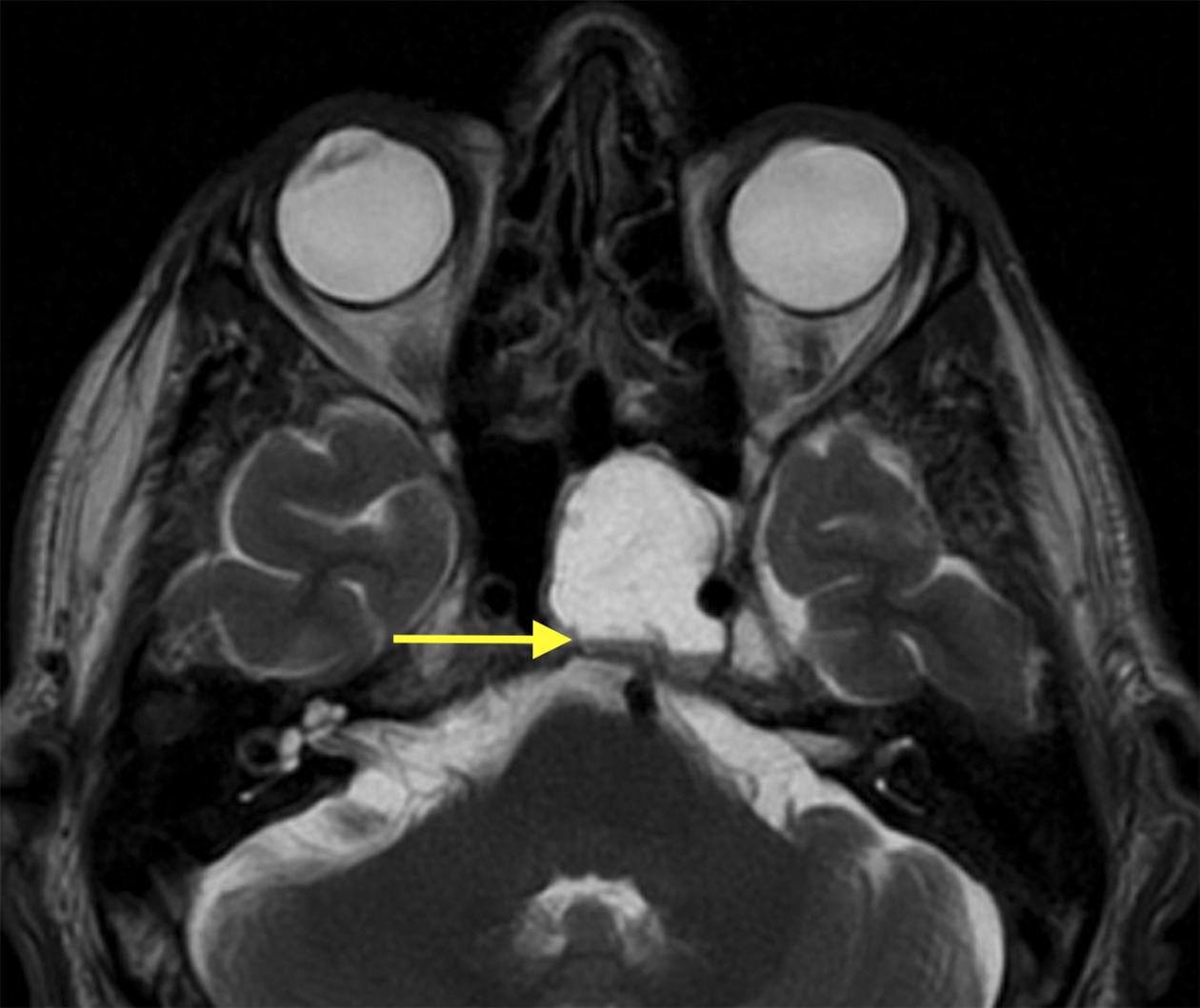
Physical exam revealed left abducens nerve palsy. An initial MRI brain scan demonstrated a cystic lesion in the left sphenoid sinus with a blood-fluid level. Follow-up CT sinus scans showed left sphenoid sinus opacification with osseous erosions and extension into the left cavernous sinus. The patient underwent septoplasty and left sphenoidotomy with partial resection of the left sphenoid sinus mass. Pathology revealed HER+ metastatic urothelial carcinoma. The residual tissue exhibited continued growth prior to initiating trastuzumab deruxtecan and radiation therapy.
Imaging Findings
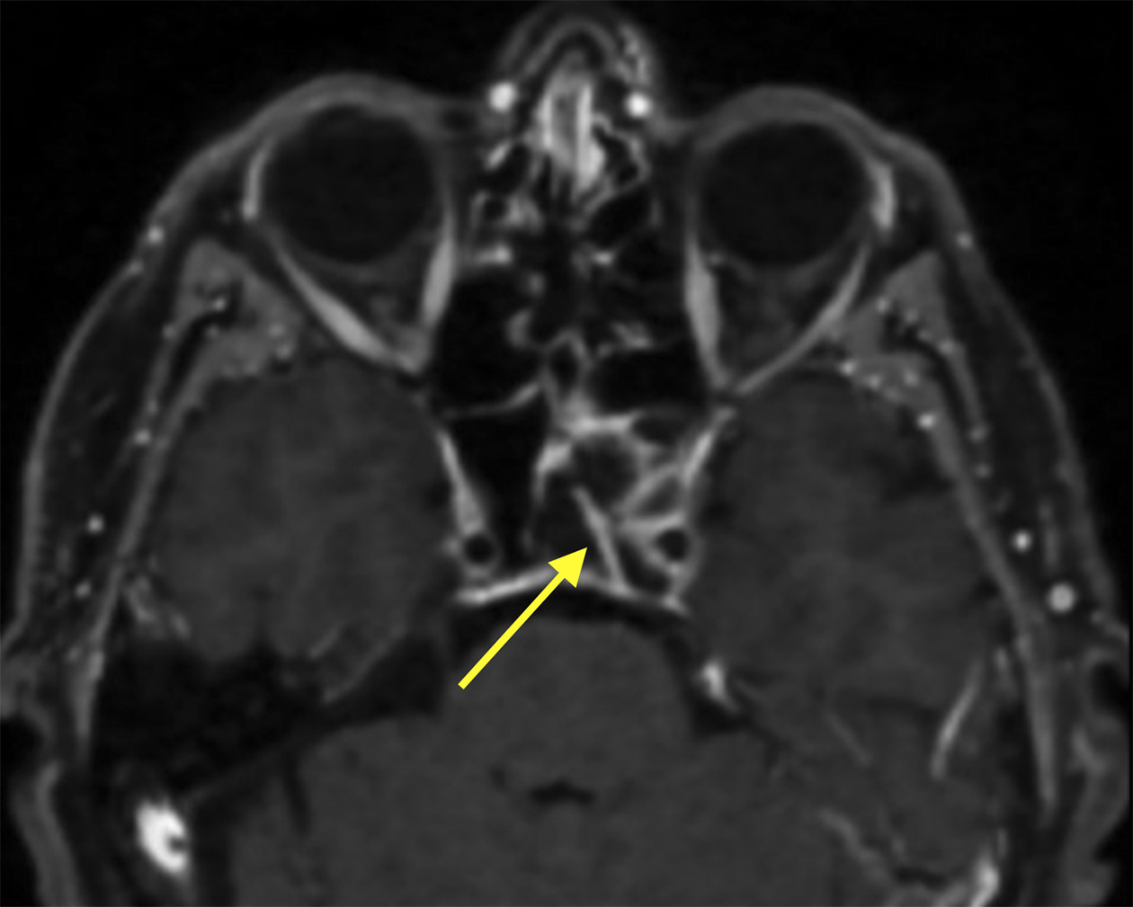
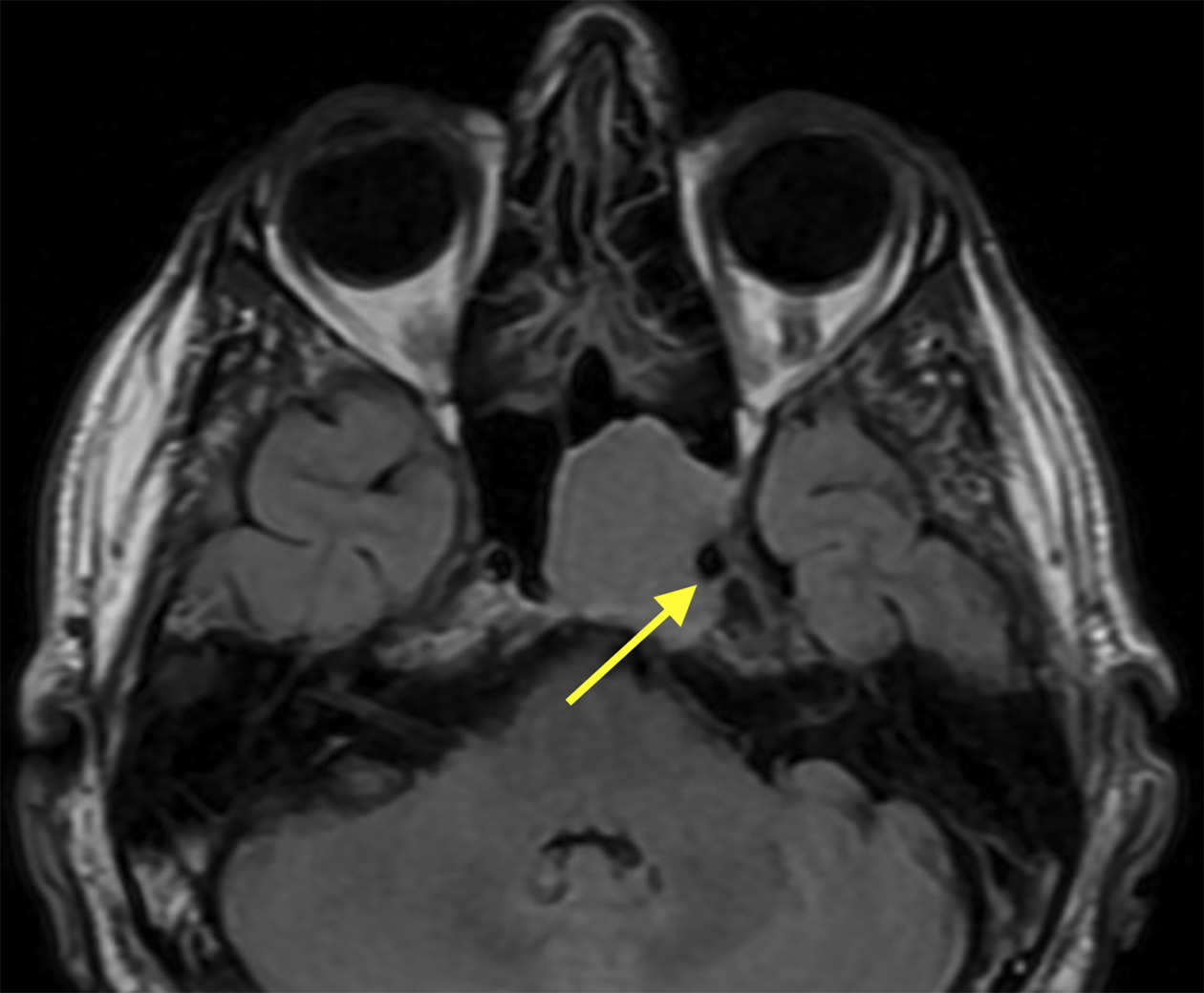
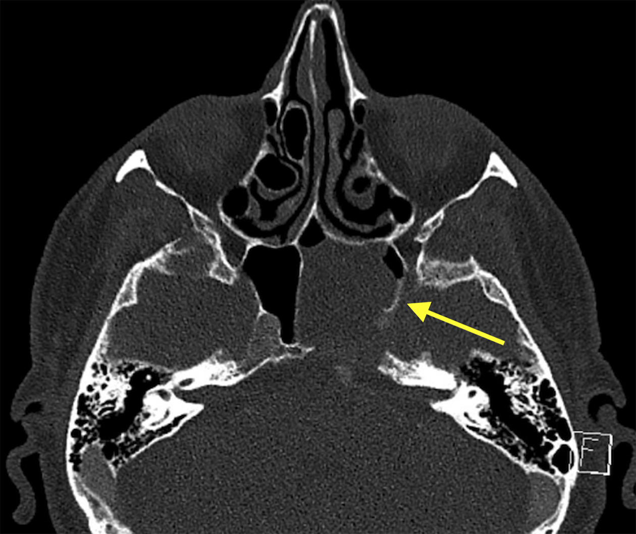
The initial axial T2 postcontrast PROPELLER MRI demonstrated a well-circumscribed expansile mass in the left sphenoid sinus exhibiting T2 hyperintensity and an internal blood-fluid level (Figure 1, arrow). On axial postcontrast T1 CUBE MRI, there was smooth peripheral enhancement and thin linear enhancement along the superior aspect that could represent a vessel or a septation (Figure 2, arrow). No nodular enhancement corresponding to this mass was noted. On axial T2 FLAIR MRI, the mass extended laterally to partially encase the left cavernous intracranial carotid artery (Figure 3, arrow). Additionally, there was thinning of the clivus dorsally without intracranial extension. Given the osseous thinning, CT sinus was recommended for further characterization. A subsequent CT sinus scan demonstrated osseous erosions of the clivus and lateral sphenoid sinus wall (Figure 4, arrow).
Diagnosis
Sphenoid sinus metastasis secondary to urothelial carcinoma.
Differential diagnosis includes sphenoid sinus mucocele, secondary aneurysmal bone cyst, and primary or secondary malignancy.
Discussion
The sphenoid sinus, one of the 4 paranasal sinuses, is situated near the optic nerve, cavernous sinus, pituitary gland, cranial nerves, and internal carotid artery. Recognizing pathologies affecting the sphenoid sinus is critical, as they can lead to compression of these structures. Patients initially present with headache, facial pain, visual changes, and cranial nerve dysfunction, most often abducens palsy.1
Common pathologies seen on MRI and CT include bacterial or fungal sinusitis and mucoceles.1 Although primary tumors in the sphenoid sinus are rare, they include squamous cell carcinoma, adenoid cystic carcinoma, and chondrosarcoma.2 Metastasis to the sphenoid sinus, although also rare, is associated with poor prognosis. Sphenoid sinus metastasis most commonly arises from breast, prostate, lung, and colon primary cancers.3 Metastasis from urothelial carcinoma, as seen in this patient, usually reaches the bones, lungs, and liver through lymphatic and hematogeneous routes. Urothelial carcinoma metastasis to the sphenoid sinus is exceedingly rare and, to our knowledge, has not been reported in the literature.4
CT and MRI are recommended for initial imaging of the paranasal sinuses. CT is better at evaluating bony changes like cortical erosion, destruction, and remodeling, while MRI is used to characterize soft-tissue components. Together, they help determine the size of the lesion, differentiate it from mucous and fluid, and assess for extension beyond the sinus walls.5 Metastases in the sphenoid can present as a homogeneous soft-tissue masses; in some cases, they extend out of the sphenoid sinus or damage the sphenoid bone. Although 18F-fluorodeoxyglucose (FDG) PET/CT is used in whole-body evaluation for metastases in patients with known malignancy,6 biopsy is used to determine the diagnosis.
Mucocele, a benign cystic lesion lined with respiratory epithelium and filled with mucoid secretions, was also considered based on this patient’s MRI. A destructive mucocele is a more aggressive form that erodes the bone and can spread to nearby structures.7 On CT, mucoceles appear as opacities with contents of varying density, as well as bowing and thinning of the osseous margins. On MRI, they can appear with high intensity on T2 imaging.8 Mucoceles and mucous retention cysts contain a mix of water, mucus, and protein, and rarely contain blood levels such as that seen on MRI in our case.7 A third diagnosis considered was a secondary aneurysmal bone cyst, which are masses with blood fluid levels. These entities often demonstrate nodular enhancement on the periphery, which was not seen on this patient’s MRI.9
Despite the well-circumscribed appearance of the mass in our patient, the presence of blood-fluid levels and osseous erosions at CT were highly suspicious for an aggressive process. Given the patient’s history of urothelial carcinoma, metastasis was the leading differential and was eventually confirmed at histopathology. Metastasis to the sphenoid sinus can be treated with surgery, but total resection is difficult owing to the region’s topography and nearby structures. In our patient, total resection was limited by the carotid artery, optic canal, and involvement of the cavernous sinus. Patients with inoperable tumors can receive radiation therapy, while recurrence can be treated with combined radiation and chemotherapy.2 Repeat imaging is critical for timely identification of lesion growth.
Conclusion
Sphenoid sinus pathologies typically present with headache, cranial nerve dysfunction, and facial pain and require CT and MRI evaluation. These modalities play a complementary role in determining the extent of the lesion, involvement of nearby structures, and presence of destructive osseous changes. They also help to narrow the differential diagnosis, which can include sinusitis, mucoceles, bone cysts, primary malignancy, and metastases, as well as aid in preoperative planning.
References
- Sethi K, Choudhary S, Ganesan P. Sphenoid sinus anatomical variants and pathologies: pictorial essay. Neuroradiology. 2023;65(8):1187-1203. doi:10.1007/s00234-023-03163-4.
- DeMonte F, Ginsberg L, Clayman G. Primary malignant tumors of the sphenoidal sinus. Neurosurgery. 2000;46(5):1084-1091. doi:10.1097/00006123-200005000-00012.
- Mickel R, Zimmerman M. The sphenoid sinus—a site for metastasis. Otolaryngol Head Neck Surg. 1990;102(6):709-716. doi:10.1177/019459989010200614.
- Shinagare A, Ramaiya N, Jagannathan J. Metastatic pattern of bladder cancer: correlation with the characteristics of the primary tumor. AJR Am J Roentgenol. 2011;196(1):117-122. doi:10.2214/AJR.10.5036.
- Kawaguchi M, Kato H, Tomita H. Imaging characteristics of malignant sinonasal tumors. J Clin Med. 2017;6(12). doi:10.3390/jcm6120116.
- Akay S, Pollard J, Saad Eddin A. PET/CT imaging in treatment planning and surveillance of sinonasal neoplasms. Cancers. 2023;15(15). doi:10.3390/cancers15153759.
- Moore W, Cohn J, Bundrick P. Destructive frontonasal mucocele following facial trauma and surgery. J Craniofac Surg. 2022;33(3). doi:10.1097/SCS.0000000000008090.
- Charles Burke M, Taheri R, Bhojwani R, Singh A. A practical approach to the imaging interpretation of sphenoid sinus pathology. Curr Probl Diagn Radiol. 2015;44(4):360-370. doi:10.1067/j.cpradiol.2015.02.002.
- Restrepo R, Zahrah D, Pelaez L, Temple H, Murakami J. Update on aneurysmal bone cyst: pathophysiology, histology, imaging and treatment. Pediatr Radiol. 2022;52(9):1601-1614. doi:10.1007/s00247-022-05396-6.
Disclosures
The authors have no conflicts of interest to disclose. None of the authors received outside funding for the production of this original manuscript and no part of this article has been previously published elsewhere.
Acknowledgments
The authors of this manuscript declare that in the writing process of this work, no generative artificial intelligence (AI) or AI-assisted technologies were used to generate content, ideas, or theories. We utilized AI solely for the purpose of enhancing readability and refining language.
Citation
. Sphenoid Sinus Metastasis Secondary to Urothelial Carcinoma. Applied Radiology. 2026;55(5). doi:10.37549/AR-D-25-0120.