Steatohepatitis
Journal of Pediatric Case Reports — Vol. 1 , Issue 7
Published: October 1, 2026
1 Renaissance School of Medicine, Stony Brook University, Stony Brook, New York
2 Department of Radiology, Phoenix Children’s Hospital, Phoenix, Arizona
3 Department of Radiology, Cincinnati Children’s Hospital, University of Cincinnati College of Medicine, Cincinnati, Ohio
* Corresponding author: Richard B. Towbin (rtowbin@gmail.com)
Abstract
Steatohepatitis in children most commonly arises in the setting of metabolic dysfunction-associated steatotic liver disease (MASLD) but can also occur in association with other conditions, including Wilson disease, Reye syndrome, cystic fibrosis, viral hepatitis, and inherited metabolic disorders. While liver biopsy remains the diagnostic gold standard, noninvasive imaging techniques such as US and MRI-proton density fat fraction play an important role in screening and monitoring disease progression. Lifestyle modification remains the cornerstone of therapy for MASLD, although adherence challenges persist. Early recognition and management are essential to prevent potential progression to advanced fibrosis, cirrhosis, and hepatocellular carcinoma.
Keywords
gastrointestinal, liver, inflammation
Categories
Case Summary
An adolescent with a heterozygous cystic fibrosis transmembrane conductance regulator mutation and a history of recurrent pancreatitis presented for imaging during workup for total pancreatectomy with islet autotransplantation.
Imaging Findings
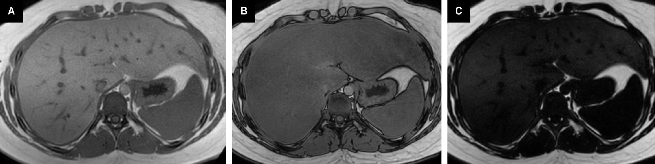
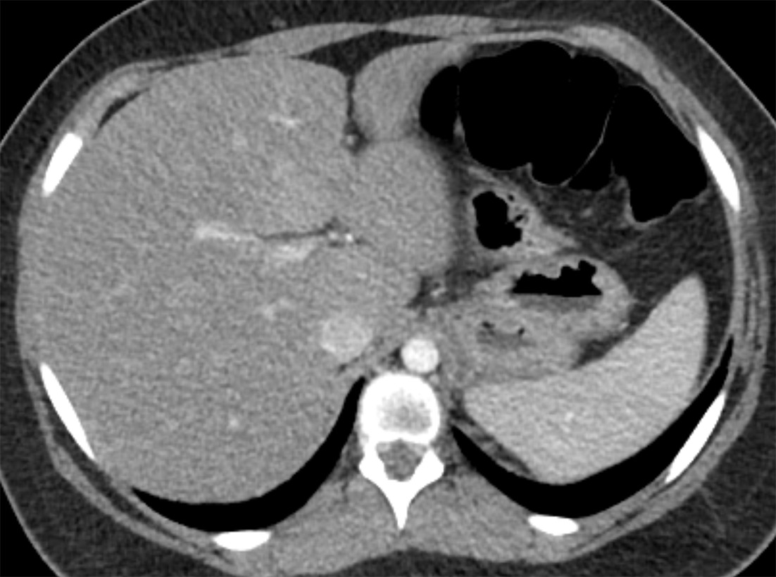
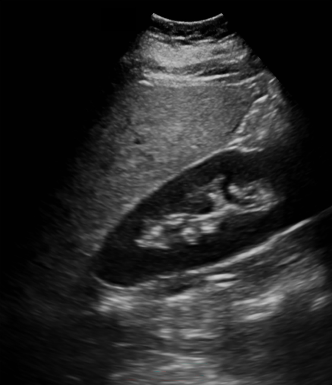
Abdominal MRI (Figure 1) showed hepatic steatosis with a fat fraction of 21%. Hepatic steatosis was also visible on CT (Figure 2) and US (Figure 3).
Diagnosis
Steatohepatitis.
The differential diagnosis is broad and includes metabolic dysfunction-associated steatotic liver disease (MASLD), linked to conditions such as type 2 diabetes, insulin resistance, hypertension, obesity, and dyslipidemia. The differential further encompasses genetic disorders such as Wilson disease, Reye syndrome, cystic fibrosis, alpha-1 antitrypsin deficiency, and hereditary hemochromatosis, as well as viral hepatitis (A, B, C), pediatric autoimmune liver diseases, drug-induced liver injury, malnutrition, and inherited metabolic disorders.
Discussion
Steatohepatitis is characterized by the accumulation of fat in the liver, leading to inflammation and hepatocellular injury. In children, the primary cause is MASLD, previously known as nonalcoholic fatty liver disease. MASLD begins with steatosis. Over time, inflammation may develop, causing steatohepatitis. In some patients, this can progress to fibrosis, cirrhosis, and ultimately hepatocellular carcinoma. The progressive form of MASLD is termed metabolic dysfunction-associated steatohepatitis (MASH).1
MASLD should be suspected in children and adolescents with persistently elevated liver enzymes and established risk factors, such as male sex, obesity, central adiposity, insulin resistance, and dyslipidemia. The diagnosis is made after exclusion of other liver diseases, including inborn errors of fatty acid metabolism, peroxisomal disorders, and lysosomal storage disorders, or it may be identified incidentally. Other chronic liver diseases that can present with steatohepatitis in children include Wilson disease, Reye syndrome, hemochromatosis, viral hepatitis, autoimmune hepatitis, and alpha-1 antitrypsin deficiency.2,3
The prevalence of MASLD has increased substantially over the past decade, making it the leading cause of chronic liver disease in children and adolescents worldwide. In the general pediatric population, MASLD affects an estimated 7% of children and adolescents, with rates exceeding 34% among children with obesity.4 The development of MASLD is influenced by a complex interplay of genetic, epigenetic, environmental, and gut microbiome factors. Additional risk factors include maternal obesity, perinatal exposure to antibiotics, and an intestinal microbiota enriched in alcohol-producing bacteria.5 Lifestyle factors, such as poor diet and physical inactivity, further contribute to disease risk.
Several noninvasive imaging modalities are available to quantify liver fat and monitor the progression of steatohepatitis.6-8 Conventional US is widely used for screening due to its accessibility, though it offers only moderate accuracy. In steatosis, the liver demonstrates increased echogenicity relative to the adjacent right kidney. As hepatic fat accumulates, there is progressive loss of visualization of the portal triads, and eventually, the right hemidiaphragm becomes less distinct. US shear-wave elastography provides a quantitative assessment of liver stiffness, with elevated shear-wave velocities indicating increased tissue stiffness that may correlate with fibrosis progression.9
MRI-derived proton density fat fraction (MRI-PDFF) quantifies the proportion of liver parenchyma composed of fat.9 A value greater than 5% is considered indicative of hepatic steatosis. It is important to note that MRI-PDFF measures the percentage of fat within the parenchyma, whereas histopathologic assessment quantifies the percentage of hepatocytes containing lipid droplets. MR elastography is also used to assess liver stiffness and evaluate for fibrosis. Prior studies have suggested a threshold of 2.71 kPa to predict significant hepatic fibrosis.10 However, the complex interplay between fat and fibrosis can variably affect liver stiffness measurements.11
Liver biopsy remains the gold standard for diagnosing MASLD and MASH, as it provides direct assessment of hepatic inflammation and fibrosis. However, it is invasive and carries limitations, including sampling variability and interobserver differences in interpretation. Given these limitations, noninvasive tools such as elastography and MRI-PDFF are increasingly used to support diagnosis and monitor disease progression.
Management of MASLD and MASH primarily relies on dietary and lifestyle modifications. Unfortunately, the success of these interventions is often hindered by low rates of adherence. Pediatric health care providers should routinely monitor body mass index z-scores and engage families in weight management strategies. A multidisciplinary approach involving dietitians, social workers, and exercise physiologists can improve adherence and outcomes. Additionally, attention to modifiable perinatal risk factors, such as preventing gestational diabetes and avoiding excessive maternal weight gain, may help reduce future risk.5
The degree of hepatic fibrosis is the strongest predictor of disease progression, hepatic decompensation, and mortality. Early identification and monitoring of fibrosis are essential for risk stratification and guiding treatment decisions.
Conclusion
Steatohepatitis in children most commonly arises in the setting of MASLD but can also occur in association with other conditions, including Wilson disease, Reye syndrome, cystic fibrosis, viral hepatitis, and inherited metabolic disorders. While liver biopsy remains the diagnostic gold standard, noninvasive imaging techniques such as US and MRI-PDFF play an important role in screening and monitoring disease progression. Lifestyle modification remains the cornerstone of therapy for MASLD, although adherence challenges persist. Early recognition and management are essential to prevent potential progression to advanced fibrosis, cirrhosis, and hepatocellular carcinoma.
References
- Xanthakos S. Nonalcoholic steatohepatitis in children. Clin Liver Dis. 2022;26(3):439-460. doi:10.1016/j.cld.2022.05.001.
- Goldner D, Lavine J. Nonalcoholic fatty liver disease in children: unique considerations and challenges. Gastroenterology. 2020;158(7):1967-1983. doi:10.1053/j.gastro.2020.01.048.
- Trandafir L, Frasinariu O, Leon-Constantin M. Pediatric nonalcoholic fatty liver disease - a changing diagnostic paradigm. Rom J Morphol Embryol. 2020;61(4):1023-1031. doi:10.47162/RJME.61.4.04.
- Stroes A, Vos M, Benninga M, Koot B. Pediatric MASLD: current understanding and practical approach. Eur J Pediatr. 2024;184(1). doi:10.1007/s00431-024-05848-1.
- Vittorio J, Lavine J. Recent advances in understanding and managing pediatric nonalcoholic fatty liver disease. F1000Res. 2020;9. doi:10.12688/f1000research.24198.1.
- Bozic D, Podrug K, Mikolasevic I, Grgurevic I. Ultrasound methods for the assessment of liver steatosis: a critical appraisal. Diagnostics (Basel). 2022;12(10). doi:10.3390/diagnostics12102287.
- Tamaki N, Ajmera V, Loomba R. Non-invasive methods for imaging hepatic steatosis and their clinical importance in NAFLD. Nat Rev Endocrinol. 2022;18(1):55-66. doi:10.1038/s41574-021-00584-0.
- Rinaldi L, Giorgione C, Mormone A. Non-invasive measurement of hepatic fibrosis by transient elastography: a narrative review. Viruses. 2023;15(8). doi:10.3390/v15081730.
- Bailey S, Youssfi M, Patel M. Shear-wave ultrasound elastography of the liver in normal-weight and obese children. Acta Radiol. 2017;58(12):1511-1518. doi:10.1177/0284185117695668.
- Xanthakos S, Podberesky D, Serai S. Use of magnetic resonance elastography to assess hepatic fibrosis in children with chronic liver disease. J Pediatr. 2014;164(1):186-188. doi:10.1016/j.jpeds.2013.07.050.
- Joshi M, Dillman J, Singh K. Quantitative MRI of fatty liver disease in a large pediatric cohort: correlation between liver fat fraction, stiffness, volume, and patient-specific factors. Abdom Radiol. 2018;43(5):1168-1179. doi:10.1007/s00261-017-1289-y.
Disclosures
The authors have no conflicts of interest to disclose. None of the authors received outside funding for the production of this original manuscript and no part of this article has been previously published elsewhere.
Citation
. Steatohepatitis. Journal of Pediatric Case Reports. 2026;1(7). doi:10.37549/JPCR-26-0118.