Stent
Applied Radiology — Vol. 35 , Issue 10 , pp. 22 -36
DOI: 10.37549/AR1458
Published: October 1, 2006
Categories
Adnexal masses are very common and will develop in every woman. The majority represent functional cysts that will involute in silence; however, approximately 1.5% of women will develop a malignant ovarian tumor.1 Benign or malignant, ovarian tumors represent a major indication for gynecologic surgery, which is performed in approximately 5% to 10% of U.S.women. Imaging plays a major role in staging and treatment planning.2
Ultrasound (US) is the primary modality for the evaluation of an adnexal mass, and magnetic resonance imaging (MRI) is generally used to further characterize indeterminate lesions, since it provides valuable information on the composition of soft tissue masses, using differences in tissue relaxation times. The reported sensitivity and specificity of US for differentiating benign from malignant adnexal masses ranges widely from 50% to 100% and 46% to 100%, respectively,3 depending on the diagnostic criteria used and the population studied. While US can readily distinguish simple cystic from complex cystic or solid lesions that require further work-up, it is important to remember that not all complex cystic or solid lesions are malignant, and MRI may be used to confidently diagnose certain lesions with specific MRI characteristics. Computed tomography (CT) is not generally recommended as a modality to primarily evaluate an adnexal mass, but these are often seen in women presenting with abdominal or pelvic pain, during work-up for suspected or staging for known malignancy, or as incidental findings. In this article, the authors will discuss features that are suggestive of malignancy within an adnexal mass, describe the appearance of common benign adnexal masses on MRI and CT, and detail the indications for MRI of a suspected adnexal mass.
Differentiation of benign from malignant adnexal masses
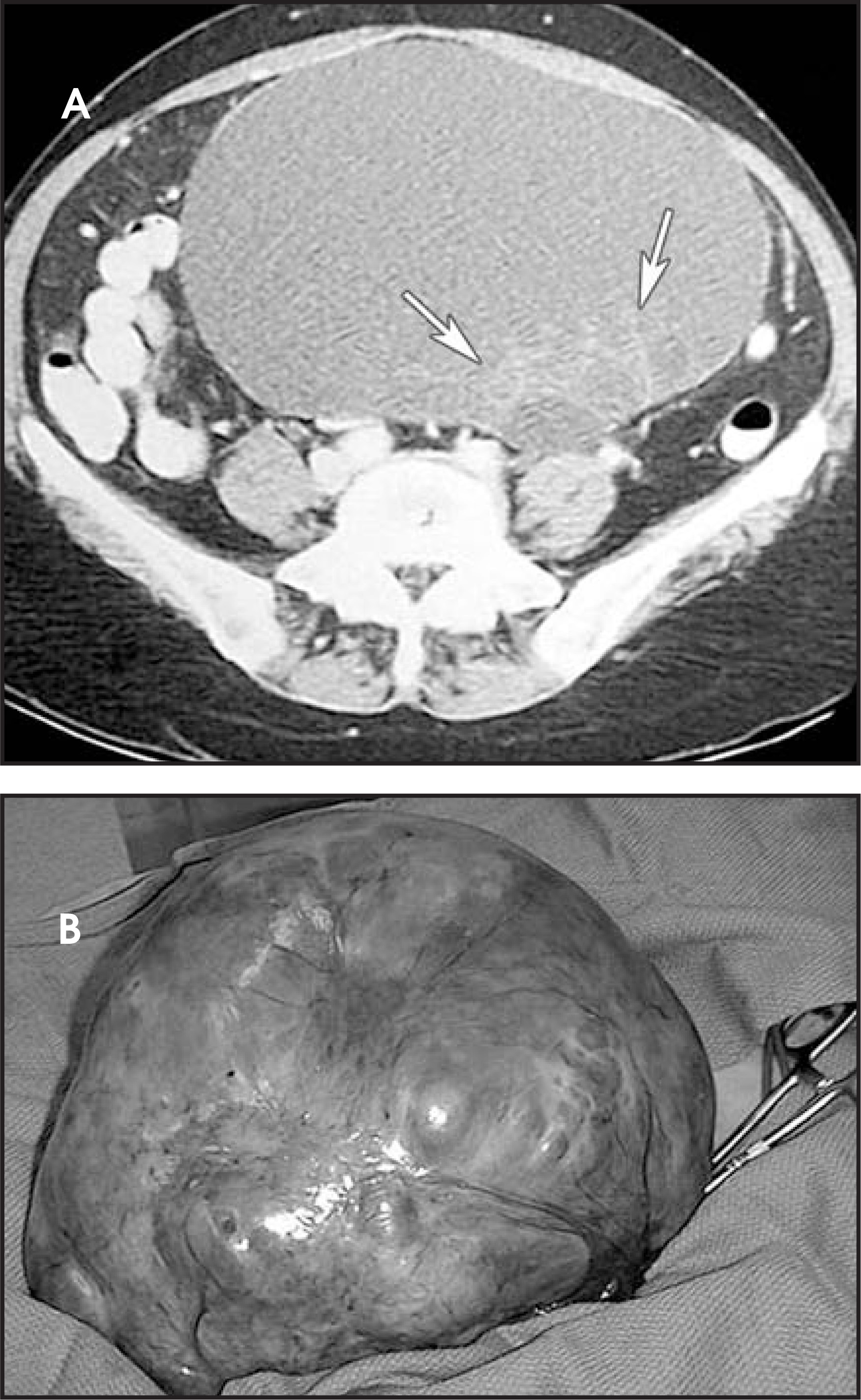
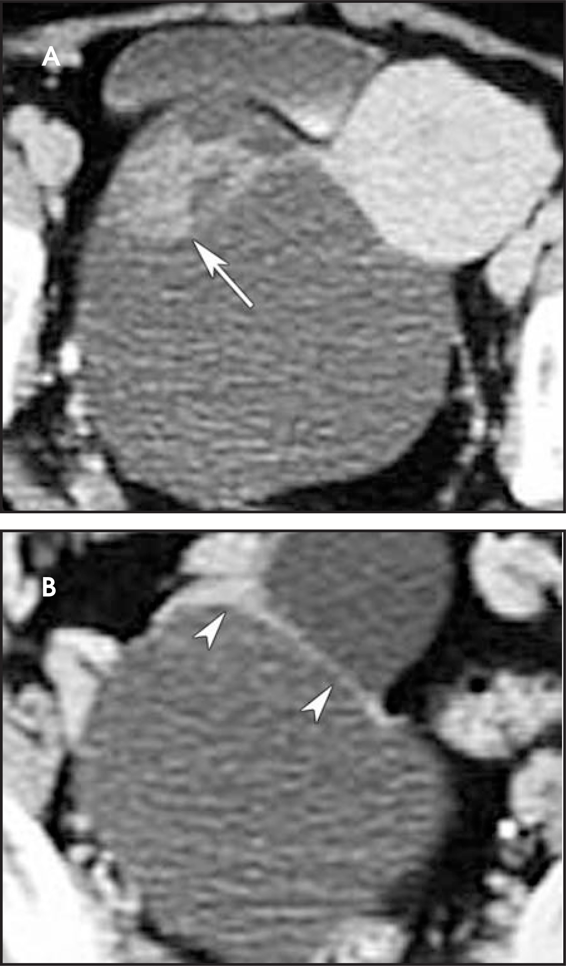
The main reason to image an adnexal mass is to distinguish between a benign and a malignant lesion. As a general rule, benign masses are cystic and have thin walls and septa (<3 mm) (Figures 1 and 2); however, there are exceptions to this rule, which will be discussed in this article. Malignant lesions are usually described as having internal complexity, which is characterized by the presence of mural nodules or thick septa. The presence of solid components, necrosis within a solid mass, and the presence of bilaeral ovarian masses also favor malignancy (Figure 3).3 Comparing the accuracy of cross-sectional modalities used to characterize ovarian masses, the common perception is that MRI is better than US, which is in turn better than CT. The Radiology Diagnostic Oncology Group study compared these modalities in the preoperative assessment of adnexal masses for possible ovarian malignancy.4 Their results showed that MRI was the most accurate technique, while there was no significant difference between US and CT. In addition, this study reported that malignancies were rarely subtle and peritoneal spread was often present at the time of imaging. Size has been described as a further feature that may be used to differentiate a benign from a malignant lesion; however, it is important to note that the majority of ovarian masses are benign, including very large tumors (Figure 2).
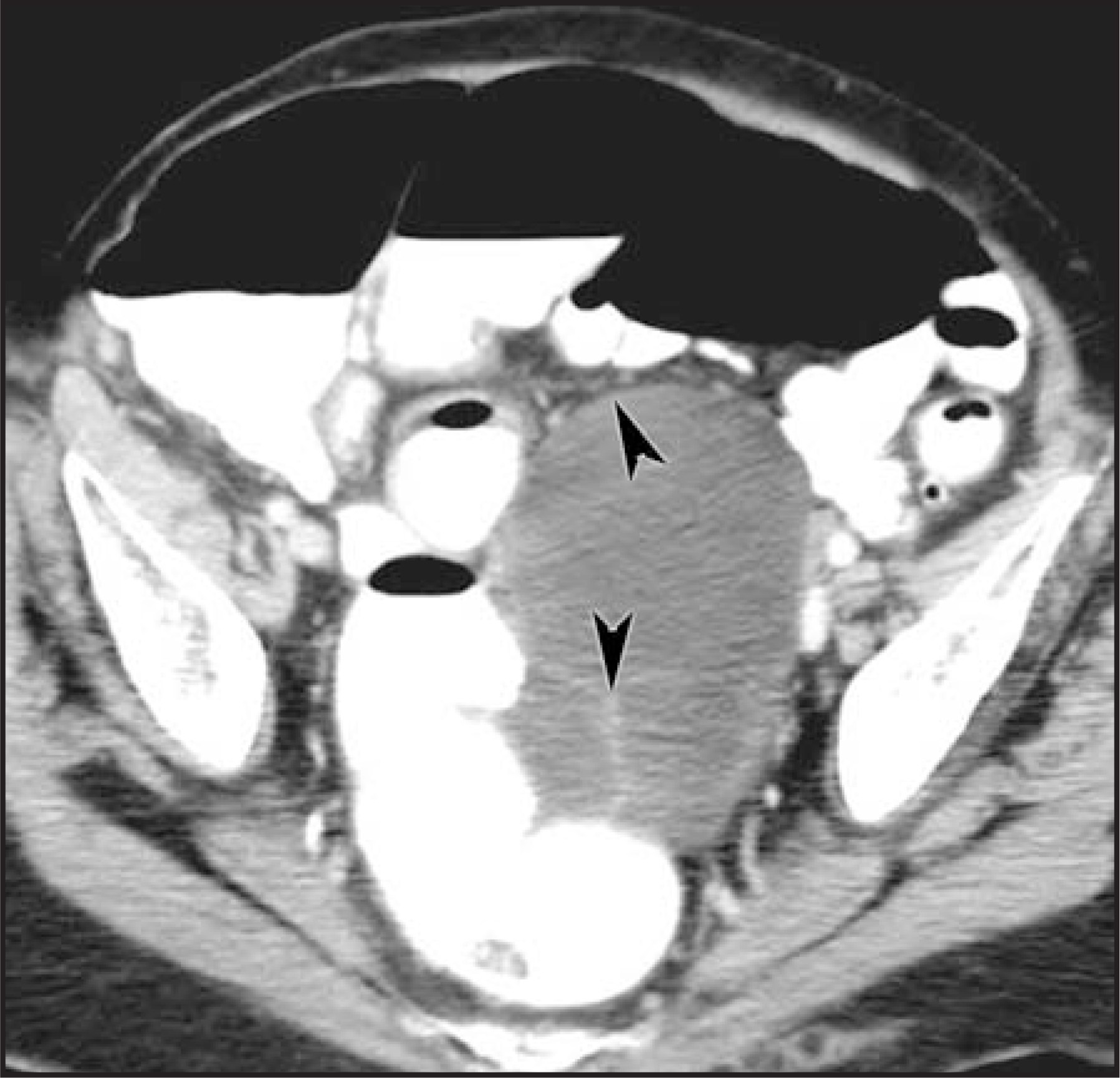
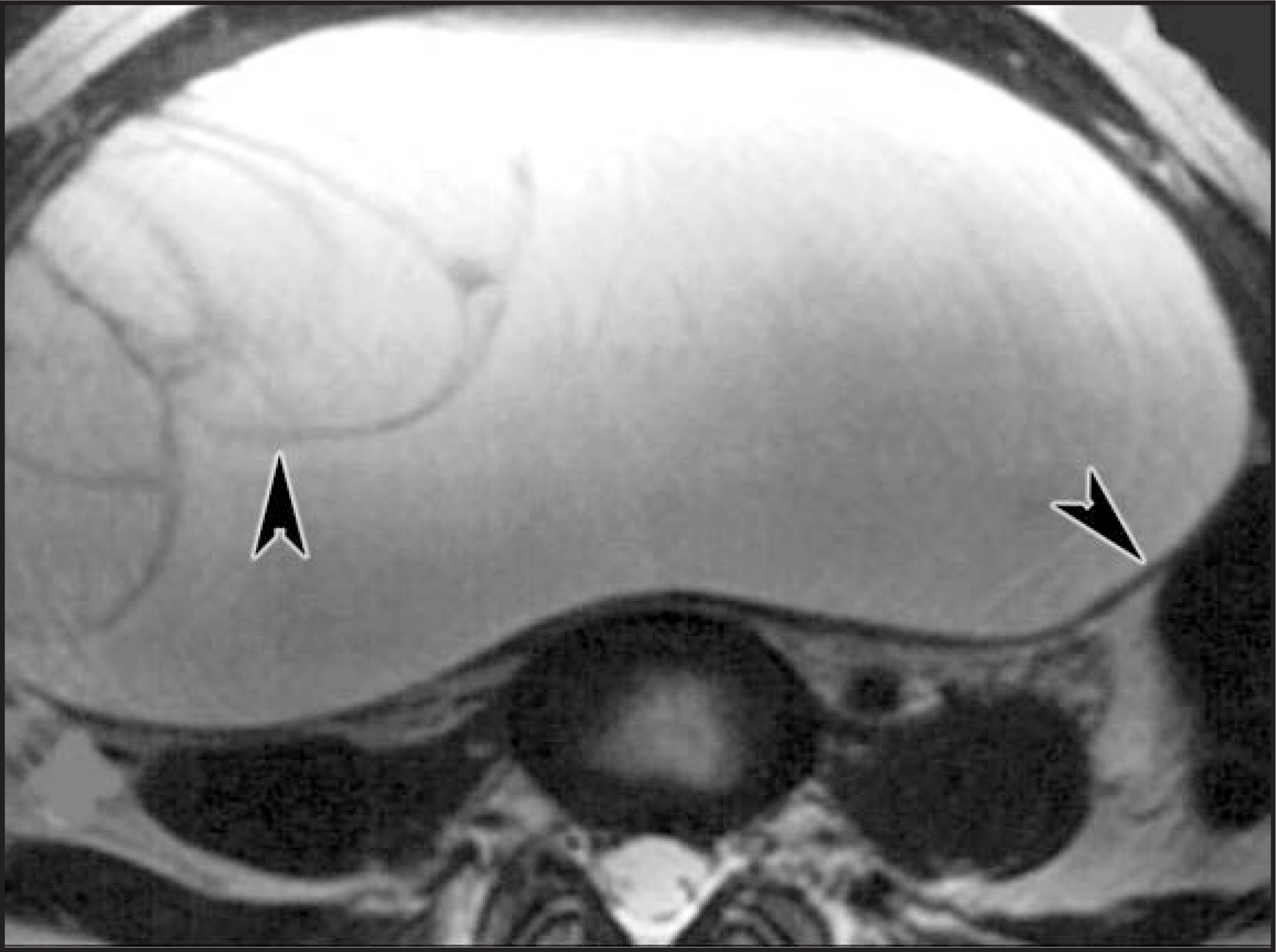
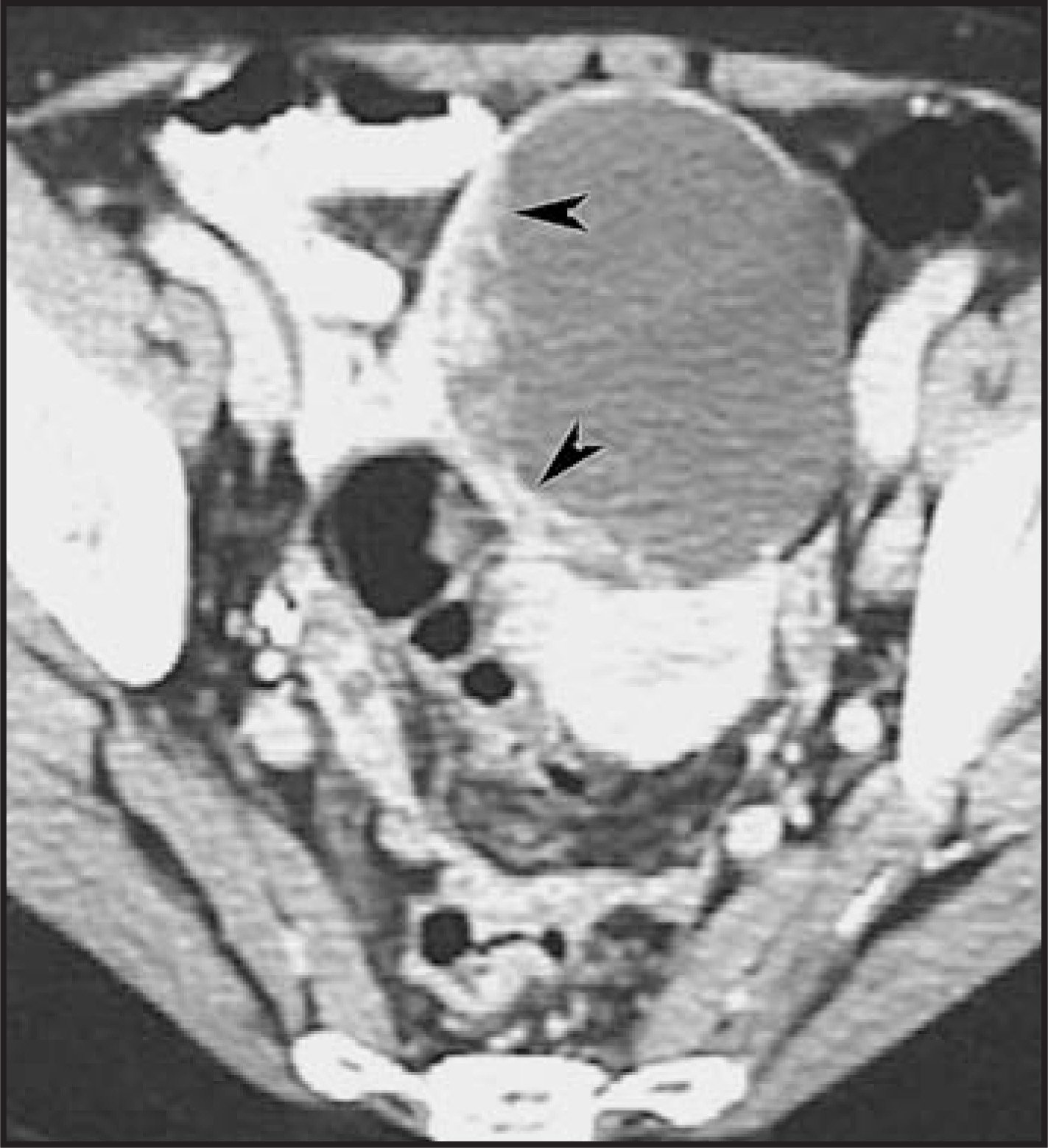
For practical purposes, adnexal masses may be categorized as: 1) lesions that have distinctive imaging characteristics that enable their diagnosis; 2) lesions that are cystic and appear to be benign; 3) lesions that are cystic and appear to be malignant; and 4) lesions that are predominantly solid.
Adnexal masses with distinctive imaging characteristics
Dermoid cyst
Also known as benign mature teratoma, these lesions are not uncommon and are more frequently identified during the first 3 decades of life. Dermoid cysts are hamartomas and characteristically contain hair, teeth, fat, and mural nodules.3,5-7 Because of the echogenicity of fat on US, they may be missed. In a series of 22 cases, Bloomfield et al5 reported that 27% of dermoid cysts were not identified by US. Conversely, fat is exactly what makes these tumors easily identified on CT and MRI, even if just a small amount is present (Figures 4 and 5). On MRI, fat within dermoids demonstrates high signal intensity on T1-weighted images that is lost with the application of fat suppression (Figure 5).3,6
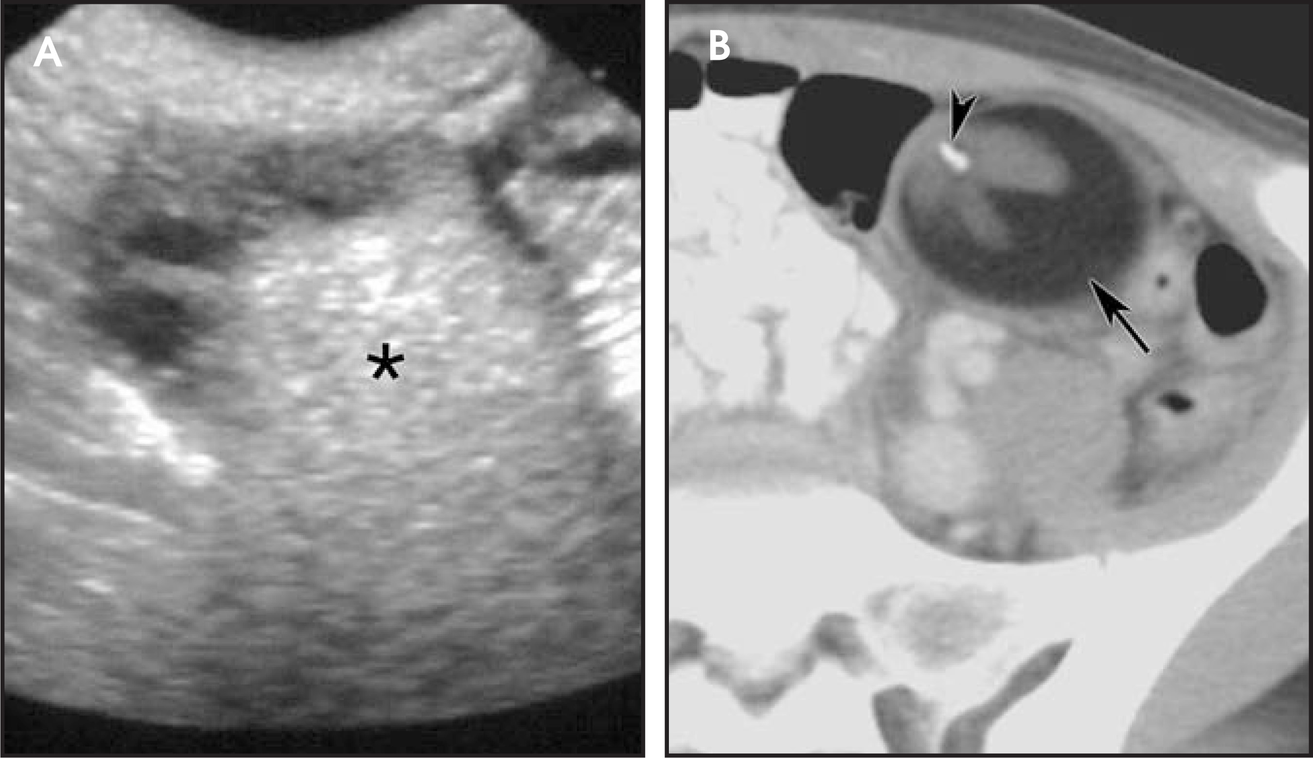
Corpus luteum cyst
The corpus luteum is a normal post-ovulation structure that is characteristically seen as a well-vascularized lesion with crenulated walls (Figure 6). It normally regresses in 14 days if fertilization does not occur, but occasionally it may develop into a cystic lesion. Follow-up typically shows regression after 1 or 2 cycles. These are commonly seen at CT in patients presenting with abdominal pain.8,9
Theca-lutein cyst
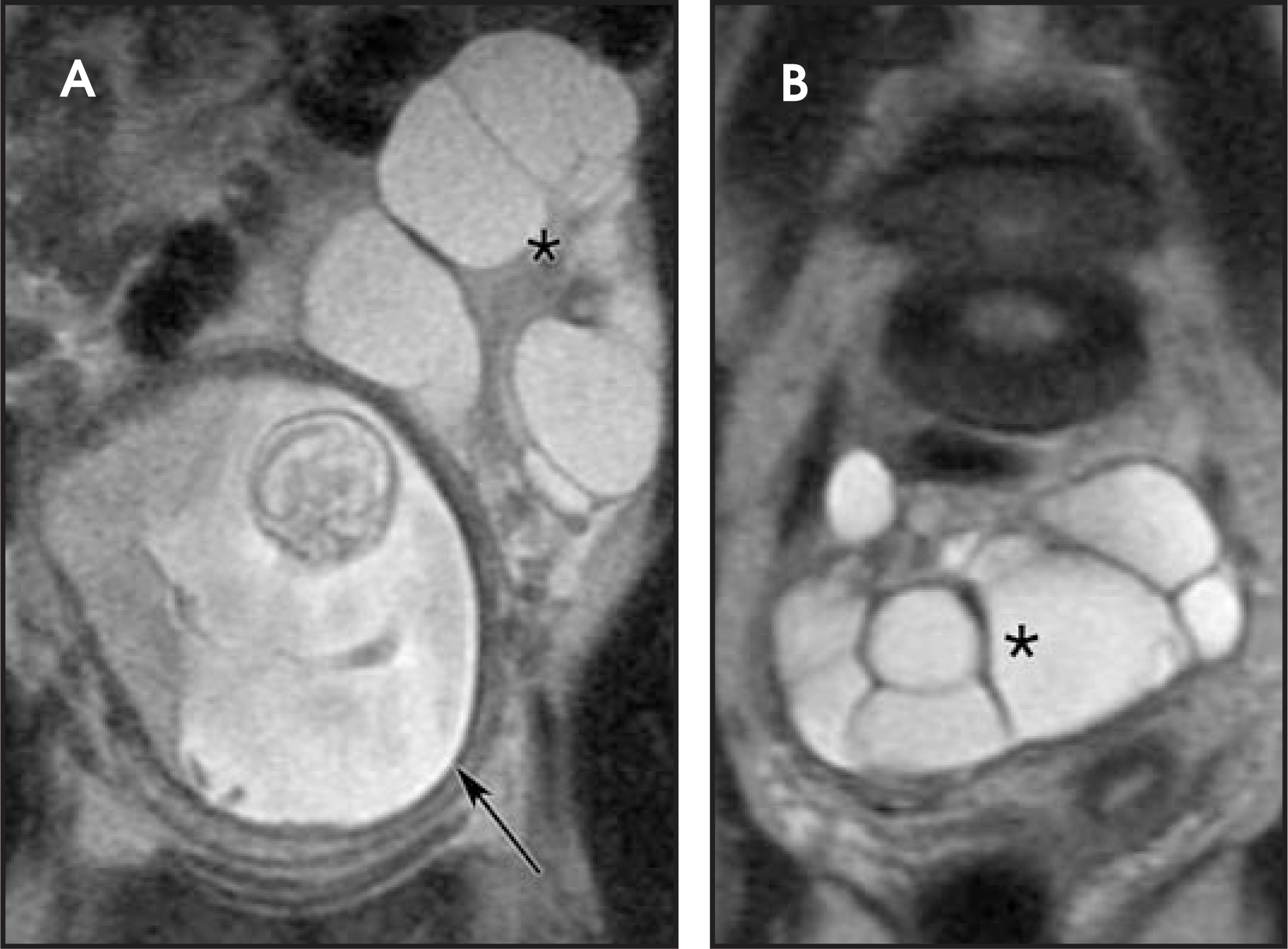
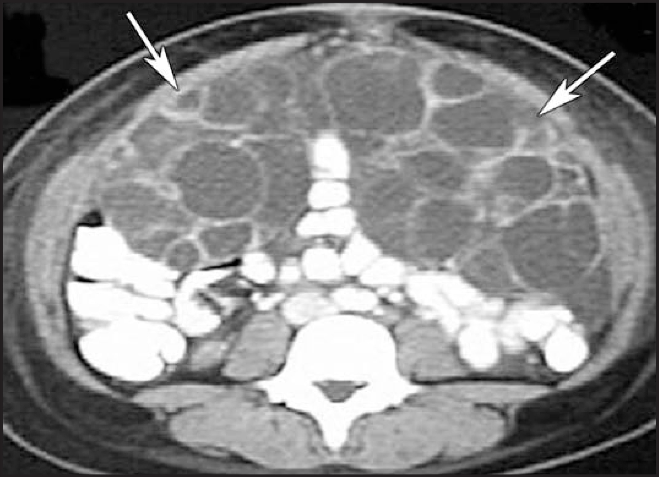
These are bilateral, large (up to 10 to 15 cm in diameter), multilocular, functional cysts that develop secondary to high levels of serum human chorionic gonadotrophin. The most common causes are gestational trophoblastic disease (hydatidiform mole or choriocarcinoma), hyperstimulation secondary to infertility treatments, and extensive luteinization of theca cells, usually in a multiple gestation. Typically, the cysts regress after the source of gonadotrophin is removed.10 The cysts are thin-walled and have fluid signal intensity at MRI and fluid attenuation at CT (Figures 7 and 8).
Blood-containing lesions
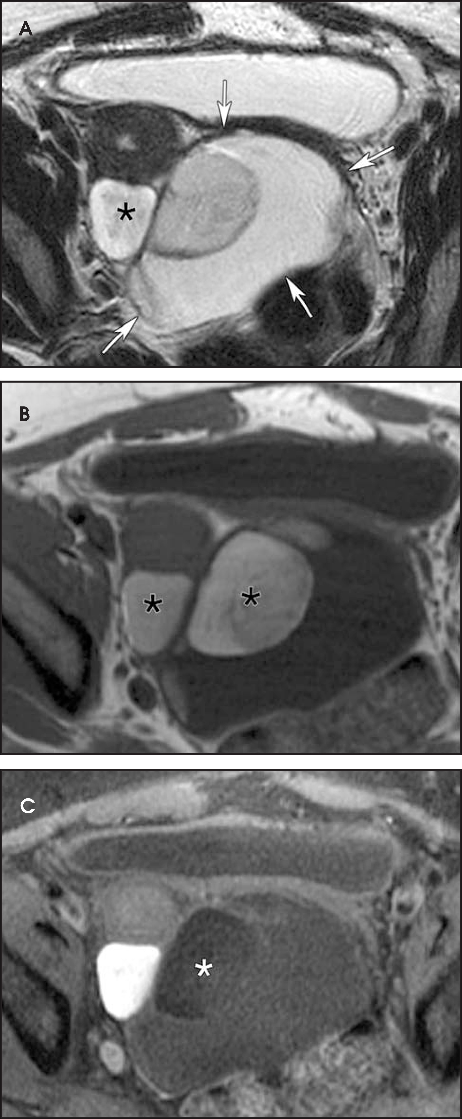
The most common lesions in this group are hemorrhagic cysts and endometriomas. MRI is usually requested when US shows failure of regression of a suspected hemorrhagic cyst in the context of endometriosis. Endometriomas may have thick irregular walls and septations and can be misdiagnosed as an ovarian malignancy at US. The wall of the endometrioma may show contrast enhancement. The acquisition of T1-weighted images with and without fat suppression is crucial to the diagnosis, since blood products will have high signal intensity on both sequences; however, fat within the mass will lose signal intensity on fat-suppressed images (Figure 5).11 On T2-weighted images, blood products may also have high signal intensity; however, a black rim may be identified surrounding the lesion, which is due to susceptibility artifact from iron secondary to degradation of blood (Figure 9). Simple hemorrhagic cysts are usually resorbed before such chronic changes can occur in red blood cells. A further characteristic of endometriomas is “T2-shading.” This is the internal layering of the blood products that appear as low signal intensity on T2-weighted sequences (Figure 10).12 Although uncommon, cancer has been reported in as-sociation with endometriosis and should be suspected in the presence of a solid nodule within the lesion (Figure 11).13 CT is of limited utility in the distinction of these entities.
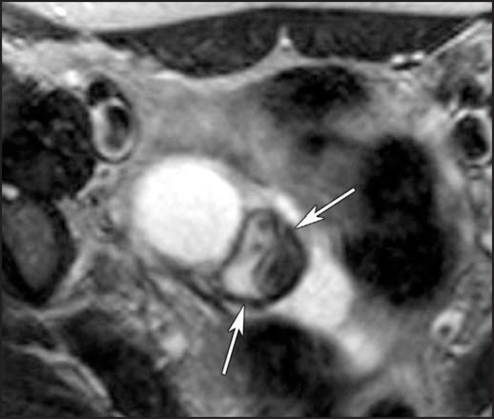
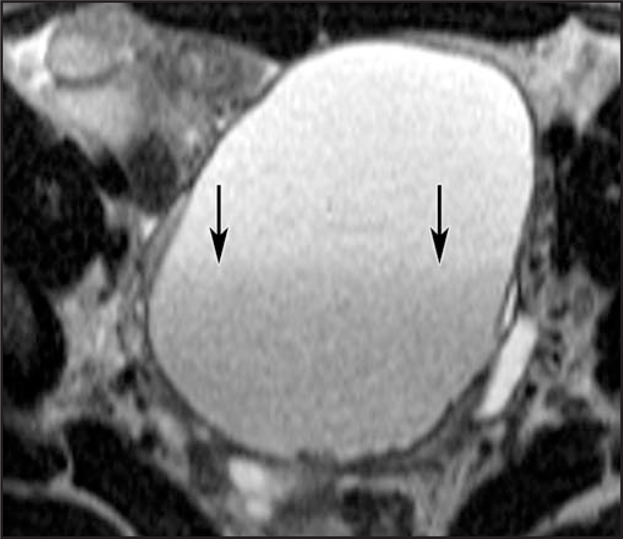
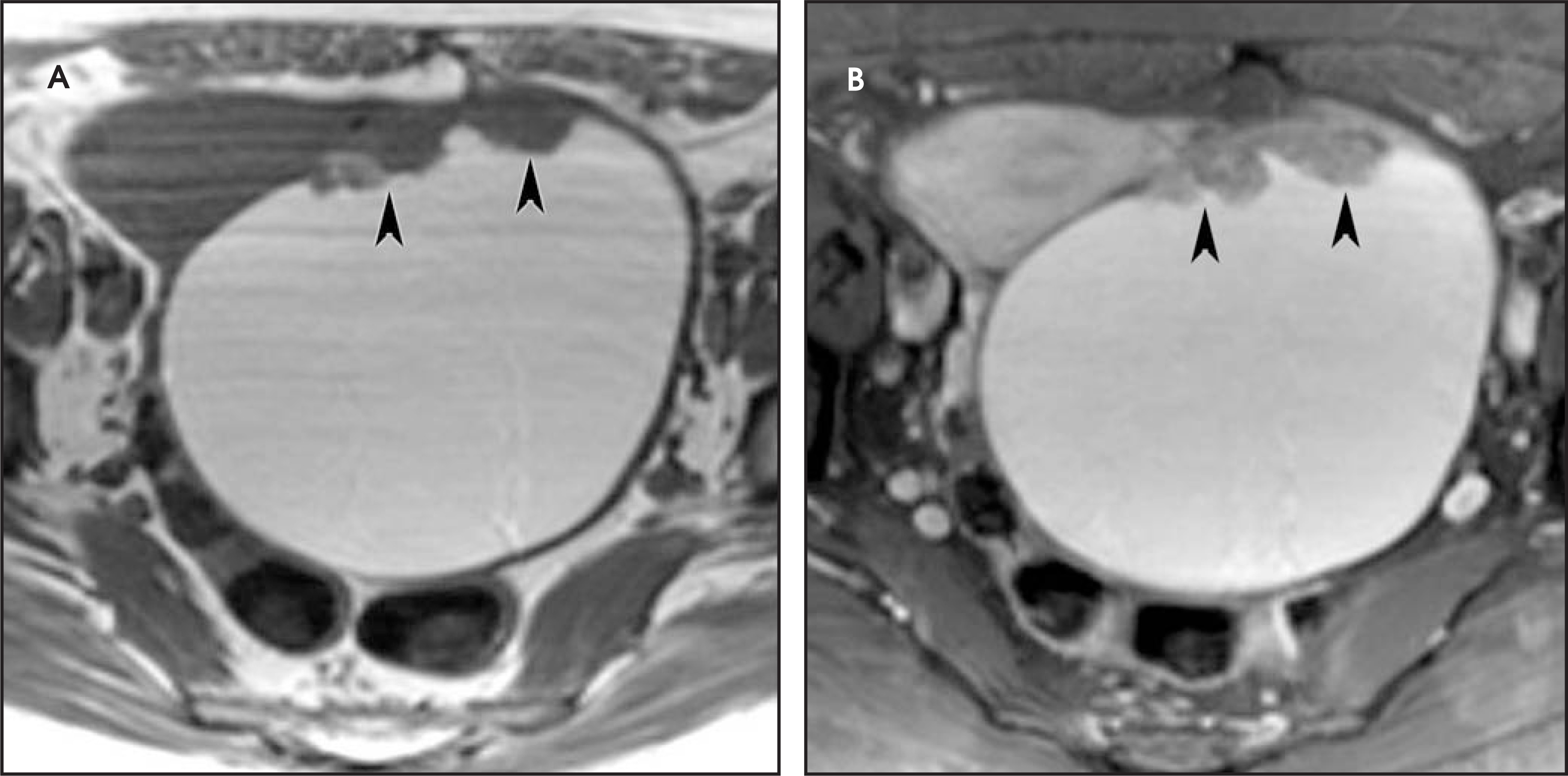
Hydrosalpinx
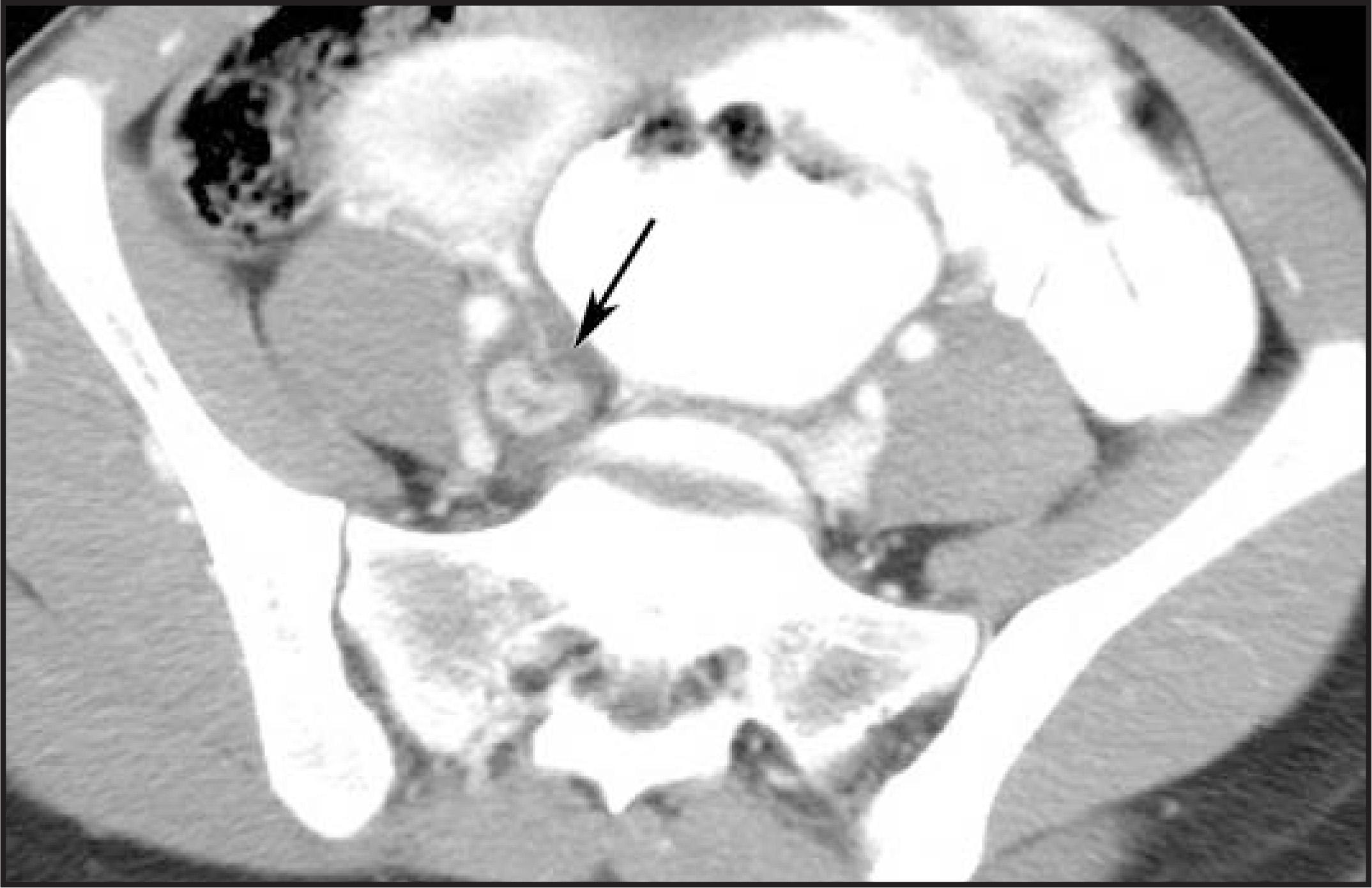
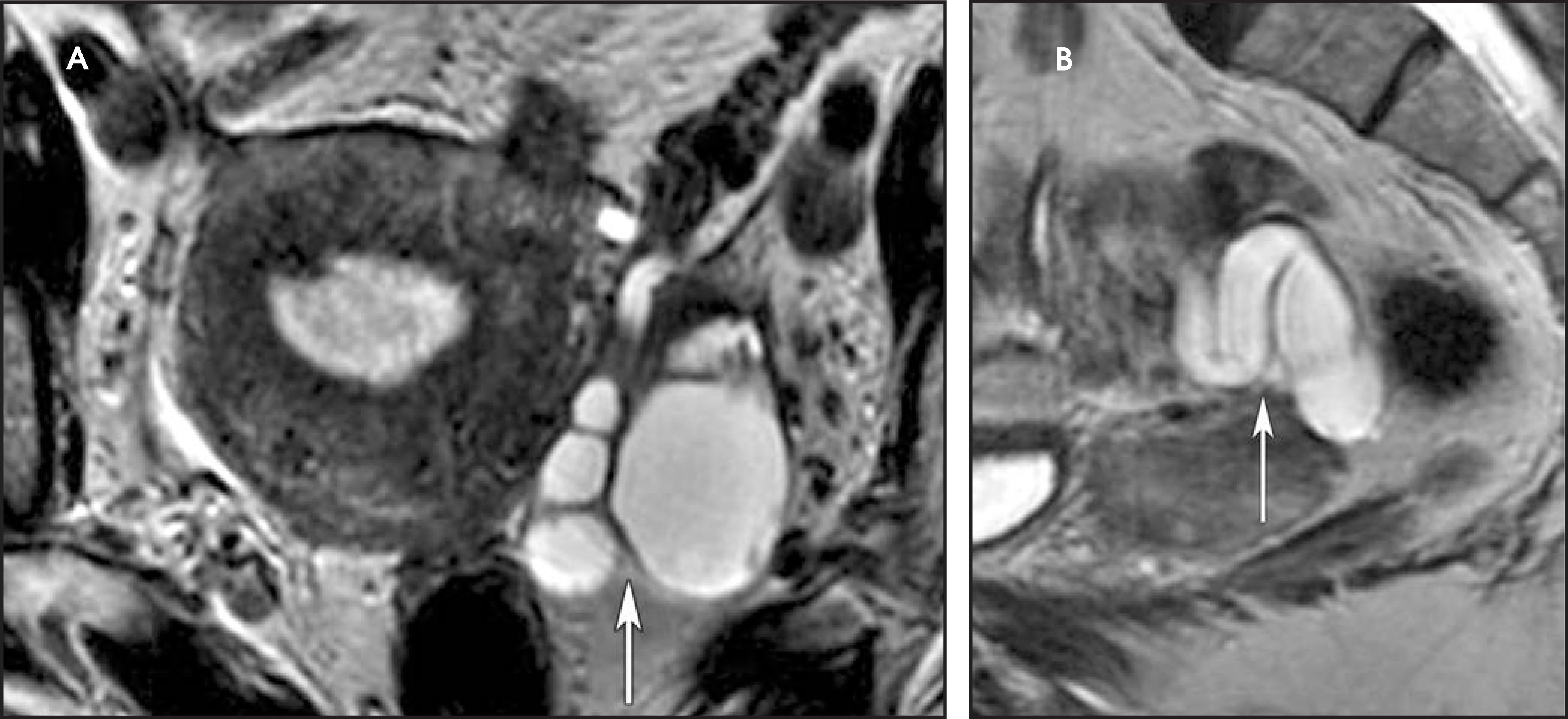
While dilated fallopian tubes may be identified at US, they can be misdiagnosed as complex cystic adnexal masses. The multiplanar capabilities of MRI often enable the identification of the serpiginous tubular structure with high T2 signal intensity that may demonstrate T2-shading or high signal intensity on T1-weighted images in the context of endometriosis (Figure 12). CT can also be used to identify these tubular structures (Figure 13).14
Pedunculated leiomyoma
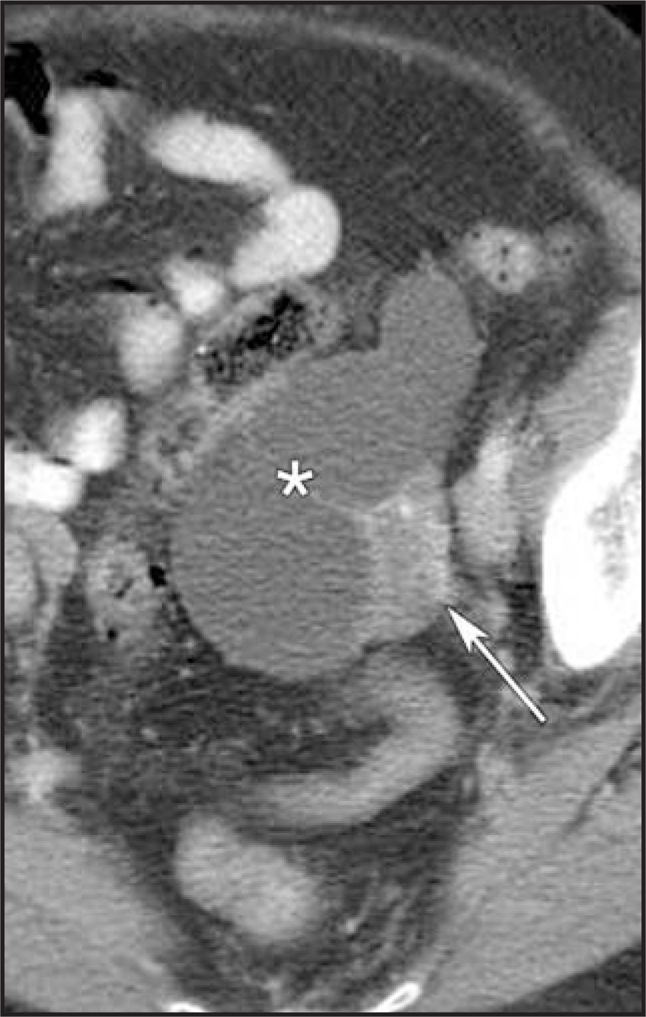
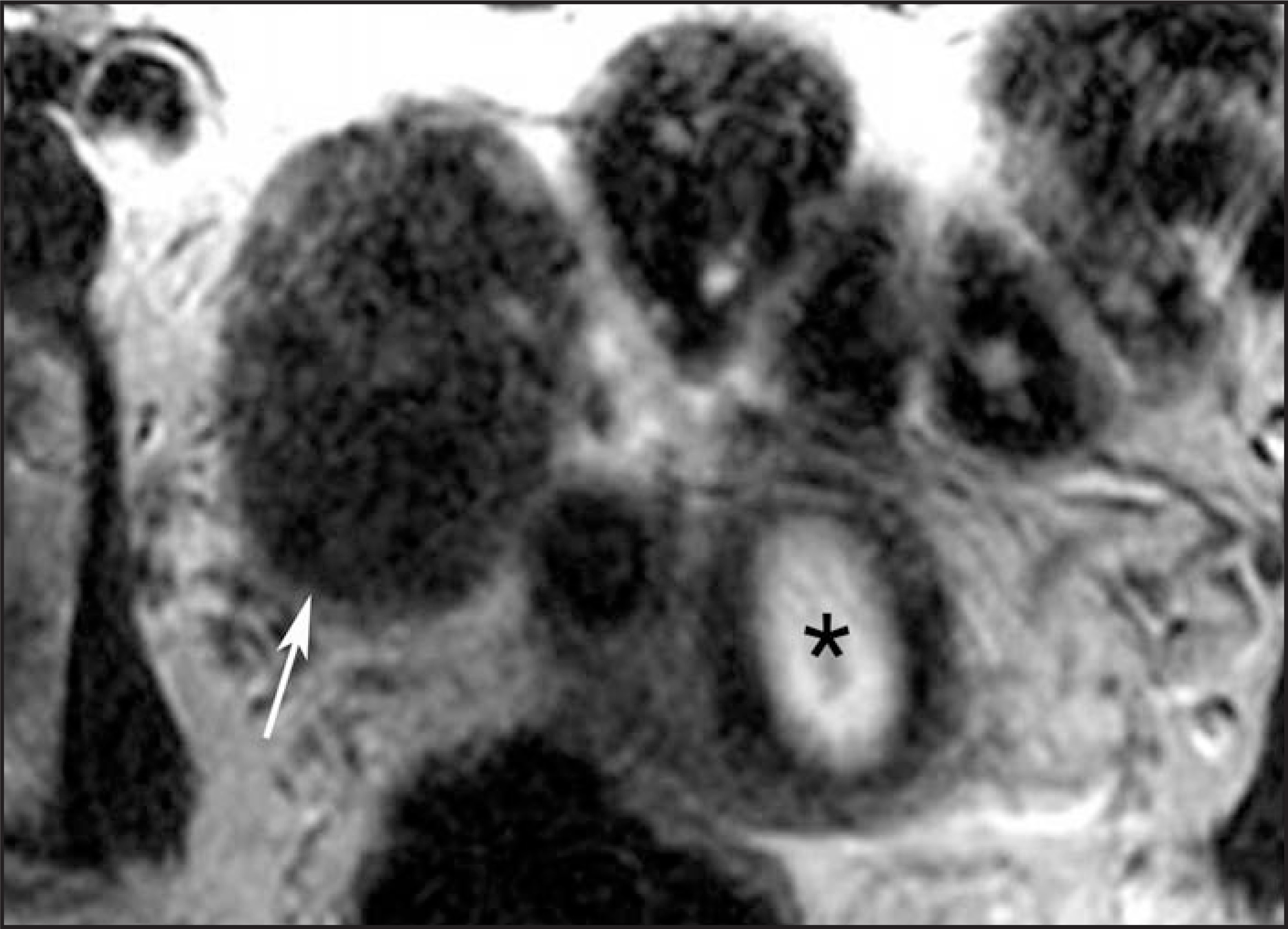
Leiomyomas are the most common neoplasms of the uterus, occurring in 20% to 30% of women over the age of 30 years.15 MRI may be used to confidently diagnose leiomyomas. Simple leiomyomas are well-circumscribed, rounded lesions of low signal intensity on T1- and T2-weighted sequences (Figure 14), and characteristically demonstrate a high-signal-intensity pseudocapsule on T2-weighted images when intramural.16,17 Most leiomyomas enhance slightly less or similar to the surrounding myometrium, although avid enhancement is frequently seen and may be helpful to distinguish pedunculated, broad-ligament, or ovarian leiomyomas from fibromas or malignant ovarian neoplasms, which generally show only slight enhancement. Degenerating fibroids typically demonstrate areas of high signal intensity on T2-weighted sequences.16,18,19 Hemorrhagic or red degeneration is due to massive hemorrhage and infarction and can occur during pregnancy, with the use of oral contraceptives or following embolization. It is recognized by the presence of high signal intensity on T1-weighted images.16 Cystic degeneration occurs in 4% of leiomyomas and is associated with edema (Figure 15).19 One unusual kind of leiomyoma is the lipoleiomyoma, which contains a considerable amount of fat. Fat is readily recognized as a region of high signal intensity on T1-weighted sequences that becomes of low signal intensity with the application of fat suppression.19
Benign-appearing adnexal cystic masses
Physiologic cysts
Physiologic cysts are a common incidental finding at MRI and CT in women of reproductive age. They are typically less than <3.0 cm in size, contain fluid, and have imperceptible nonenhancing walls (Figure 16).3
Cystadenomas
Cystadenomas are usually large cystic masses (particularly mucinous cystadenomas), with thin walls and may have a few thin internal septations (Figure 17). The presence of extensive or nodular septations or solid components is suggestive of malignancy.3,6
Peritoneal inclusion cyst
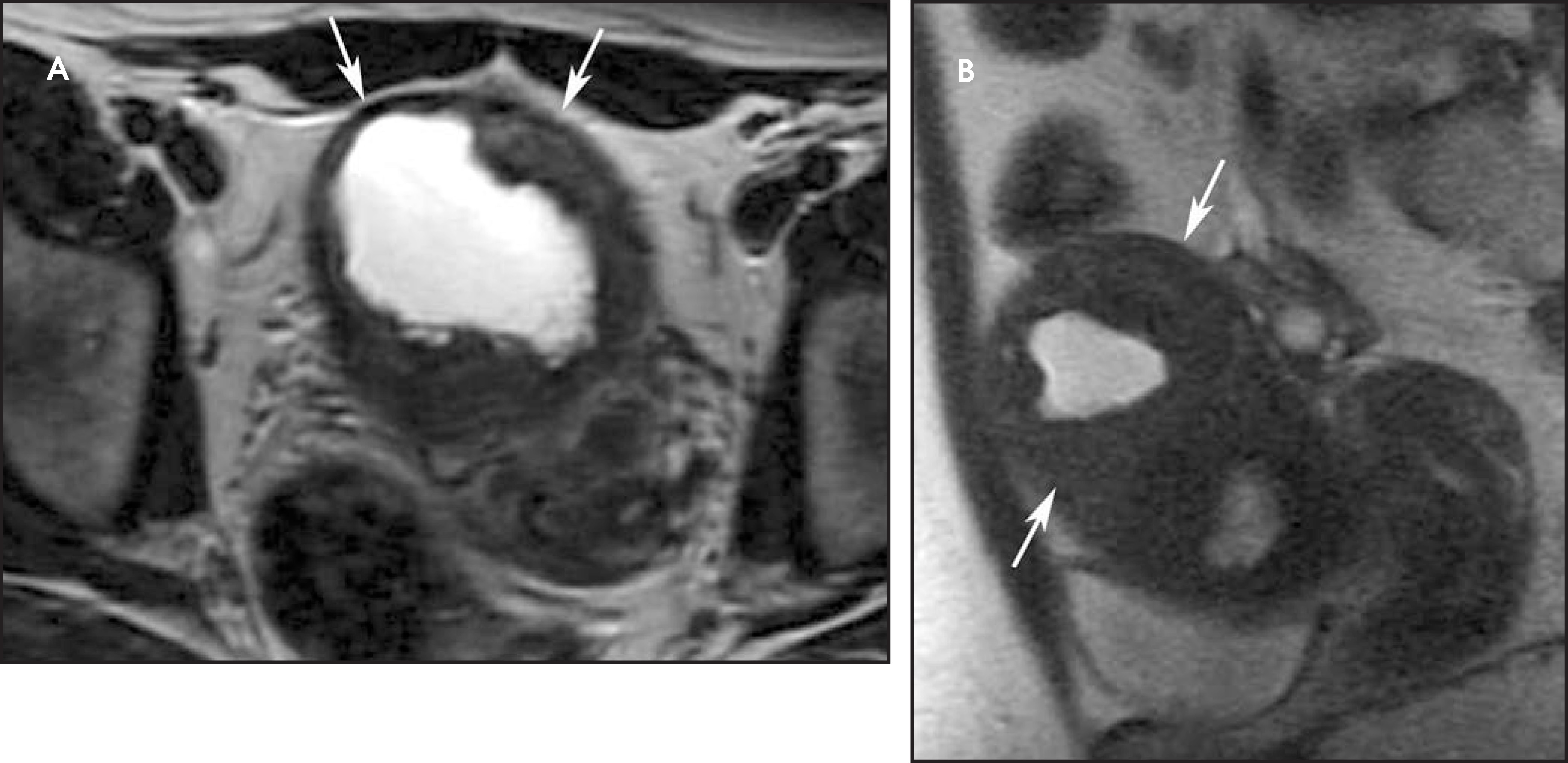
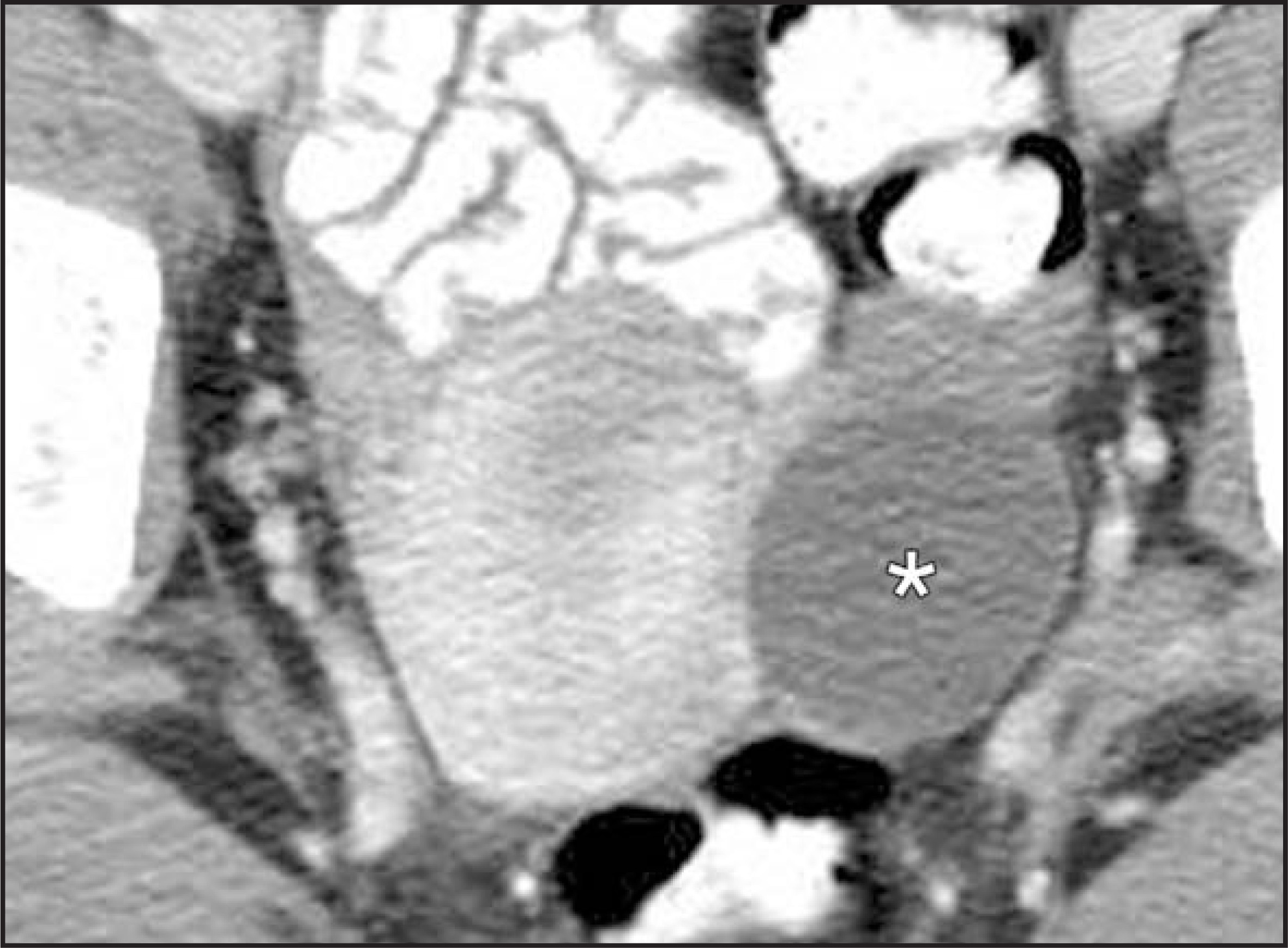
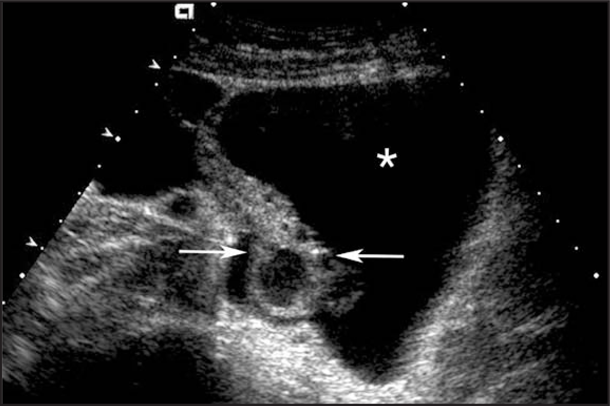
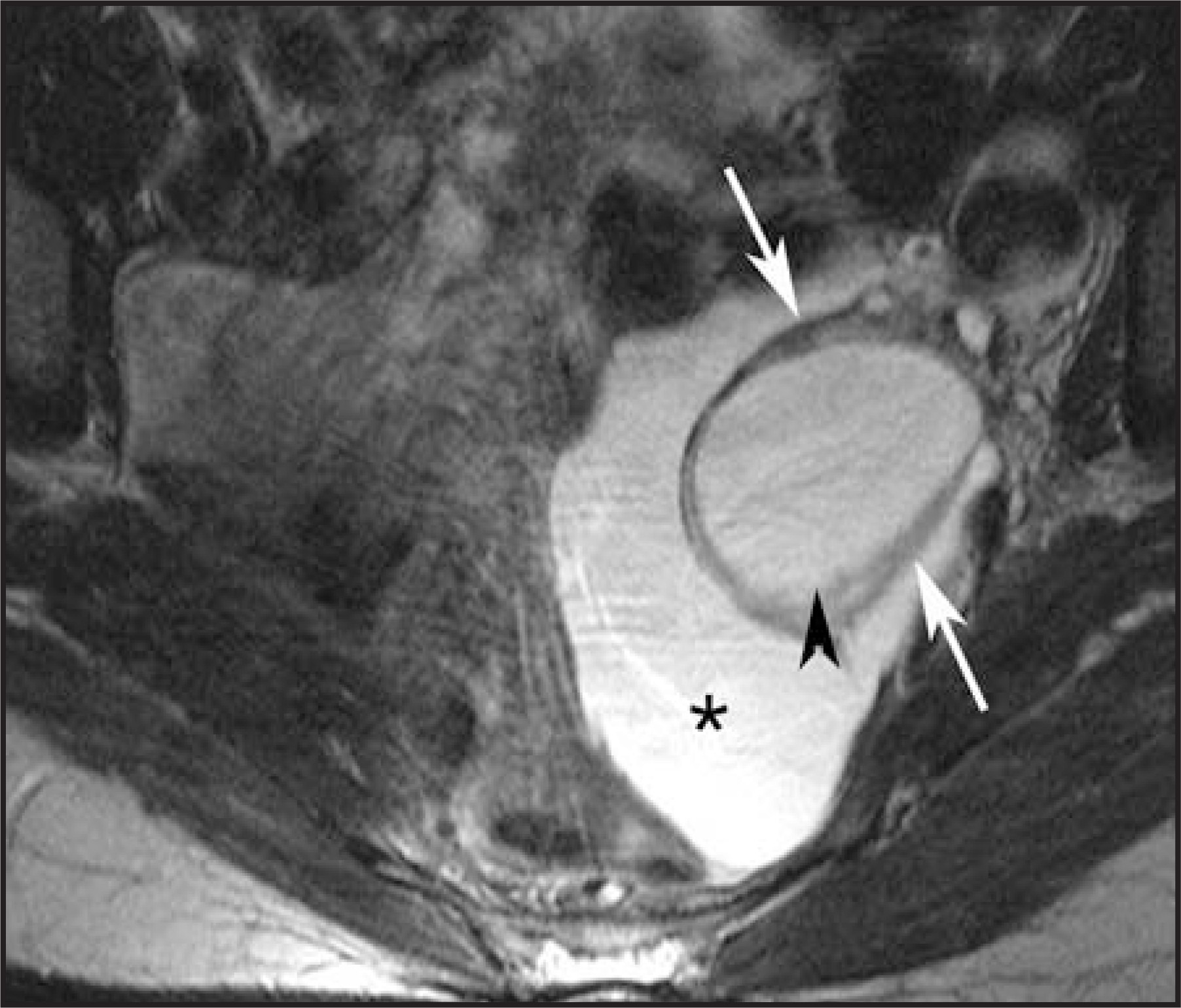
Peritoneal inclusion cysts are the result of entrapment of ovarian fluid by adhesions, and are more frequently seen in premenopausal woman with a history of pelvic surgery, pelvic inflammatory disease, or endometriosis. They are likely to slowly increase in size as fluid is constantly secreted by the ovaries but not reabsorbed by the injured peritoneum. Peritoneal inclusion cysts are extraovarian lesions that are adherent to the ovaries, commonly completely surrounding them. This results in the typical appearance of an ovary within a cyst (Figures 18 and 19). Seldom can they be identified at some distance from the ovaries. They may have thin or thick septations, may have nodular projections, and may contain debris and blood.20,21
Paraovarian cyst
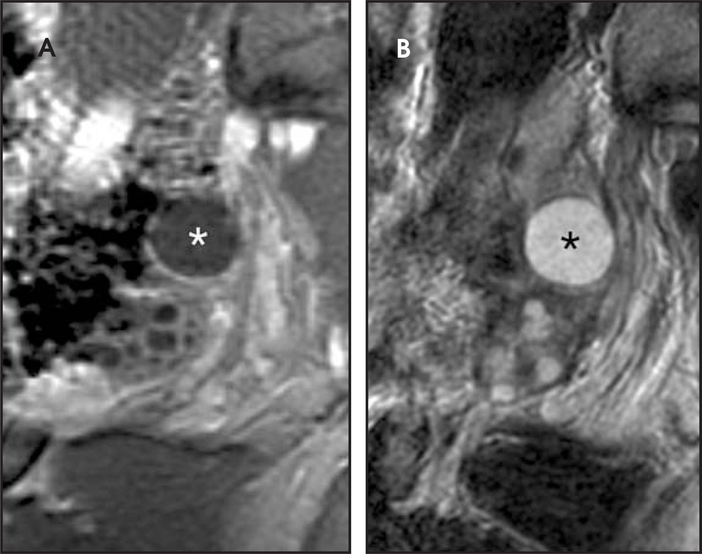
Paraovarian cysts are benign lesions that arise from wolffian duct remnants and are usually seen along the broad ligament. They may represent up to 10% to 20% of adnexal masses. A clue to their diagnosis is identification of a normal ovary separate from the adnexal cyst; however, this is not always easily accomplished (Figure 20). They usually do not regress in time and may be complicated by torsion, hemorrhage, rupture, and infection.22
Malignant-appearing cystic adnexal masses
Malignant-appearing cystic adnexal masses are generally epithelial tumors, usually a cystadenocarcinoma or a borderline tumor. Both have thickened walls or septations and some solid components (Figure 21). It may be difficult to distinguish between these 2 entities, but in the absence of invasion or metastatic disease, a borderline tumor may be considered. Ascites is frequently considered a sign of more aggressive disease.23
Predominantly solid adnexal masses
Fibroma
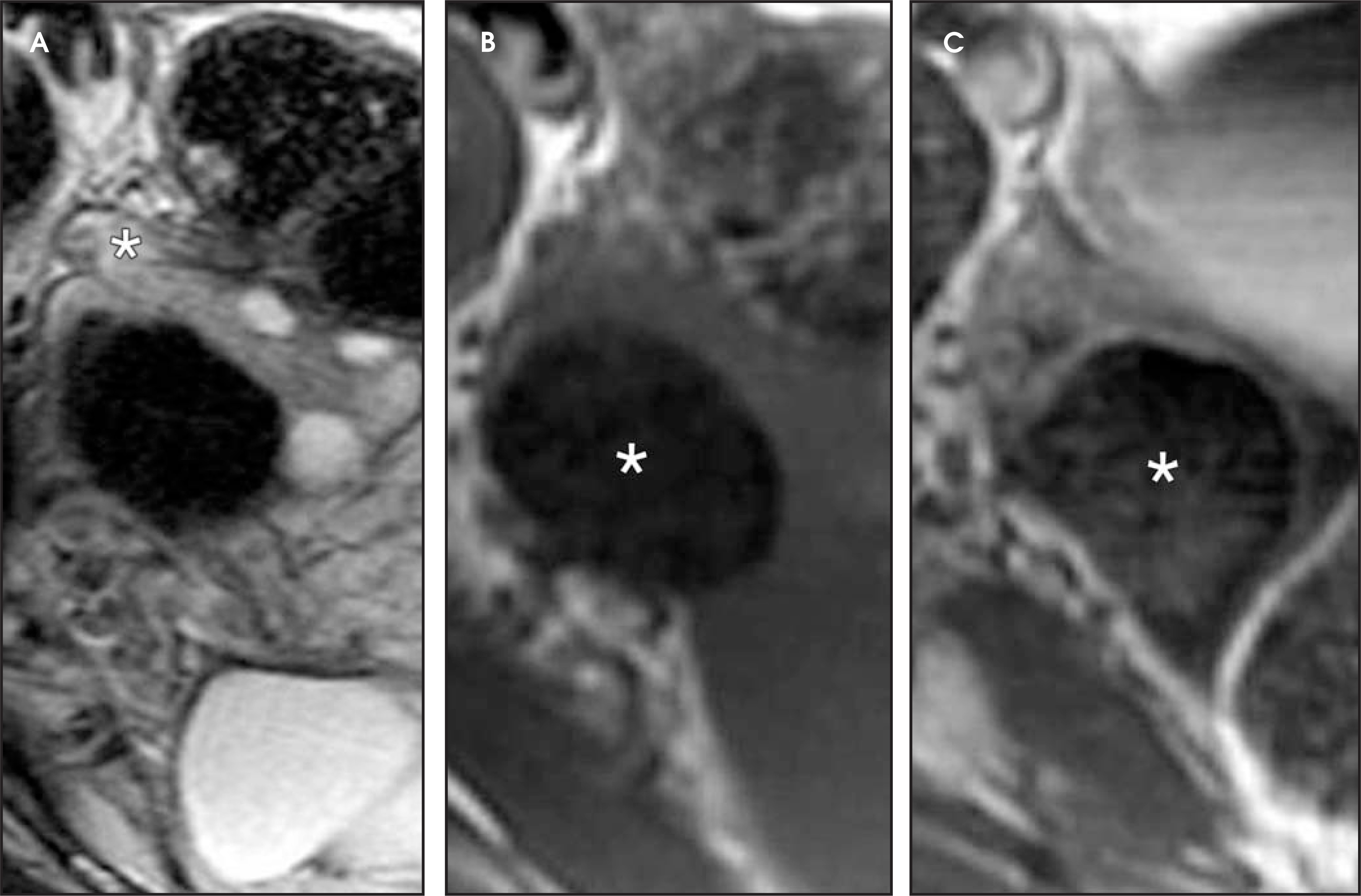
Fibromas are the most common sex cord tumors and represent 4% of all ovarian neoplasms. Because of their solid appearance, they can be misdiagnosed as ovarian cancer at US. On MRI, however, fibromas have a very characteristic appearance with low signal intensity on both T2- and T1-weighted sequences and only slight enhancement after gadolinium administration (Figure 22). Edema and cystic formations (central or eccentric) are recognized and fibromas may be mistaken for cystadenofibromas. Ascites may be present in 40% of cases, particularly if the lesion is large. Fibromas are also associated with pleural effusions (Meigs’ syndrome).3,6,23
Metastases to the ovaries
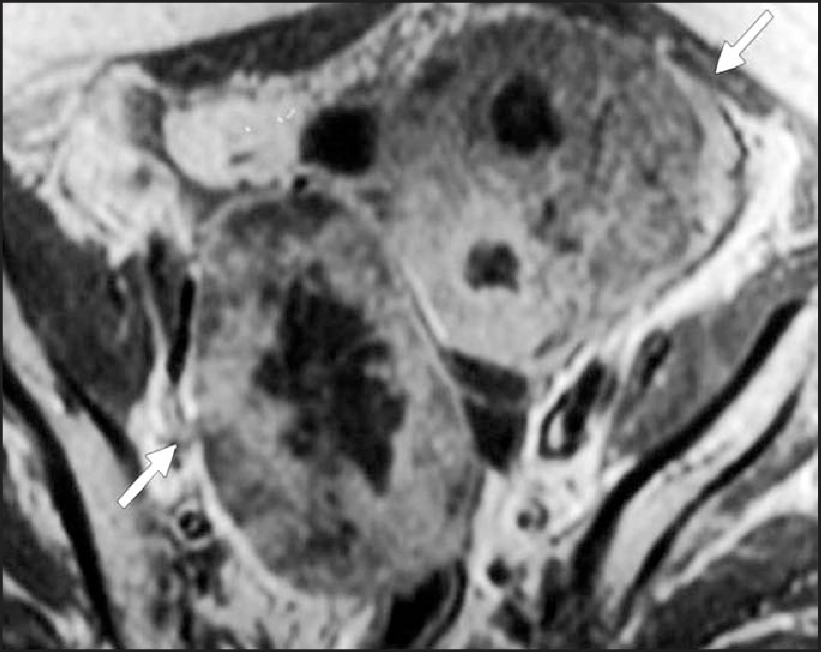
The ovaries are the most common site of metastasis to the female reproductive tract. Gastric, colorectal, and breast cancer are the most frequent extragynecological causes of ovarian metastasis, while primary endometrial and cervical cancers are the usual gynecologic sites of origin.24 Although frequently used as a synonym for ovarian metastases, Krukenberg tumor refers only to metastases composed of mucin signet-ring cells, usually from gastric cancer. Ovarian metastases have no specific appearance, but are commonly bilateral, solid, and strongly enhancing lesions. Cystic and necrotic areas are commonly identified (Figure 23). The primary tumor is usually clinically overt.27-27
Lymphoma
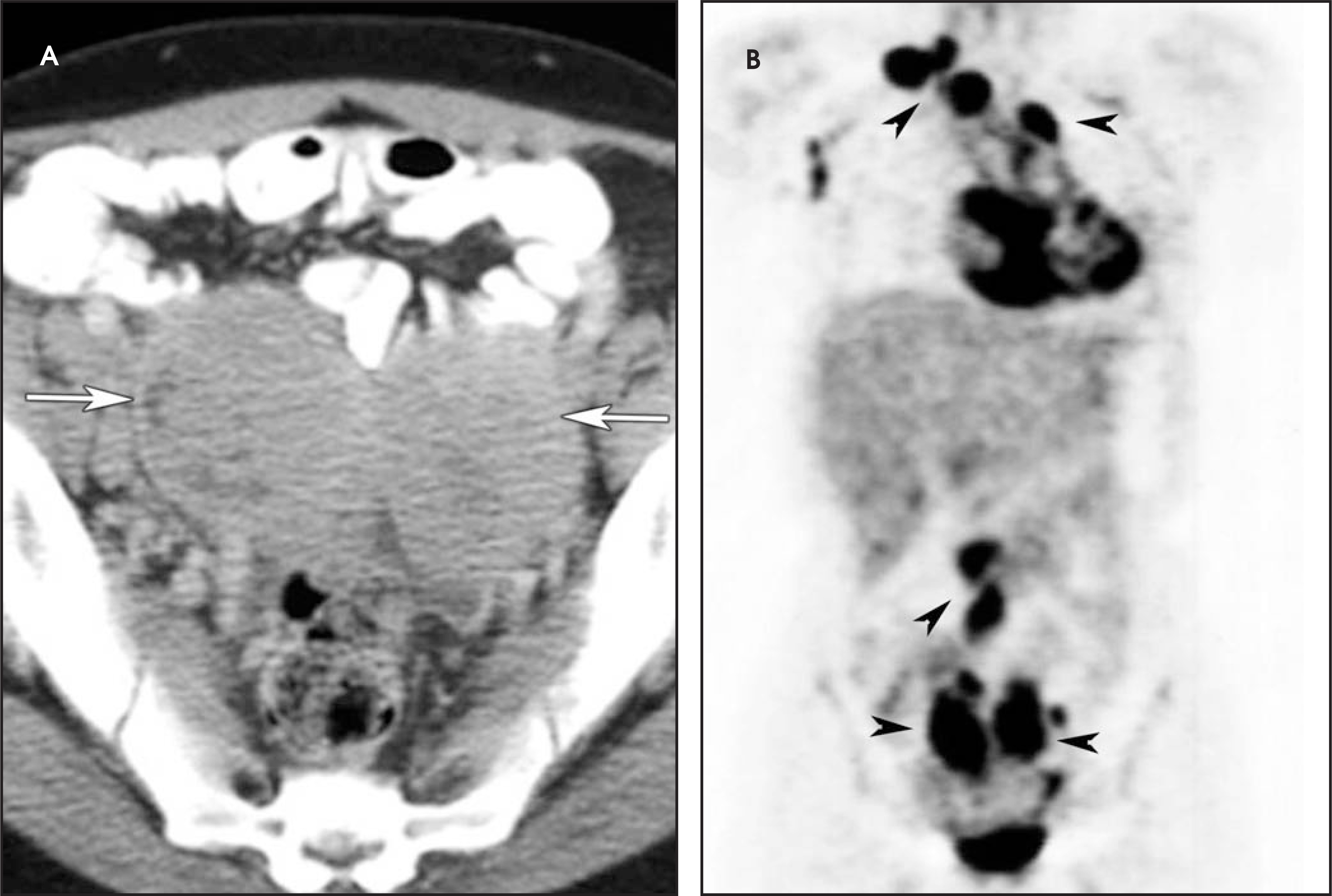
Ovarian lymphoma is exceedingly rare and does not have specific imaging features, but may be suspected in the presence of extensive nodal disease with encasement of vessels (Figure 24).28
Dysgerminoma
Dysgerminomas represent 1% of all ovarian cancers but 20% to 30% of those diagnosed during pregnancy. Eighty percent occur during the first 2 decades of life. Most are unilateral, solid, bosselated tumors with an average size of 15 cm in diameter. Cysts, hemorrhage, and necrosis may be identified. Although dysgerminomas are always malignant and spread to lymph nodes is common, most of these cancers are diagnosed at an early stage and may be treated conservatively.29 A disproportionate amount of adenopathy is sometimes noted, relative to the size and appearance of the ovarian lesion. In these cases, a dysgerminoma should be considered in a young woman and poorly differentiated adenocarcinoma in an elderly patient.
Granulosa cell tumor
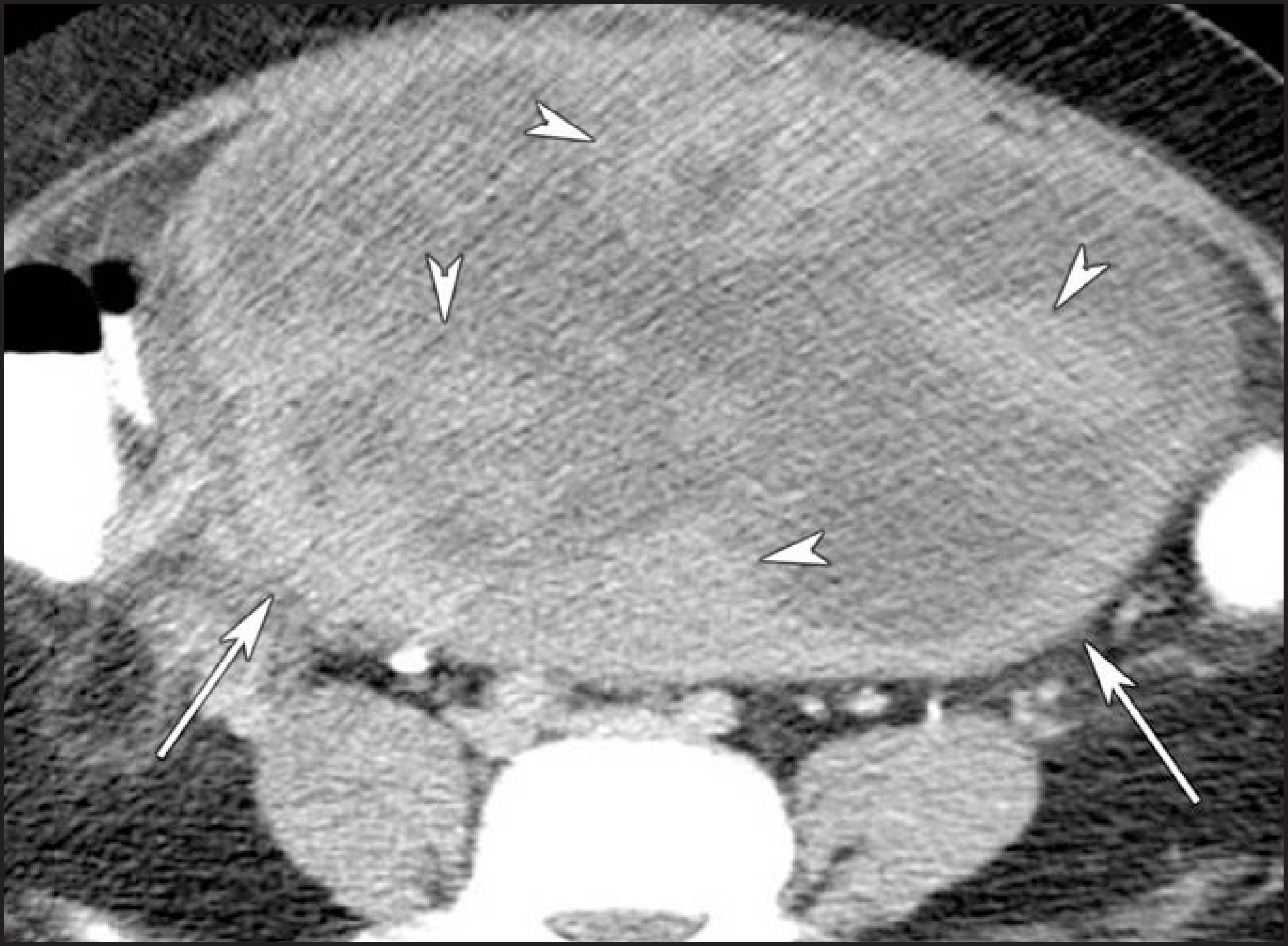
These lesions have variable morphology; they may present with cystic, solid, or a mixture of both components; and they are prone to hemorrhage. Estrogen production is common, and is usually the reason for an early-stage diagnosis. Granulosa cell tumors have a bimodal age distribution: postmenopausal women and those <30 years of age. In the younger age group, recurrence tends to occur within the first 3 years after treatment, but in postmenopausal patients, if the tumor recurs, it happens later in the course of disease. Granulosa cell tumors should be suspected if a solid-appearing ovarian mass has extensive hemorrhage (Figure 25).23,30,31
Sertoli-Leydig cell tumor
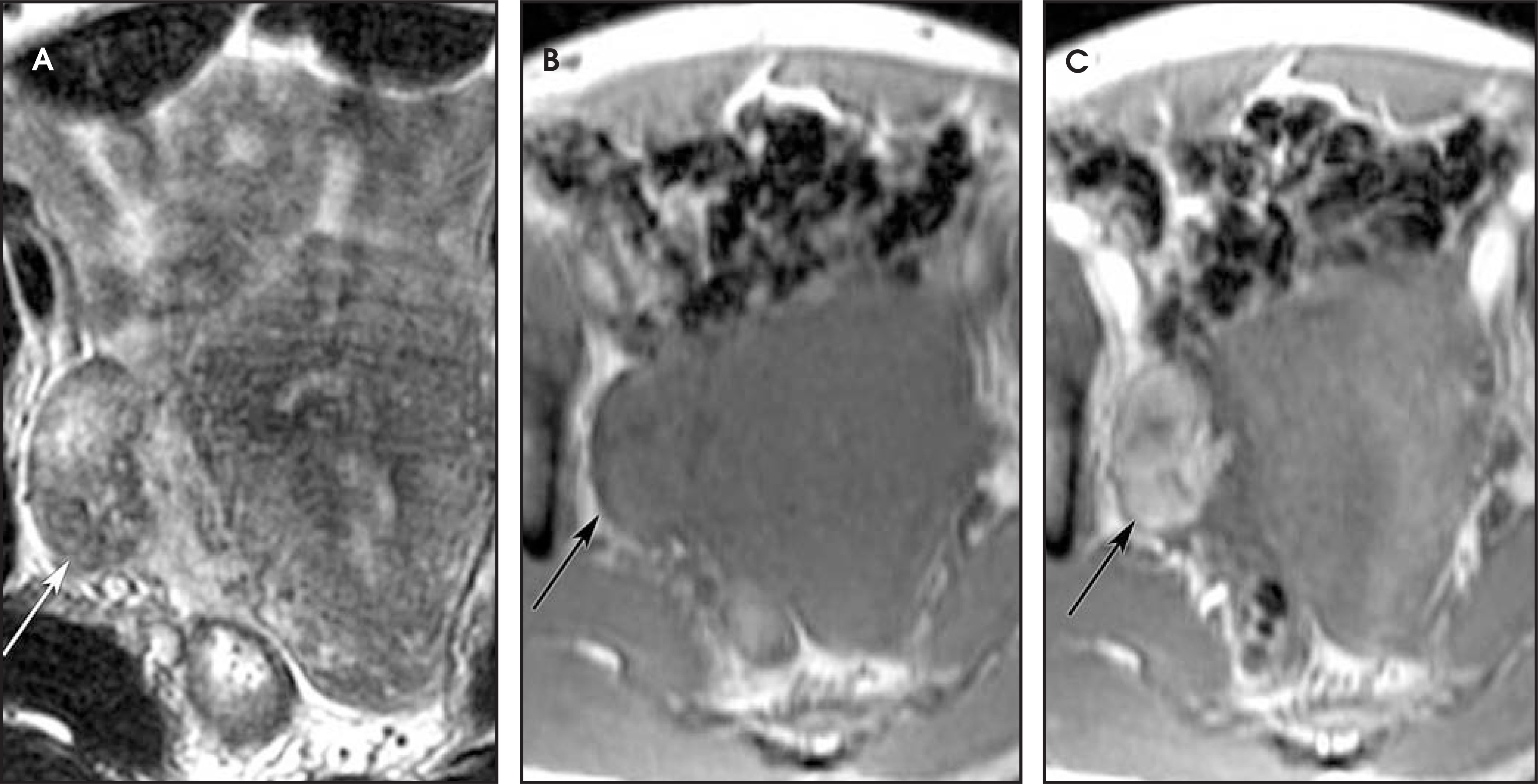
Also known as androblastomas and arrhenoblastomas, these rare lesions represent <0.5% of all ovarian tumors. Most of them are diagnosed in women younger than 30 years old, but rarely before puberty. Androgen secretion causes virilization in >50% of patients. Sertoli-Leydig cell tumors are usually benign and diagnosed at an early stage. They are typically unilateral solid masses, although cystic areas may be identified (Figure 26).23,32 At MRI, these tumors demonstrate high T2 signal intensity, low T1 signal intensity, and enhancement after gadolinium administration.
Conclusion
CT and US have similar sensitivity for differentiating benign from malignant ovarian masses. Although MRI is more accurate than either US or CT, it is usually reserved as a “problem-solving” tool.
Some ovarian masses have specific, distinctive characteristics that allow a diagnosis to be established with a greater degree of certainty. Among these lesions are the dermoid cysts, corpus luteum cysts, theca-lutein cysts, endometriomas and hemorrhagic cysts, hydrosalpinx, and pedunculated leiomyomas.
When issuing a report, it is important to try to emphasize whether imaging findings favor a benign, borderline, or malignant etiology since this is crucial in determining the next step in patient management.
References
- Cancer Stat Fact Sheets: Cancer of the Ovary. 2005.
- .
- Jeong Y, Outwater E, Kang H. Imaging evaluation of ovarian masses. RadioGraphics. 2000;20:1445-1470.
- Kurtz A, Tsimikas J, Tempany C. Diagnosis and staging of ovarian cancer: Comparative values of Doppler and conventional US, CT, and MR imaging correlated with surgery and histopathologic analysis– report of the Radiology Diagnostic Oncology Group. Radiology. 1999;212:19-27.
- Bloomfield T. Benign cystic teratomas of the ovary: A review of seventy-two cases. Eur J Obstet Gynecol Reprod Biol. 1987;25:231-237.
- Saini A, Dina R, McIndoe G. Characterization of adnexal masses with MRI. AJR Am J Roentgenol. 2005;184:1004-1009.
- Comerci J, Licciardi F, Bergh P. Mature cystic teratoma: A clinicopathologic evaluation of 517 cases and review of the literature. Obstet Gynecol. 1994;84:22-28.
- Novak E, Woodruff J, Novak E, Woodruff J. Novak’s Gynecologic and Obstetric Pathology with Clinical and Endocrine Relations. 1979:355-374.
- Borders R, Breiman R, Yeh B. Computed tomography of corpus luteal cysts. J Comput Assist Tomogr. 2004;28:340-342.
- Wajda K, Lucas J, Marsh W. Hyperreactio luteinalis. Benign disorder masquerading as an ovarian neoplasm. Arch Pathol Lab Med. 1989;113:921-925.
- Woodward P, Sohaey R, Mezzetti T. Endometriosis: Radiologic-pathologic correlation. RadioGraphics. 2001;21.
- Glastonbury C. The shading sign. Radiology. 2002;224:199-201.
- Wu T, Coakley F, Qayyum A. Magnetic resonance imaging of ovarian cancer arising in endome triomas. J Comput Assist Tomogr. 2004;28:836-838.
- Outwater E, Siegelman E, Chiowanich P. Dilated fallopian tubes: MR imaging characteristics. Radiology. 1998;208:463-469.
- Silverberg S, Kurman R. Tumors of the Uterine Corpus and Gestational Trophoblastic Disease. 1992.
- Murase E, Siegelman E, Outwater E. Uterine leiomyomas: Histopathologic features, MR imaging findings, differential diagnosis, and treatment. RadioGraphics. 1999;19:1179-1197.
- Mittl R, Yeh I, Kressel H. High-signal– intensity rim surrounding uterine leiomyomas on MR images: Pathologic correlation. Radiology. 1991;180:81-83.
- Karasick S, Lev-Toaff A, Toaff M. Imaging of uterine leiomyomas. AJR Am J Roentgenol. 1992;158:799-805.
- Ueda H, Togashi K, Konishi I. Unusual appearances of uterine leiomyomas: MR imaging findings and their histopathologic backgrounds. Radio-Graphics. 1999;19(Special issue):S131-S145.
- Jain K. Imaging of peritoneal inclusion cysts. AJR Am J Roentgenol. 2000;174:1559-1563.
- Kim J, Lee H, Woo S, Lee T. Peritoneal inclusion cysts and their relationship to the ovaries: Evaluation with sonography. Radiology. 1997;204:481-484.
- Kim J, Woo S, Suh S, Morettin L. Sonographic diagnosis of paraovarian cysts: Value of detecting a separate ipsilateral ovary. AJR Am J Roentgenol. 1995;164:1441-1444.
- Jung S, Rha S, Lee J. CT and MRI findings of sex cordstromal tumor of the ovary. AJR Am J Roentgenol. 2005;185:207-215.
- McCluggage W, Wilkinson N. Metastatic neoplasms involving the ovary: A review with an emphasis on morphological and immunohistochemical features. Histopathology. 2005;47:231-247.
- Ha H, Baek S, Kim S. Krukenberg’s tumor of the ovary: MR imaging features. AJR Am J Roentgenol. 1995;164:1435-1439.
- Hann L, Lui D, Shi W. Adnexal masses in women with breast cancer: US findings with clinical and histopathologic correlation. Radiology. 2000;216:242-247.
- Kim S, Kim W, Park K. CT and MR findings of Krukenberg tumors: Comparison with primary ovarian tumors. J Comput Assist Tomogr. 1996;20:393-398.
- Ferrozzi F, Tognini G, Bova D, Zuccoli G. Non-Hodgkin lymphomas of the ovaries: MR findings. J Comput Assist Tomogr. 2000;24:416-420.
- Russel P, Farnsworth A, Russel P, Farnsworth A. Surgical Pathology of the Ovaries. 1997:485-494.
- Schumer S, Cannistra S. Granulosa cell tumor of the ovary. J Clin Oncol. 2003;21:1180-1189.
- Kim S, Kim S. Granulosa cell tumor of the ovary: Common findings and unusual appearances on CT and MR. J Comput Assist Tomogr J Comput Assist Tomogr.
- Russel P, Farnsworth A, Russel P, Farnsworth A. Surgical Pathology of the Ovaries. 1997:435-457.
Citation
. Stent. Applied Radiology. 2006;35(10):22-36. doi:10.37549/AR1458.