Stomach-Containing Paraumbilical Hernia with Obstruction
Applied Radiology — Vol. 55 , Issue 3
Published: December 22, 2025
1 University of Arkansas for Medical Sciences, Little Rock, Arkansas
2 Baylor College of Medicine, Houston, Texas
* Corresponding author: Wayne Ngo (hngo@uams.edu)
Categories
Case Summary
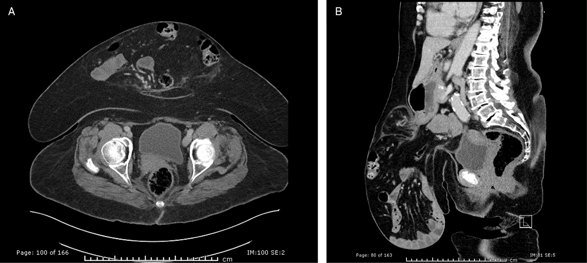
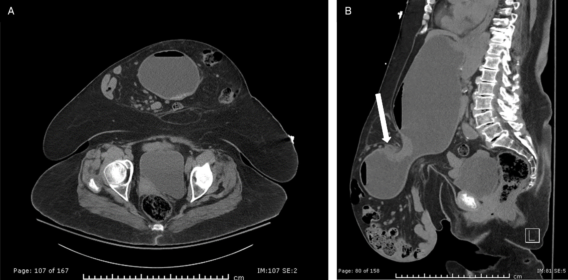
An elderly patient presented to the emergency department with abdominal pain, nausea, and vomiting. The patient had a known, large ventral abdominal paraumbilical hernia that was diagnosed 1 month prior on CT (Figure 1). This initial CT showed a large hernia sac containing small and large bowel loops without signs of bowel obstruction or strangulation. 1 month later, she returned with worsened abdominal pain, nausea, and vomiting. A repeat abdominal and pelvis CT examination was performed. This showed interval herniation of the distal stomach and proximal bowel into the patient’s known large paraumbilical hernia, with resultant gastric outlet obstruction. After receiving intravenous fluid and nasogastric tube placement, the patient underwent surgery. The operative report described a large, ventral abdominal hernia with viable stomach, proximal duodenum (D1), cecum, ascending and transverse colon, and multiple loops of small bowel. There was no evidence of strangulation. The patient successfully recovered after surgery.
Imaging Findings
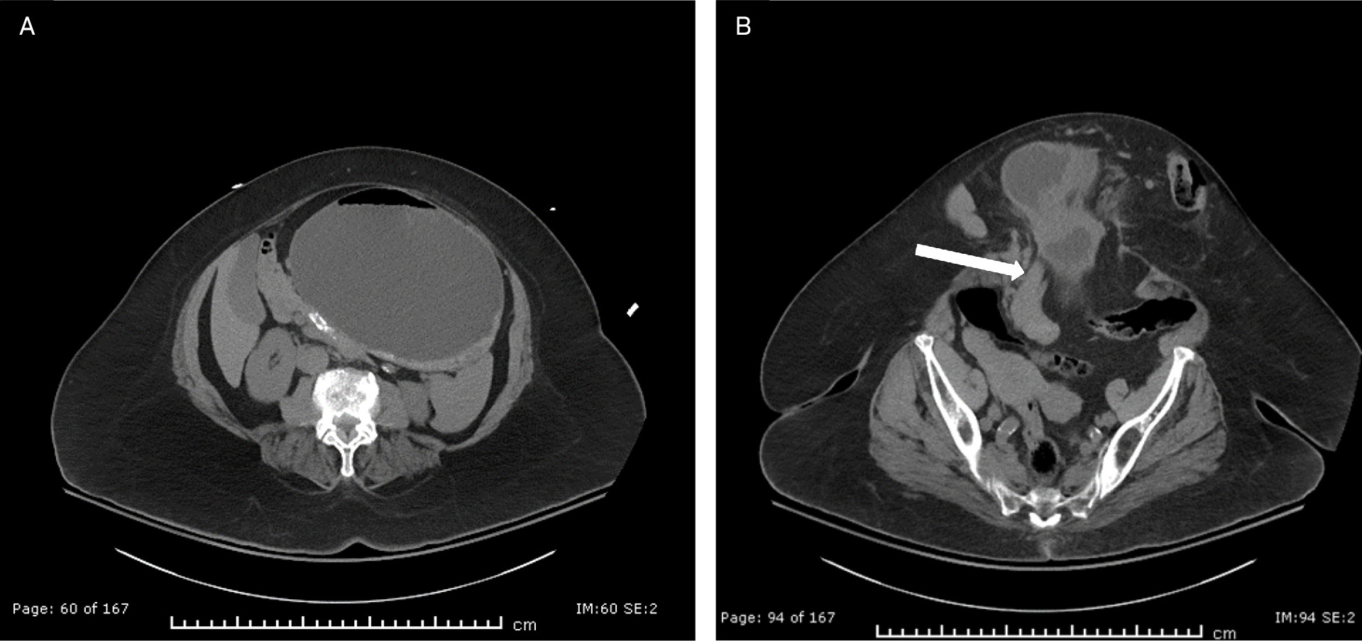
Prior comparison of abdomen and pelvis CT with intravenous contrast (axial and sagittal reconstructed images) (Figure 1) showed a large paraumbilical hernia containing small and large bowel loops as well as the appendix. There was no evidence of bowel obstruction or strangulation. Repeat abdomen and pelvis CT with intravenous contrast (axial and sagittal images) obtained 1 month later (Figure 2) showed interval herniation of distal stomach and proximal duodenum (D1 segment) into the known large paraumbilical hernia with resultant gastric outlet obstruction. There was no evidence of strangulation. Serial axial images from cranial to caudal (Figure 3) again show interval herniation of distal stomach and the proximal duodenum into the large paraumbilical hernia.
Diagnosis
Stomach within paraumbilical hernia.
Differential diagnoses could include incisional hernia, epigastric hernia, rectus abdominis diastasis, and abdominal wall neoplasm.
Discussion
Abdominal hernias are abnormal protrusions of peritoneal lined organs through the abdominal wall. They are commonly encountered and have an estimated prevalence of 25% in adults. 1 Although abdominal hernias can be asymptomatic, surgical treatment may be required due to the risk of complications such as incarceration and strangulation. 1,2
In this case report, we specifically focus on midline abdominal hernias, which include epigastric, paraumbilical, umbilical, and hypogastric hernia types. Among these types, umbilical/paraumbilical hernias are the most frequently encountered and account for about 13.9% of all hernias. They occur in and around the umbilicus as a result of a defect in the linea alba and/or abdominal rectus muscle diastasis. 2 Patients typically present with an abdominal bulge that can be asymptomatic or tender to palpation. Ventral abdominal hernia contents are typically a combination of peritoneal fat, omentum, and small bowel. 3 Larger-sized ventral abdominal hernias may contain large bowel as well. Stomach herniation into a pre-existing ventral abdominal hernia, however, is rare and has only been described a few times in the past century. 4-8
Few similar previous case reports were found in a literature review. Thomas et al was the only other study to identify the stomach in a paraumbilical hernia on CT. 8 In their study, a 91-year-old woman with a long-standing paraumbilical hernia presented with repeated vomiting and dehydration. Endoscopy showed external gastric compression, and CT revealed a hernia sac containing the distal stomach, pylorus, and proximal duodenum. Sampaio et al used a barium swallow to visualize herniation of the gastric antrum and duodenal bulb in a 59-year-old woman. 7 Orr et al. used a similar technique to demonstrate stomach incarceration in a 49-year-old female with chronic vomiting. 4 Bryk et al used barium swallow to show gastric outlet obstruction in a 63-year-old woman with large umbilical hernia. 5 In another published case involving a 62-year-old woman, US demonstrated a herniated stomach appearing as a cystic mass with internal echoes protruding through the linea alba. 6
The clinical findings in our case are consistent with those described in prior reports. The most frequently observed symptom is vomiting, which carries a risk of dehydration and electrolyte disturbances. These hernias tend to occur in multiparous women in middle to later adulthood. Pregnancy is considered a risk factor due to the increased intra-abdominal pressure, which can stretch and weaken the abdominal wall muscles. 8,9 Although the stomach is secured by multiple ligaments, these attachments may become increasingly lax with age. 5,8 This laxity may contribute to the development of stomach herniation later in life, as seen in our patient.
The initial diagnosis of paraumbilical hernias is made clinically and can be assisted by plain radiography, barium studies, ultrasonography, or CT. 2,9,10 Although previous cases have used barium studies and endoscopy, CT is currently the preferred imaging modality because it provides higher accuracy for identifying hernia contents and complications such as obstruction or incarceration. 2 US may also be useful because it allows real-time assessment of motion to identify herniated organs. In our case, CT provided high anatomical resolution, enabling rapid identification of the hernia contents and assessment for bowel obstruction or strangulation.
Definitive treatment of paraumbilical/umbilical hernias typically requires surgical repair with sutures or mesh reinforcement. 1 Prophylactic surgical treatment is often recommended to prevent complications even in asymptomatic patients. 9 Our patient successfully underwent ventral abdominal surgical repair and abdominoplasty with complex repair. A clinical implication from our case is that prompt surgical treatment at the initial encounter may have prevented her gastric herniation.
Conclusion
Gastric herniation into an existing paraumbilical/umbilical hernia is a rare complication. Patients may present with vomiting and gastric outlet obstruction. CT should be ordered to quickly identify herniated content and other complications such as bowel obstruction, strangulation, and incarceration. Multiparous women in middle to late adulthood are at higher risk of stomach herniation due to weakened abdominal muscles from pregnancy and age-related laxity of stomach ligaments. Patients fitting this demographic should be educated about their risk of herniation and referred to surgical services for prophylactic surgical treatment.
References
- Liang M, Holihan J, Itani K. Ventral hernia management: expert consensus guided by systematic review. Ann Surg. 2017;265(1):80-89. doi:10.1097/SLA.0000000000001701.
- Aguirre D, Santosa A, Casola G, Sirlin C. Abdominal wall hernias: imaging features, complications, and diagnostic pitfalls at multi-detector row CT. Radiographics. 2005;25(6):1501-1520. doi:10.1148/rg.256055018.
- Kulaçoğlu H. Current options in umbilical hernia repair in adult patients. Ulus Cerrahi Derg. 2015;31(3):157-161. doi:10.5152/UCD.2015.2955.
- Orr T. Incarceration of the stomach in an umbilical hernia. Am J Surg. 1947;74(1):96. doi:10.1016/0002-9610(47)90101-3.
- Bryk D. Gastric involvement in abdominal wall hernias. Gastrointest Radiol. 1984;9(4):311-314. doi:10.1007/BF01887858.
- Saggar K, Goyal S, Goyal R, Sodhi K. Ultrasonographic detection of herniation of stomach in paraumbilical hernia. Indian J Gastroenterol. 1999;18(2):87-88.
- Sampaio R, Ferreira M. Umbilical hernia of stomach. Eur Radiol. 1998;8(4):568-570. doi:10.1007/s003300050435.
- Thomas J, Monaghan T, Saeed A, Khan F. Umbilical herniation of the stomach. QJM. 2012;105(10):1025-1026. doi:10.1093/qjmed/hcr163.
- Coste A, Jaafar S, Parmely J. In: StatPearls. 2023.
- Ahn S, Moon S, Lee D. Sonography of gastrointestinal tract diseases: correlation with computed tomographic findings and endoscopy. J Ultrasound Med. 2016;35(7):1543-1571. doi:10.7863/ultra.15.09038.
Disclosures
The authors have no conflicts of interest to disclose. None of the authors received outside funding for the production of this original manuscript and no part of this article has been previously published elsewhere.
Citation
. Stomach-Containing Paraumbilical Hernia with Obstruction. Applied Radiology. 2025;55(3). doi:10.37549/AR-D-25-0101.