Supratentorial and infratentorial brain neoplasms
Applied Radiology — Vol. 33 , Issue 11 , pp. 23 -33
DOI: 10.37549/AR1294
Published: November 1, 2004
Categories
Neoplasms of the brain constitute a long and often confusing list of entities. The aim of this article is to review this list within a logical framework, which should help improve understanding and retention of the possible lesions. When evaluating any intracranial mass, the following three distinguishing criteria are easily determined, yet vital to arrive at an appropriate differential diagnosis: 1) adult or pediatric patient; 2) supratentorial or infratentorial lesion; 3) intra-axial or extra-axial lesion. The differential diagnostic possibilities using these criteria are outlined in Table 1.
SUPRATENORIAL NEOPLASMS Adult neoplasms
In adults, metastasis and the various subtypes of glioma account for the vast majority of intra-axial neoplasms. Neoplasms of glial origin include glioblastoma multiforme (50%), anaplastic astrocytoma (25%), low-grade astrocytoma (15%), oligodendroglioma (10%), and gliosarcoma (rare). Giant cell astrocytoma occurs in association with tuberous sclerosis. Lastly, primary cerebral (intra-axial) lymphoma has markedly increased in incidence secondary to the widespread acquired immunodeficiency syndrome (AIDS) epidemic. In the extra-axial compartment, meningioma is the most common lesion, but metastatic carcinomatous meningitis (usually of similar primary origin as intra-axial metastatic disease) and secondary lymphoma or leukemia should also be considered.
Intra-axial neoplasms
Metastasis—Metastasis is the most common supratentorial mass in adults and represents 40% of all intracranial neoplasms. While most metastases appear in multiples, solitary metastasis occurs in 30% to 50% of cases. The most common primary malignancies include breast carcinoma, lung carcinoma, melanoma, renal cell carcinoma, thyroid carcinoma, and gastrointestinal adenocarcinoma.
Metastases are typically well-defined lesions located at the junction of gray and white matter. Severe vasogenic edema is associated with most metastases. Cortical metastases are the exception, where edema may be minimal or absent. Metastases are typically hypodense on non-contrast computed tomography (CT). Magnetic resonance (MR) imaging typically reveals low signal on T1-weighted imaging and variable signal on T2-weighted imaging. Hemorrhage, necrosis, and cyst formation account for the variability in imaging findings. Nevertheless, enhancement is the rule for metastases, and, in particular, ring enhancement due to the usual central necrosis (Figure 1). As a result, contrast imaging with CT or MR is essential.1 Additionally, lesion conspicuity can be improved with double- or triple-dose contrast administration with delayed imaging.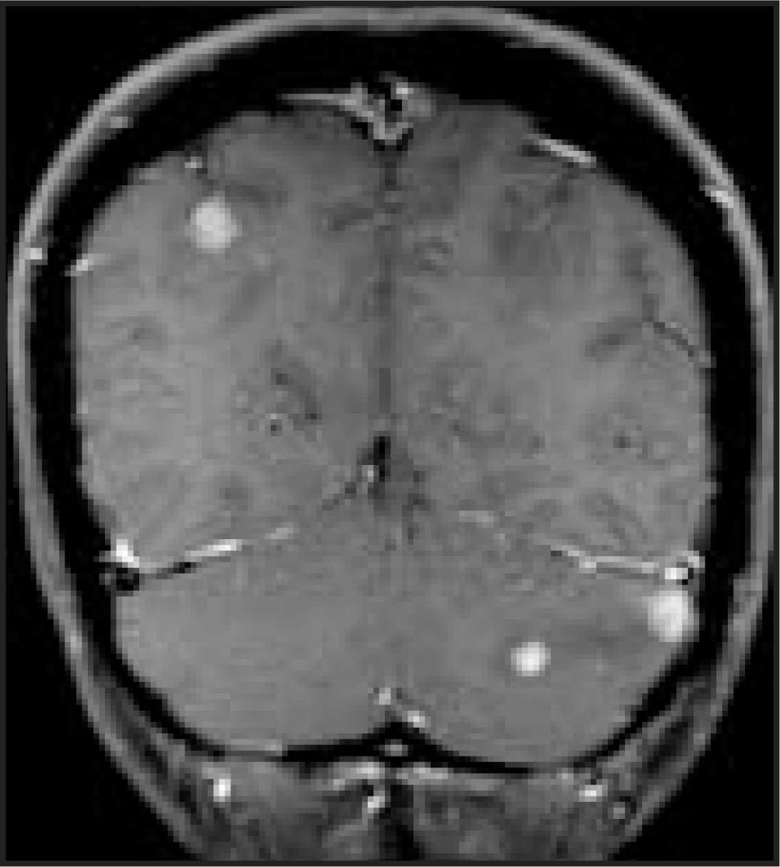
Glioma—Gliomas represent half of all solitary supratentorial tumors. The three primary intracranial gliomas, from lowest to highest grade, are (low-grade) astrocytoma, anaplastic astrocytoma, and glioblastoma multiforme (GBM). Gliomas are graded by the following pathologic criteria: number of mitoses, presence of necrosis, vascular endothelial proliferation, nuclear pleomorphism, and cellular density.
Low-grade astrocytoma (World Health Organization [WHO] Grade II): Low-grade astrocytomas represent 10% to 15% of all gliomas. While categorized pathologically as low grade, 50% evolve into anaplastic astrocytoma or GBM. These are nonhemorrhagic, diffusely infiltrating neoplasms with minimal or absent edema. Enhancement is uncommon. Calcification is seen in 15% to 20% of cases.
Anaplastic astrocytoma (WHO Grade III): Anaplastic astrocytomas represent 25% of all gliomas. They are usually seen in the frontal and temporal lobes, showing moderate mass effect, edema, neovascularity, and, most importantly, enhancement (Figure 2).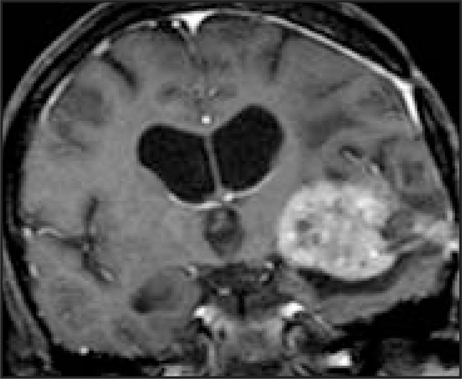
Glioblastoma multiforme (WHO Grade IV): Glioblastoma multiforme is the most common and most aggressive of all gliomas. Necrosis and intratumoral hemorrhage are common findings. Tumor spread occurs across white matter tracts, such as the corpus callosum, anterior or posterior commissures, corona radiata, and cerebral peduncles (Figure 3). Tumor spread also occurs along the ependyma and ventricular surface, leptomeninges, under the pia, and throughout the neuraxis via subarachnoid seeding. Dural invasion and extracerebral metastases are rare. Given the extensive cerebral infiltration, surgical resection is almost always sub-total, and, therefore, the 2-year survival rate is only approximately 10% to 15%. Contrast imaging should be performed within 24 hours after surgery to distinguish residual enhancing tumor from the subsequent development of enhancing postoperative granulation tissue, as well as to establish a baseline for surveillance of future tumor recurrence.1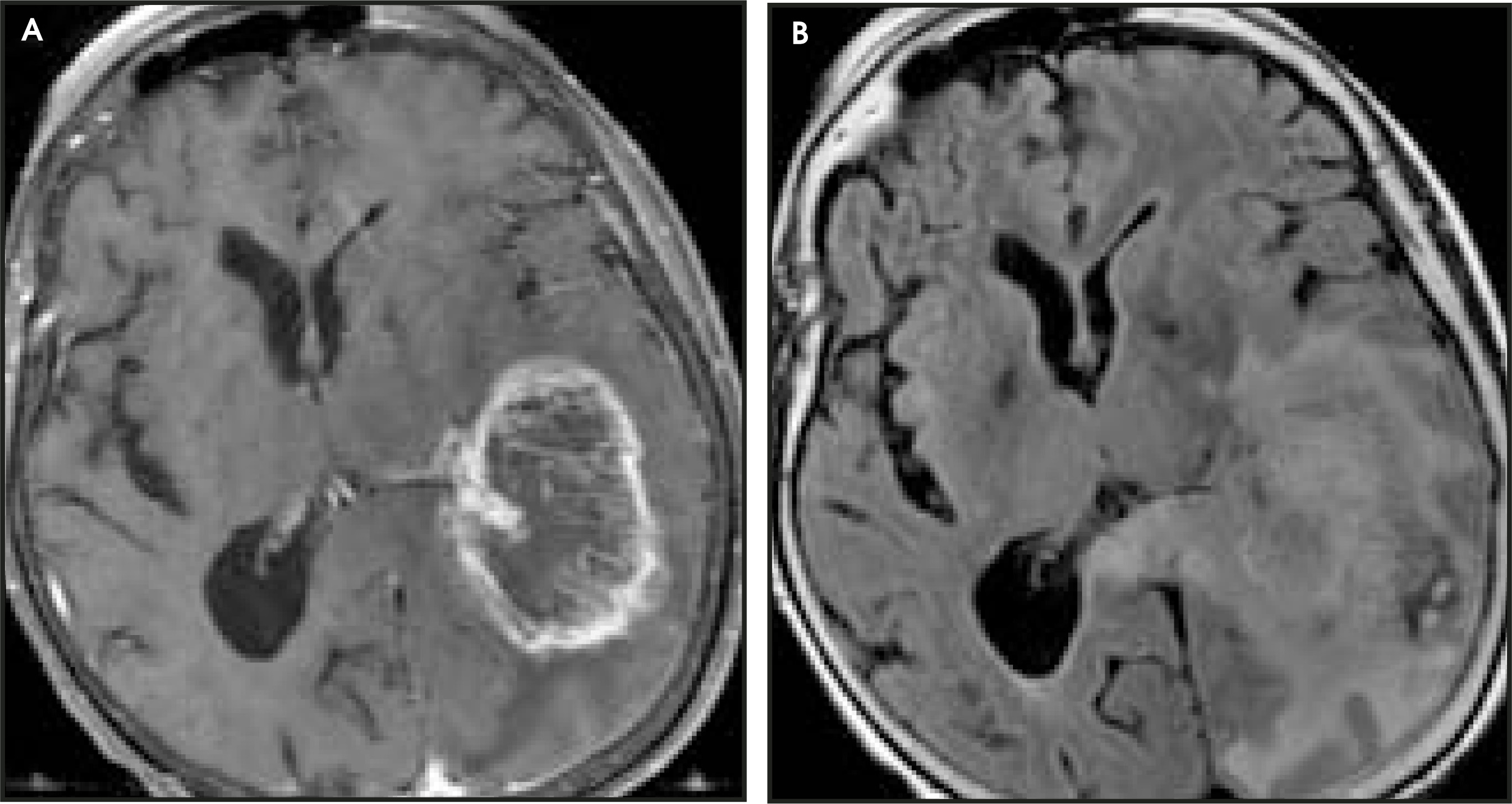
Gliomatosis cerebri: Gliomatosis cerebri is a form of glioma in which a large portion of the cerebral hemisphere is infiltrated. Mass effect and contrast enhancement are typically absent, making this entity easy to miss on CT. MR imaging demonstrates diffusely increased signal on T2-weight-ed or fluid-attenuated inversion recovery (FLAIR) imaging with minimal, if any, gadolinium enhancement. While their appearance is low-grade, given their lack of mass effect and enhancement, these lesions behave similarly to a high-grade glioma and the prognosis is poor.2
Giant cell astrocytoma: Giant cell astrocytoma is a neoplasm that occurs in association with tuberous sclerosis. It arises near the foramen of Monro and, although it is of very low grade, it may enlarge over time, potentially resulting in obstructive hydrocephalus (Figure 4). Tuberous sclerosis also demonstrates subcortical and subependymal tubers (hamartomas). Approximately 30% to 50% of subependymal lesions may enhance on MR imaging; however, this finding does not denote neoplastic transformation (as previously thought) to giant cell astrocytoma.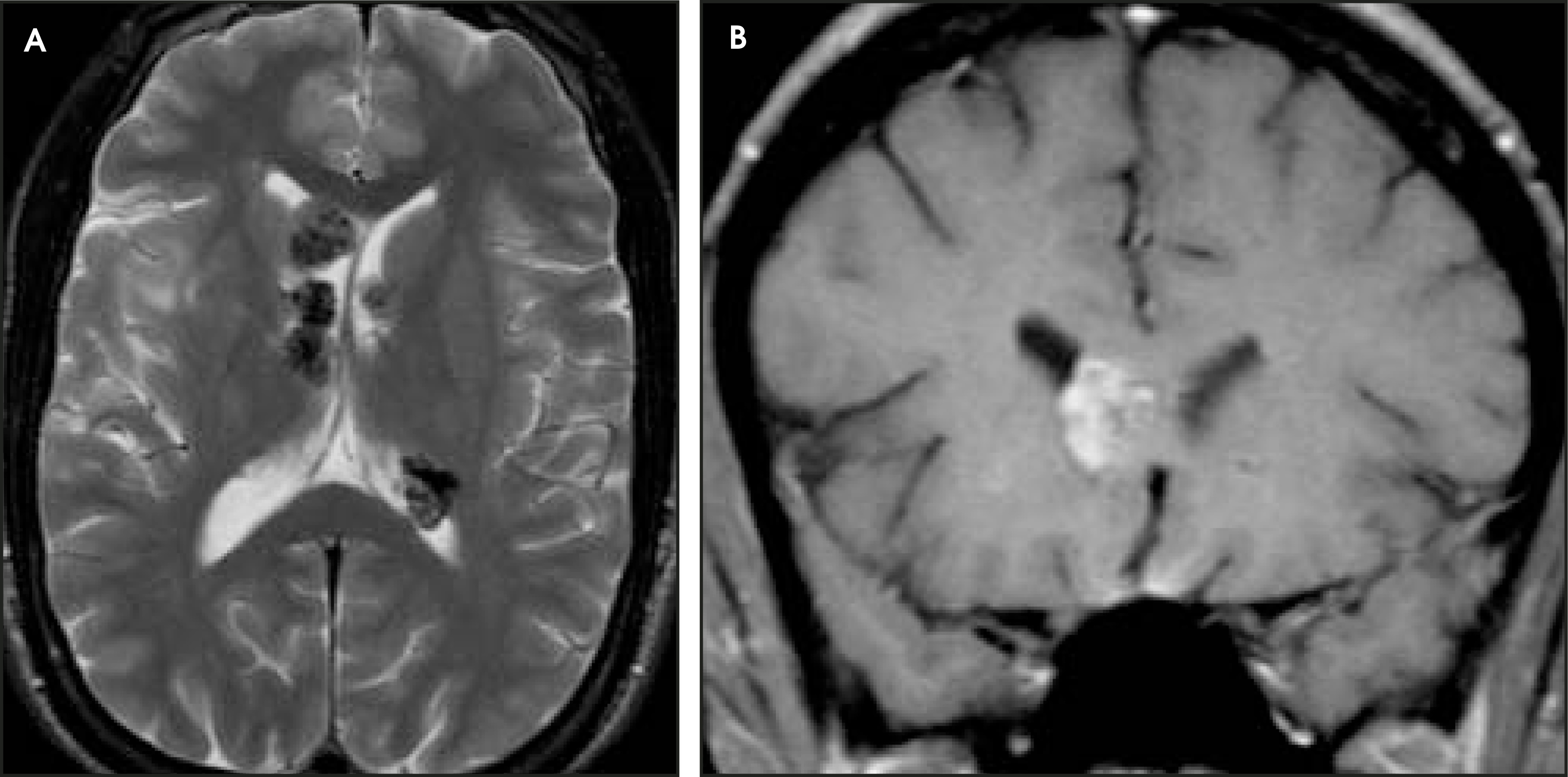
Therefore, serial imaging is required to document growth of the lesion over time and, thus, to confirm the diagnosis of giant cell astrocytoma.3
Gliosarcoma: Gliosarcoma consists of neoplastic neuronal glial cells and spindle cell mesenchymal sarcomatous elements, and carries a prognosis poorer even than GBM. The mix of cell types of this aggressive neoplasm results from marked dedifferentiation of the neoplastic neuronal cells, rather than from sarcomatous transformation of the vascular elements within GBM, as previously thought. Unlike GBM, extracranial metastases and metastases to visceral organs via hematogenous spread are common, occurring in 15% to 30% of cases. Gliosarcoma is typically located peripherally and exhibits dural invasion. Direct contact with the skull is common. An important distinction from meningioma is the lack of a broad dural attachment.4
Oligodendroglioma: Oligodendrogliomas represent 10% to 15% of all gliomas. Their histology is more commonly mixed (containing multiple gliomatous elements of astrocytomas and oligodendrogliomas) rather than pure neoplastic oligodendroglial cells. MR imaging demonstrates moderate enhancement with heterogeneous T2-signal due to calcification and cyst formation, with hemorrhage and necrosis being less common. Seen in 70% to 90% of cases, nodular clumped calcification (more easily identified on CT) is very common (Figure 5). Nevertheless, since the incidence of low-grade glioma is far higher than that of oligodendroglioma, a calcified glioma will more likely represent low-grade glioma despite the greater propensity of oligodendroglioma to calcify. Survival depends on the grade of the lesion; 10-year survival rate is 46% for low-grade lesions and 20% for higher grades.1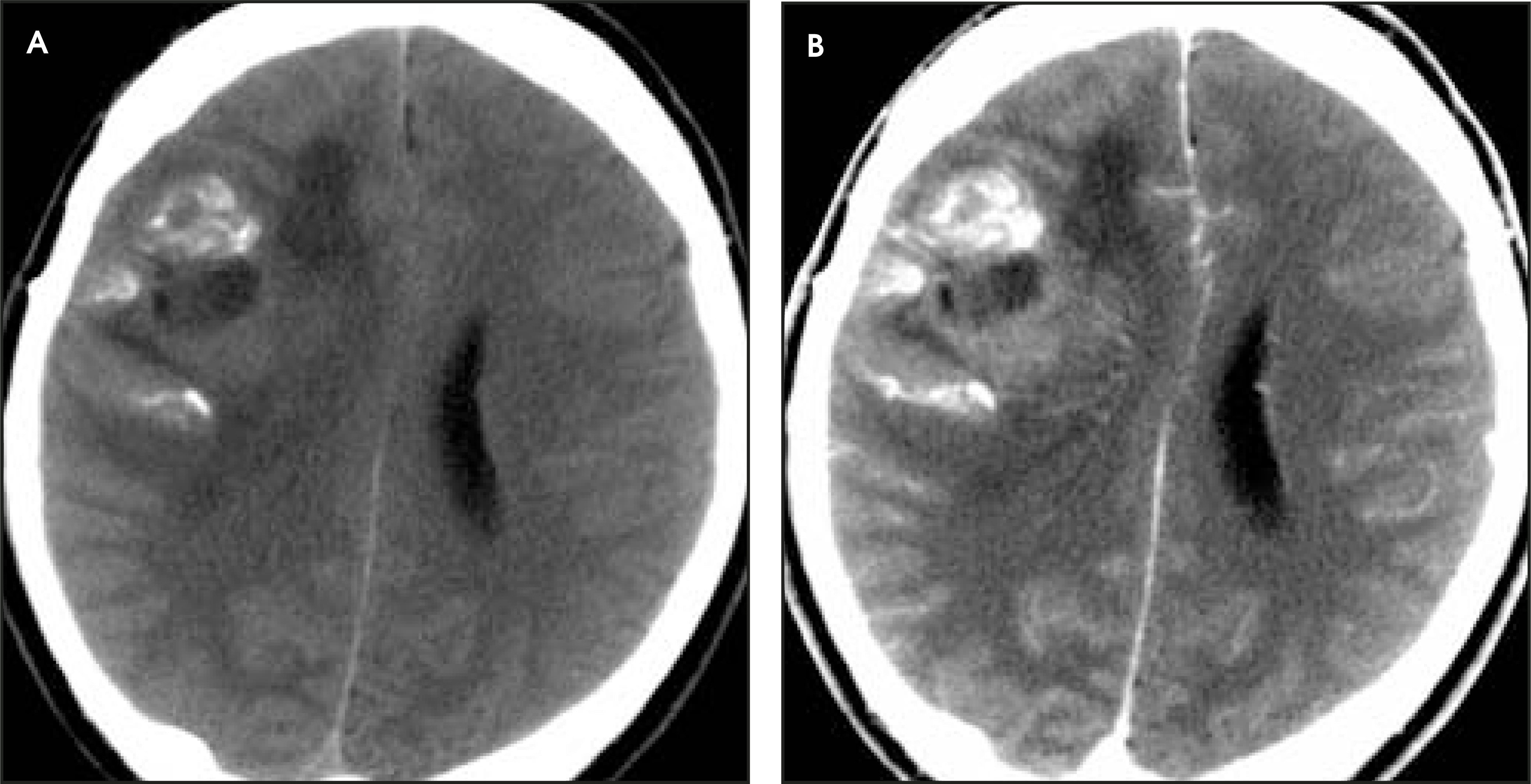
Primary central nervous system lymphoma—Primary central nervous system (CNS) lymphoma is now more common than secondary lymphomatous spread to the intracranial cavity. Occurring in 6% of AIDS patients, it is projected to become the most common primary brain malignancy. While typically seen in immunocompromised patients, its incidence is rising in immunocompetent patients as well. Interestingly, 20% to 40% of cases present with multiple lesions. Lesions are most commonly of the B-cell type and are located deep within the brain parenchyma: basal ganglia, periventricular white matter, and corpus callosum. Due to its high cellularity, CNS lymphoma is characterized by a high nuclear-to-cytoplasmic ratio and, as a result, is commonly hyperdense on CT. Consequently, it is also isointense or hypointense on T2-weighted MR imaging (Figure 6). It demonstrates prominent, usually uniform, enhancement in immunocompetent patients, although the use of steroids may suppress this characteristic. In the immunosuppressed population, in particular AIDS patients, a more aggressive type of lymphoma is present, which results in rapid tumor growth, central necrosis, and subsequently demonstrates ring enhancement. Lesions are highly sensitive to radiotherapy but frequently recur.5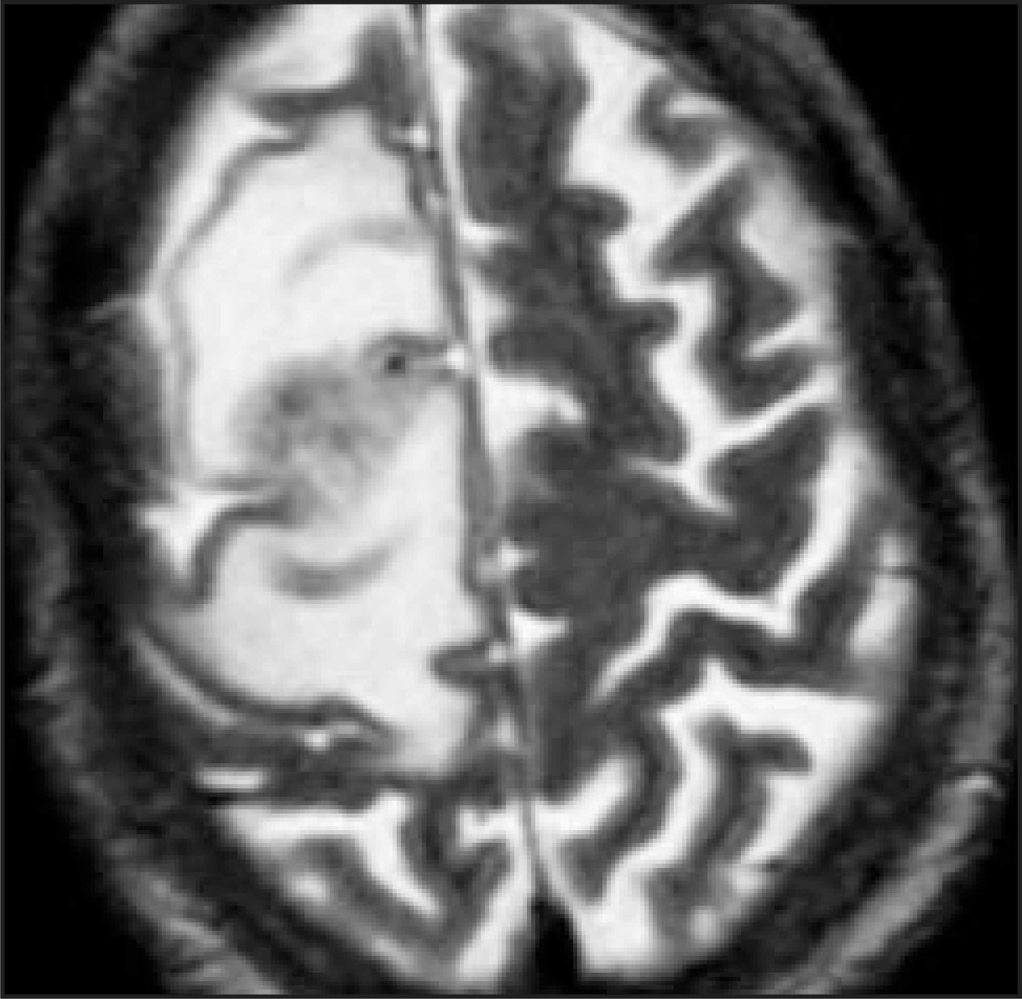
Extra-axial neoplasms
Meningioma—Meningioma represents the most common extra-axial supratentorial neoplasm. It originates from arachnoid cap cells and exhibits transitional, fibroblastic, angioblastic, and syncytial histologies. It more commonly affects middle-aged women, though is also very often seen in men. Meningioma is associated with neurofibromatosis type 2 (NF-2) and basal cell nevus syndrome. About 90% of meningiomas are supratentorial in location. The parasagittal dura is the most common location, followed by the convexities, sphenoid wing, cerebellopontine angle, foramen magnum, olfactory groove, and, finally, the planum sphenoidale.
Due to its extra-axial location, meningioma can exhibit a “cleft” of cerebrospinal fluid (CSF) between the mass and adjacent brain parenchyma. This is an easily identified radiographic sign confirming the extra-axial nature of the neoplasm. Other common manifestions of meningioma include buckling of the adjacent gray and white matter, displacement and/or encasement of adjacent vessels, and widening of the adjacent cistern. Meningioma is hyperdense on noncontrast CT in approximately 60% of cases due to its hypercellular nature. Calcification occurs in 20% of cases. The mass is relatively isointense on T1- and T2-weighted MR imaging compared with cortical signal imaging characteristics. It enhances avidly following contrast administration (Figure 7). An enhancing dural tail, adjacent parenchymal edema, and bony hyperostosis and/or osteolysis may be present in approximately 20% of cases. Of note, while the dural tail may represent neoplastic extension, it may instead only represent fibrovascular proliferation.6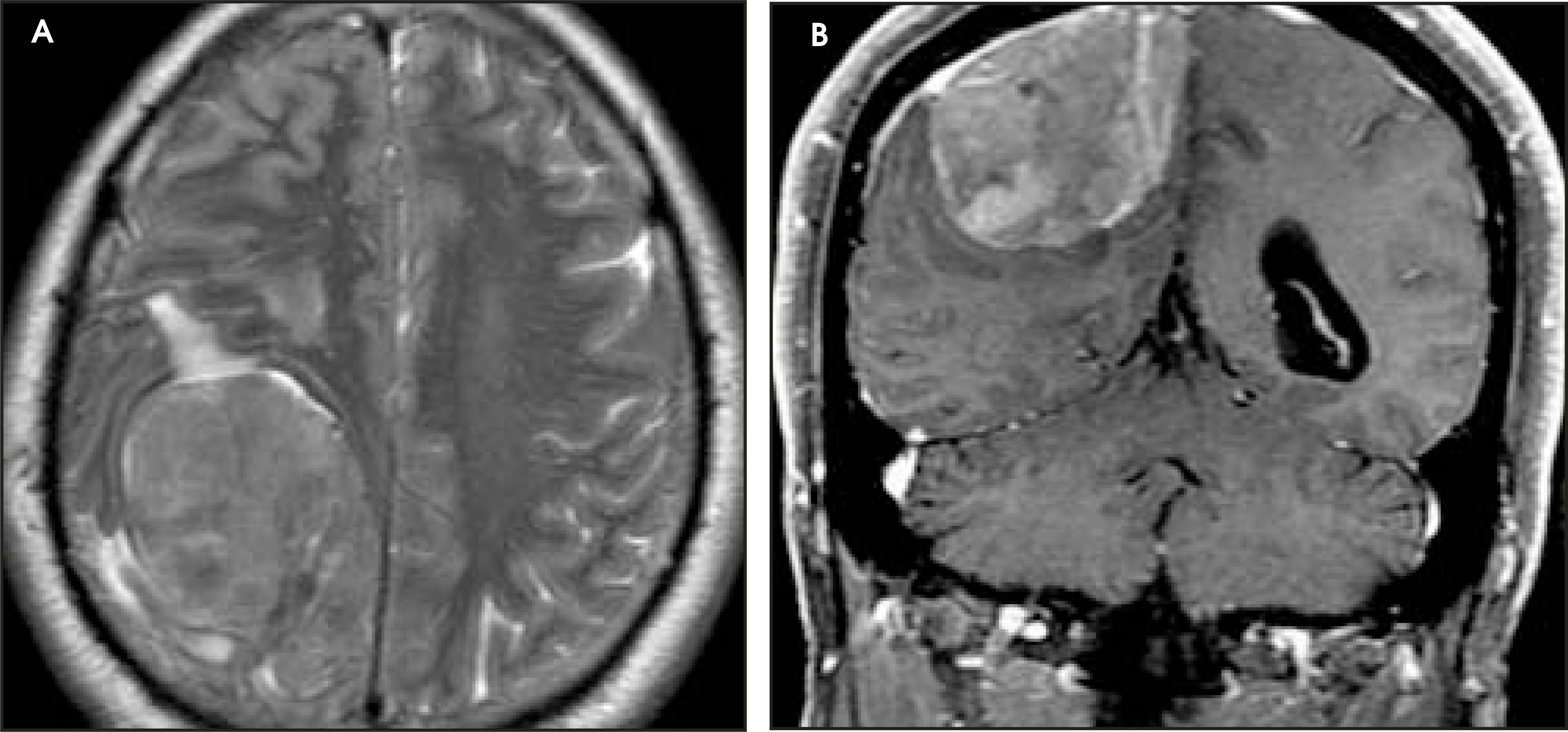
Secondary CNS lymphoma—Secondary CNS lymphoma involves the leptomeninges, more commonly than pachymeninges, resulting in meningeal enhancement. Hydrocephalus is commonly observed due to obstruction of the pacchionian granulations. Non-Hodgkin’s B-cell lymphoma is much more common than is Hodgkin’s lymphoma. Imaging characteristics are similar to those of primary CNS lymphoma, showing hyperdensity on CT, decreased signal intensity on T2-weighted MR imaging, and avid enhancement (Figure 8).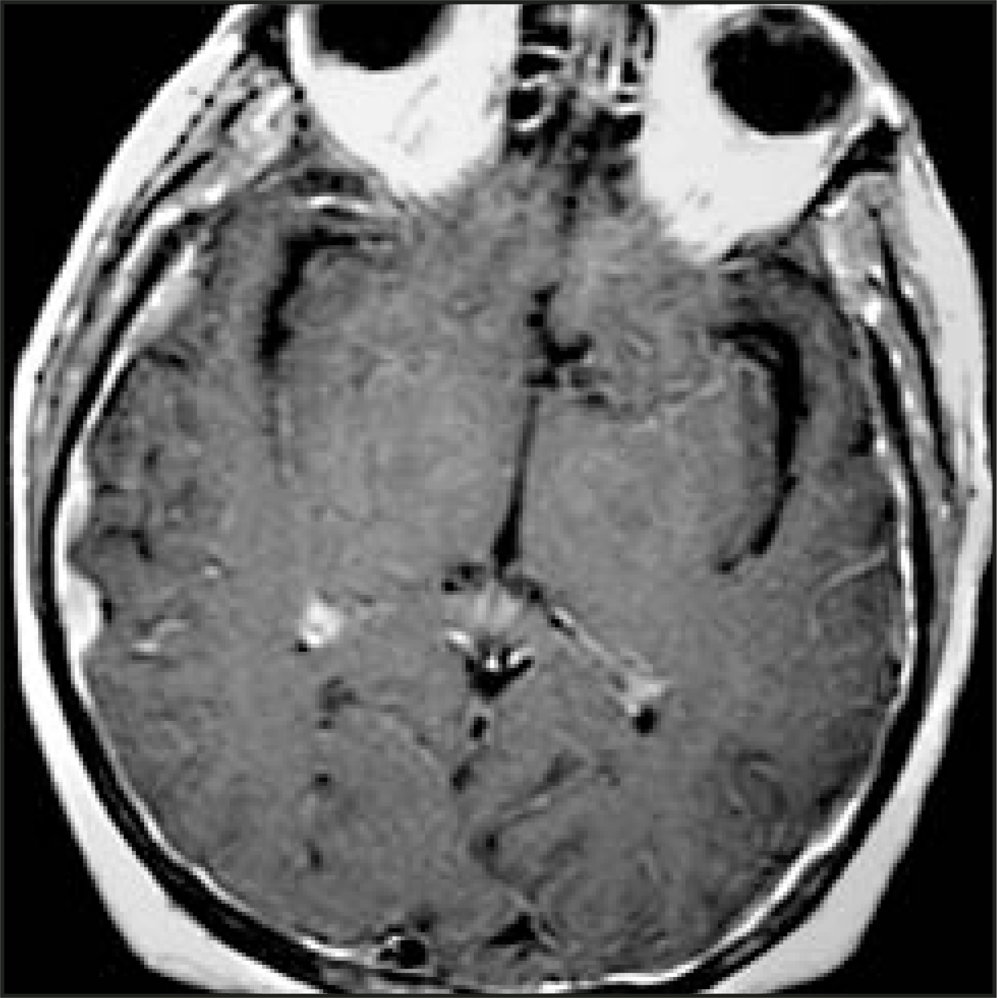
Pediatric neoplasms
Gliomas are the most commonly observed supratentorial neoplasms in the pediatric population. Unlike those in adults, pediatric gliomas are usually of the astrocytic variety and in children (in particular, those >2 years of age) they are typically low in grade. Imaging findings are similar to those described in the adult section, and, therefore, will not be discussed further in this section.
A distinct group of lesions occurring almost exclusively in children is worthy of individual discussion. Ganglioglioma, gangliocytoma, primary cerebral neuroblastoma, and pineal tumors account for most supratentorial neoplasms in the pediatric population. Dysembryoplastic neuroepithelial tumor (DNET) and pleomorphic xanthoastrocytoma are additional, less common, possibilities. Since extra-axial pediatric neoplasms are exceedingly rare, only intra-axial lesions will be discussed.1
Intra-axial neoplasms
Ganglioglioma—Ganglioglioma is a benign, slowly growing neoplasm that contains both neoplastic neuronal and glial components and, therefore, can undergo malignant degeneration. Approximately 60% to 80% of cases occur in individuals under the age of 30. In particular, this diagnosis should be considered in a patient with long-standing seizure disorder and a cortically based lesion in the temporal lobe. Forty percent to 50% of cases are cystic, and 30% contain calcification. MR imaging characteristics are variable, with approximately half demonstrating mild peripheral, focal, or nodular enhancement. Scalloping of the overlying calvarium may be seen, related to pressure erosion by the slowly growing mass.
Gangliocytoma—Gangliocytomas lack a glial component, and cannot undergo malignant degeneration. These lesions typically occur in the cerebral cortex or cerebellum. When in the cerebellum, the abnormality is known as Lhermitte-Duclos disease. Rather than a true neoplasm, it likely represents dysplasia. This lesion is isointense on T1- and T2-weighted MR imaging, and hyperintese on FLAIR or proton-density– weighted imaging.7
Primary cerebral neuroblastoma— Also called primitive neuroectodermal tumor (PNET), this lesion is typically a large hemispheric mass that usually involves the frontal and parietal lobes. Neuroblastoma, medulloblastoma, and pineoblastoma arise from similar primitive cells and are, thus, in this family of lesions. Neuroblastoma demonstrates calcification, necrosis, hemorrhage, and cyst formation. Subarachnoid seeding may also occur. Most arise in children under the age of 10 years. CT and MR imaging demonstrate a heterogeneous mass with moderate contrast enhancement. Since the primitive cells that comprise this neoplasm have a high nuclear-to-cytoplasmic ratio, the lesion is characteristically of low signal intensity on T2-weighted MR imaging. This neoplasm is associated with a recurrence rate of approximately 40%.8
Pleomorphic xanthoastrocytoma— Pleomorphic xanthoastrocytoma is a benign, indolent tumor found in children and young adults. It is partially cystic with an avidly enhancing mural nodule found in a superficial cortical location. It frequently occurs in the temporal lobe.9
Dysembryoplastic neuroepithelial tumor—Dysembryoplastic neuroepithelial tumor (DNET) is a benign cortical tumor found classically in the temporal lobe causing complex partial seizures. It demonstrates low signal on T1-weighted MR imaging and high signal on T2-weighted sequences and may enhance with gadolinium. It is frequently associated with cortical dysplasia and may resemble a benign cyst (Figure 9).7,10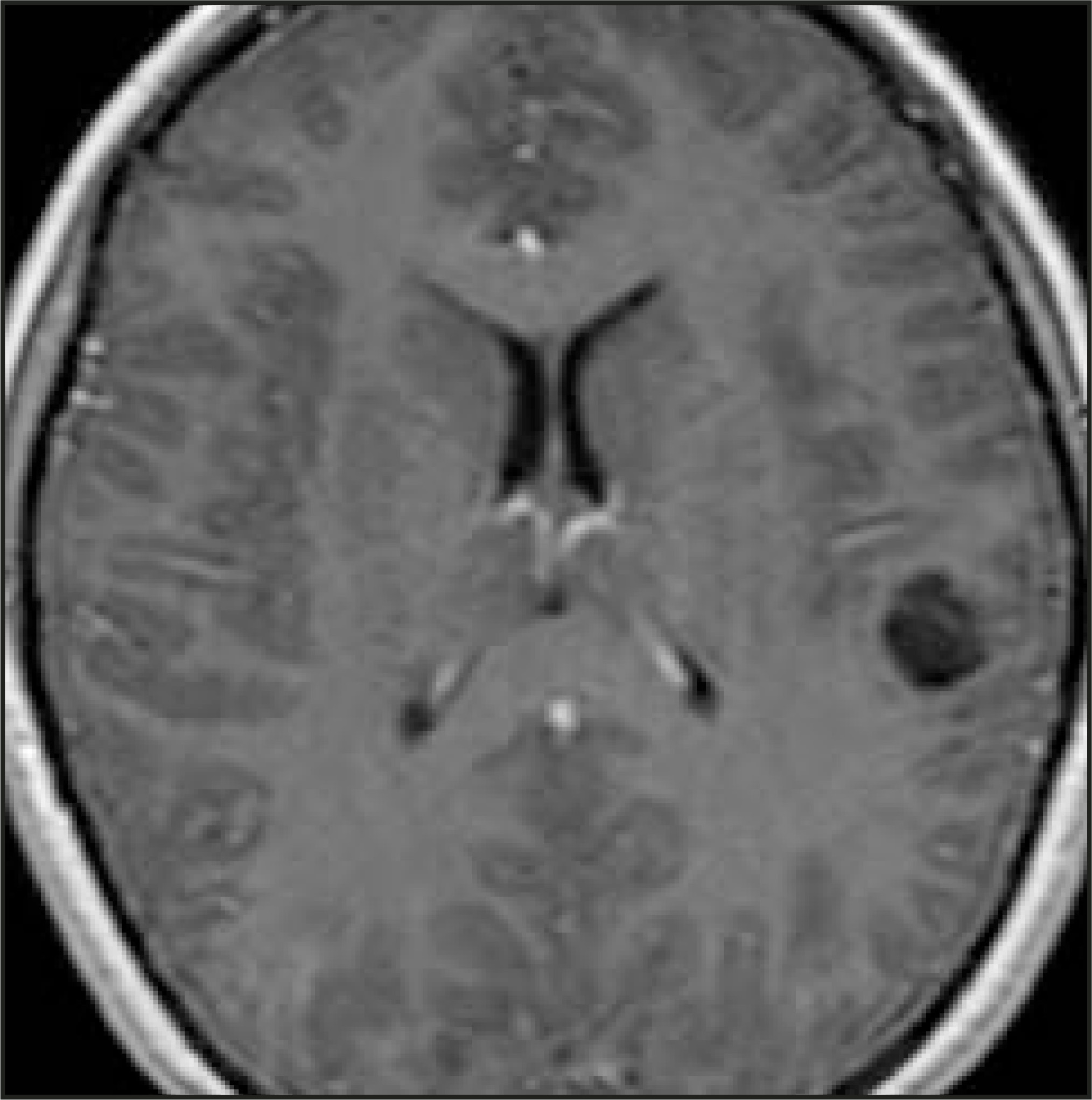
Pineal region masses—Pineal tumors are of germ cell origin (germinoma, choriocarcinoma, embryonal cell carcinoma, endodermal sinus tumor, teratoma) or pineal cell origin (pineoblastoma, pineocytoma). Other masses within this region include pineal and tectal glioma, cavernous hemangioma, meningioma, and benign cysts. Hydrocephalus is caused by compression on the posterior midbrain (tectum) and obstruction of the cerebral aqueduct. The tectal involvement can result in Parinaud’s syndrome, which is paralysis of upward gaze.
Pineal germ cell tumors: Pineal germ cell tumors are the most common neoplasm in the pineal gland; germinoma accounts for nearly 70% of all types of pineal germ cell tumors and 40% of all pineal region masses. Young men are almost exclusively affected. Due to its hypercellularity, this neoplasm is dense on CT and slightly hypointense on T2-weighted MR imaging. It demonstrates marked enhancement. Associated calcification of the pineal gland is frequently seen. Germinoma is sensitive to both radiation and chemotherapy. Due to its propensity for CSF seeding, contrast-enhanced MR imaging of the brain and entire spine should be performed to locate possible foci of metastatic subarachnoid spread.
Teratoma: Teratoma is the second most common pineal region tumor. It contains elements of fat, bone, calcification, and cysts. These components yield distinctive imaging characteristics on CT or MR imaging. Choriocarcinoma, embryonal cell carcinoma, and endodermal sinus tumor are less common pineal germ cell tumors. They are hemorrhagic neoplasms that occur more frequently in men than in women. They carry a poorer prognosis than do germinomas. Serum markers are useful in characterizing pineal germ cell tumors. These markers are summarized in Table 2.
Pineal cell tumors: Pineal cell tumors include pineoblastoma and pineocytoma. Pineoblastoma is a PNET, representing 15% of pineal region tumors. It demonstrates a similar incidence in men and women. Pineoblastoma occurs in a slightly younger age group than does pineocytoma, is slightly more invasive, and exhibits a higher rate of CSF seeding. Their imaging characteristics, however, are generally similar. On CT, both are hyperdense, demonstrate marked contrast enhancement, and engulf the pineal calcification. Pineoblastoma is typically larger in size than pineocytoma, and occasionally the pineal calcification may appear “exploded.” Being composed of primitive cells, pineoblastoma shows distinctive low signal intensity on T2-weighted MR imaging (Figure 10). Melatonin is a serum marker for pineoblastoma.11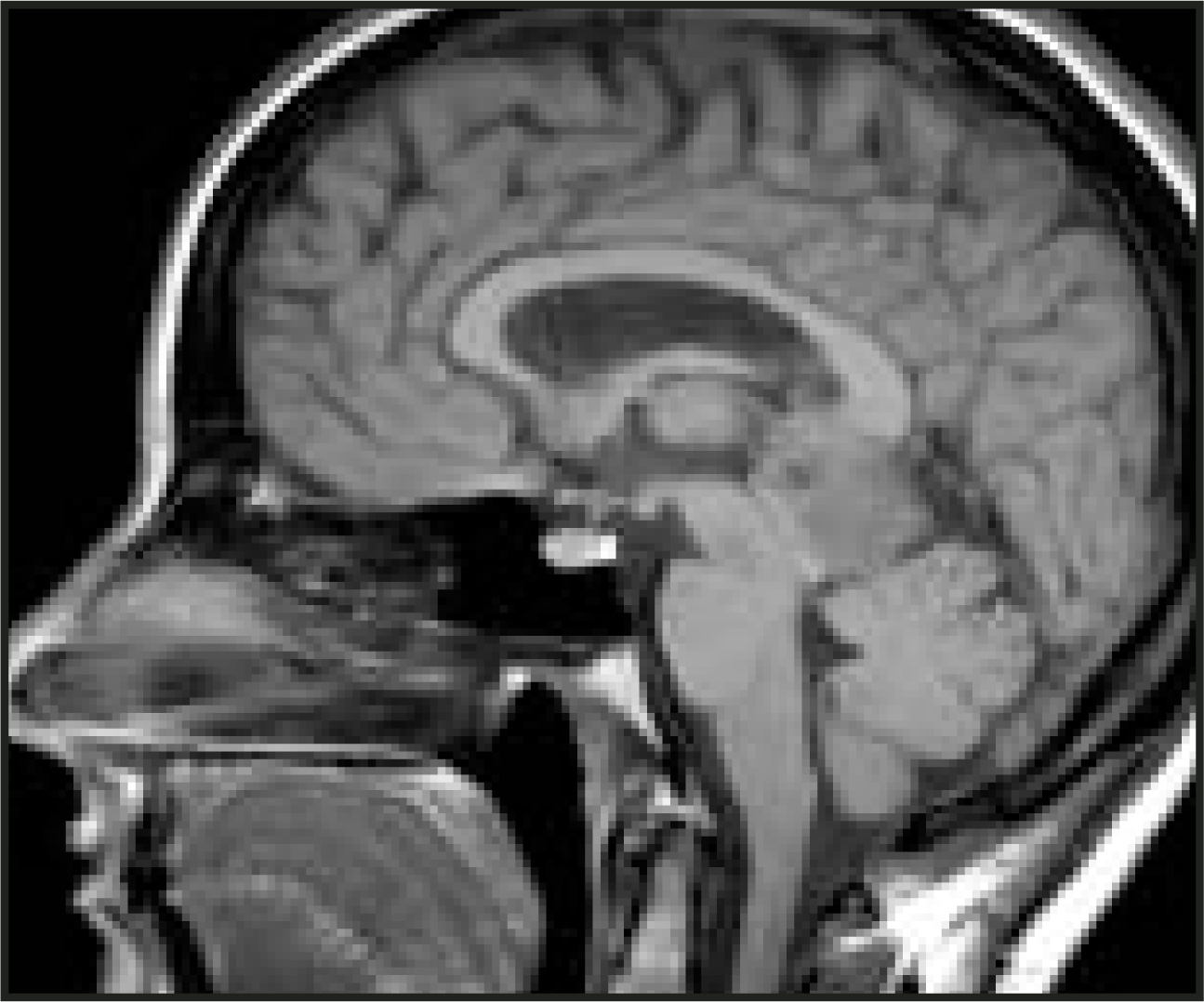
INFRATENTORIAL NEOPLASMS Adult neoplasms
In the adult population, infratentorial neoplasms occur less frequently than do supratentorial lesions. Within this compartment, extra-axial masses are most common, and intra-axial infratentorial neoplasms are somewhat rare; therefore, the extra-axial neoplasms will be discussed first.
Extra-axial neoplasms
Extra-axial infratentorial neoplasms can be separated into those that involve the foramen magnum and those that involve the cerebellopontine angle. Neoplasms that affect the foramen magnum are limited to meningioma and schwannoma. Cerebellopontine angle masses additionally include epidermoid, arachnoid cyst, metastasis, and lipoma. As previously discussed, lymphoma and carcinomatous meningitis are considerations as well.
Schwannoma—Schwannoma originates from perineural Schwann cells. It may be divided histologically into two tissue types: Antoni type A and Antoni type B. Antoni type A displays densely packed neural and fibrous tissue, while Antoni type B displays a looser tissue structure. As a result, Antoni type A is hypointense on T2-weighted MR imaging, while Antoni type B is hyperintense. Schwannoma is sporadic and multiple in NF-2, affecting cranial nerve (CN) VIII far more frequently than CN V, VII, or other cranial nerves. Bilateral CN VIII schwannomas are pathognomonic for NF-2. While schwannoma usually shows enhancement, it demonstrates heterogeneous enhancement in approximately one third of cases, and peritumoral edema is noted in only one third of cases.
Vestibular schwannoma is the most common neoplasm in the cerebellopontine angle, accounting for 75% of masses in this region. Since the internal auditory canal (IAC) is involved (80%), flaring of the porous acousticus, expansion of the IAC, and an acute angle with the dura can be seen (Figure 11). A coexistent arachnoid cyst may be present in 7% to 10% of cases.12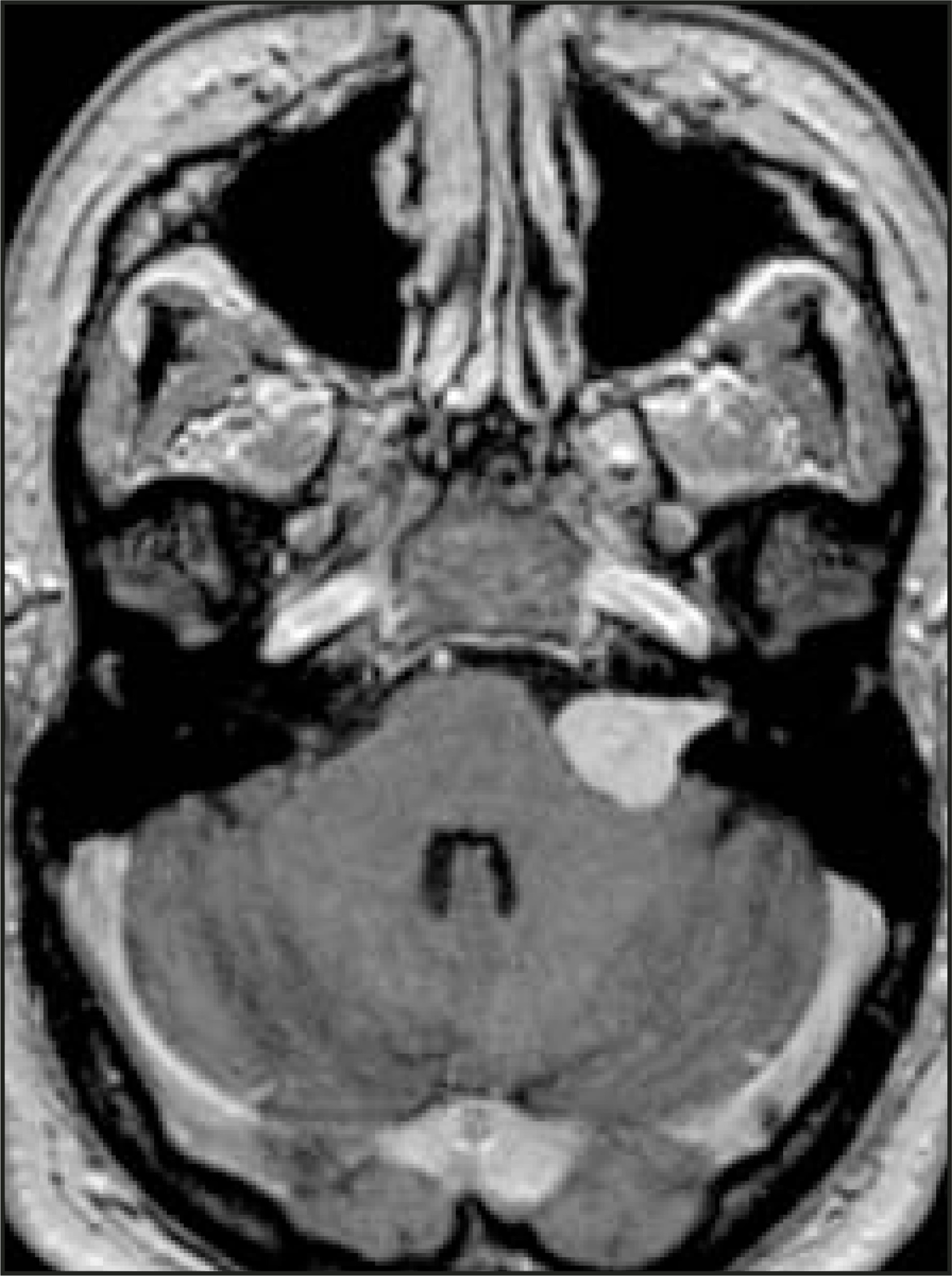
Meningioma—In the posterior fossa, meningioma is the second most common extra-axial neoplasm (10%), schwannoma being the most common (75%). It is usually located at the cerebellopontine angle; however, it can also be found at Meckel’s cave, along the clivus, or at the foramen magnum. While benign, this neoplasm can result in significant morbidity due to its location adjacent to vital primitive neural structures. Imaging characteristics are similar to the supratentorial lesions. Occasionally, differentiation from schwannoma at the cerebellopontine angle can be challenging; however, specific imaging characteristics can help identify the appropriate etiology of the mass. A dural tail, the presence of bony reaction, and an obtuse angle with the dura, as well as the center of the lesion being located away from the IAC, suggest meningioma (Figure 12). Meningioma is also frequently hyperdense on noncontrast CT imaging.6 These and additional differentiating features are shown in Table 3.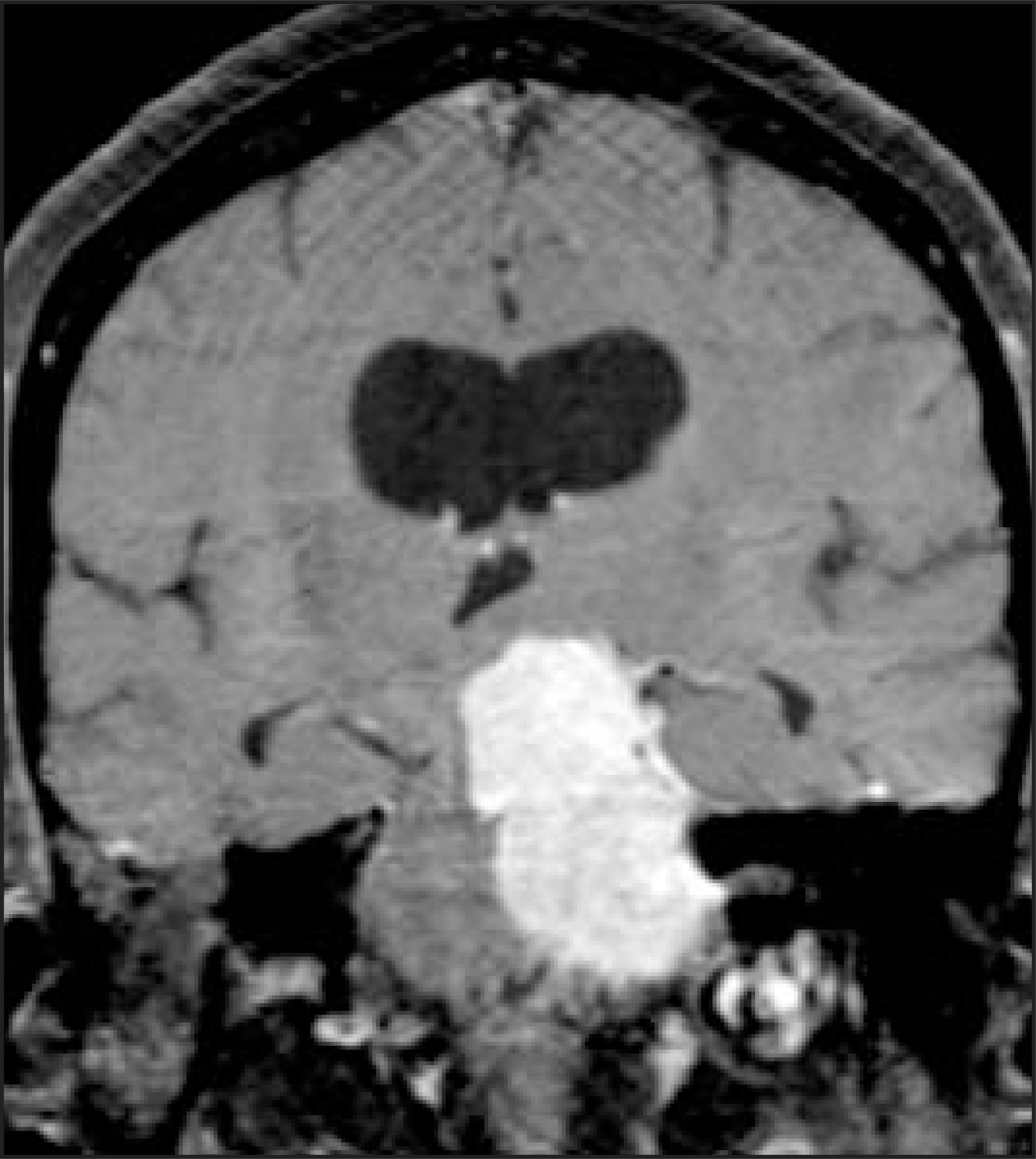
Epidermoid—Epidermoid is an inclusion cyst from ectodermal origin, which is lined by squamous epithelium and contains keratin. It may present with cranial neuropathy in adulthood, due to its slow growth. It is seen in men and women with equivalent frequency, and should be differentiated from schwannoma and meningioma due to its lack of enhancement. While epidermoid could be confused with arachnoid cyst (since both these entities are of low density on CT, are low T1- and high T2-weighted signal intensity on MR imaging, and do not show enhancement), epidermoid can be differentiated from arachnoid cyst on the basis of several imaging features.
Epidermoid demonstrates low density on unenhanced CT but is usually slightly more dense than CSF. Calcification is present in 25% of cases. It is characterized more clearly with MR imaging, which demonstrates scalloped margins and vascular encasement, unlike arachnoid cyst, which demonstrates smooth, rounded margins and vascular displacement. Furthermore, arachnoid cyst follows CSF signal on all sequences on MR imaging, while epidermoid deviates from CSF signal on proton-density–weighted or FLAIR images, showing hyperintensity on these sequences. Epidermoid, being composed of solid tissue, characteristically demonstrates restricted diffusion on diffusion-weighted MR imaging, unlike arachnoid cyst, which is purely liquid and does not demonstrate restricted diffusion.13 These differences are summarized in Table 4.
Intra-axial neoplasms
In the adult population, intra-axial infratentorial neoplasms primarily involve the cerebellar hemispheres, with the most common lesions being metastasis and hemangioblastoma. Glioma and medulloblastoma occur rarely, accounting for 1% of posterior fossa tumors in the adult population. Infrequently, lesions may arise in the fourth ventricle, including subependymoma, ependymoma, and choroid plexus papilloma.
Metastases—As in the supratentorial compartment, metastases are the most common infratentorial intra-axial neoplasms in the adult population. Similar to the supratentorial lesions, they are usually well-defined round masses, often with nodular or ring enhancement (due to central necrosis) (Figure 13). Multiple masses are usually seen and help confirm the diagnosis. However, even if solitary, the lesion is still more commonly metastasis than a primary intracranial neoplasm. Lung and breast carcinoma account for the majority of metastatic disease, followed by melanoma, renal cell carcinoma, thyroid carcinoma, and gastrointestinal malignancies. Metastases are typically hypodense on CT; they are hypointense on T1-weighted and hyperintense on T2-weighted MR imaging. Hemorrhage and calcification are two common variations that can yield variable signal on T2-weighted MR imaging. Enhancement is the rule, and contrast should be administered in all suspected cases.1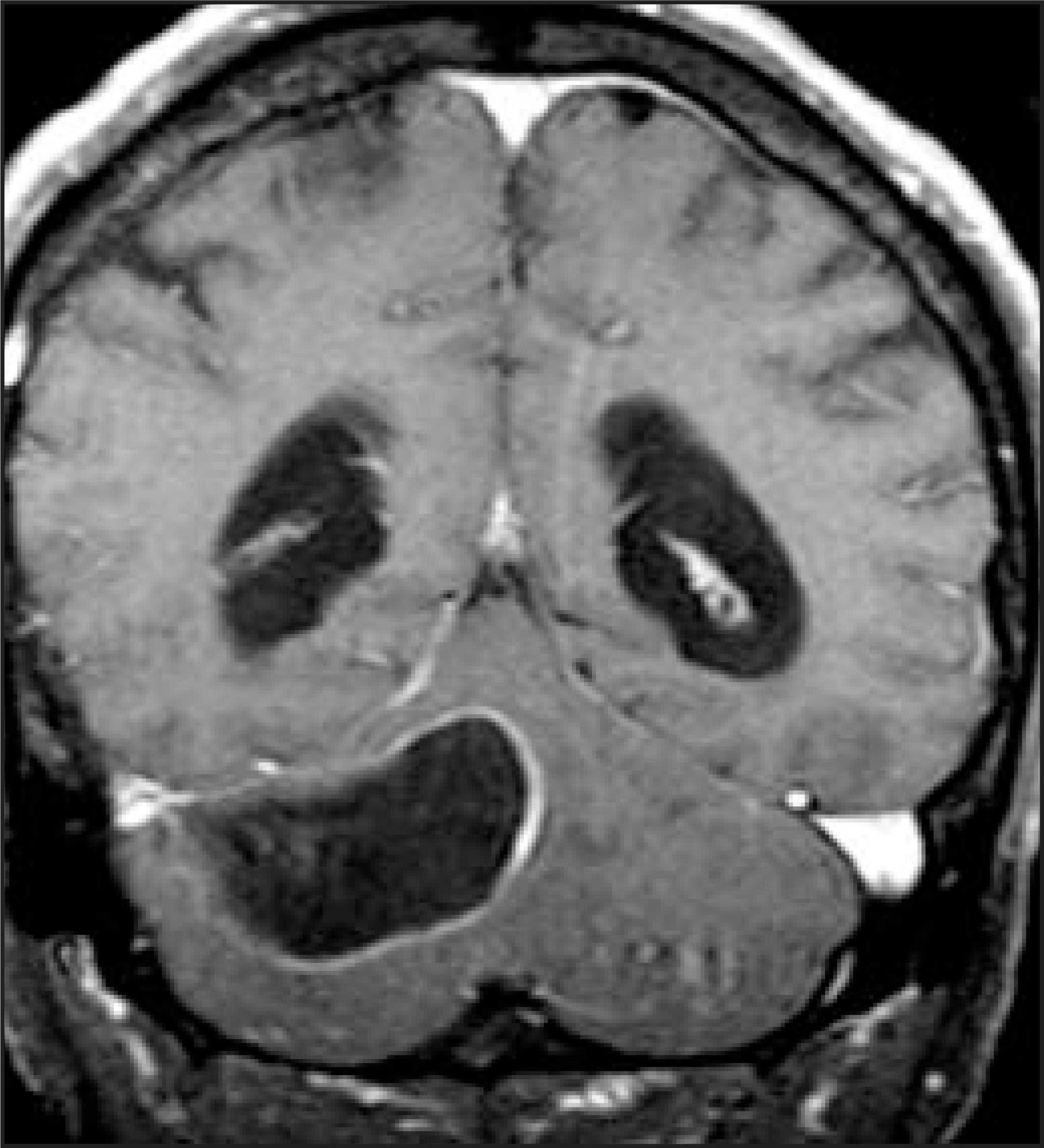
Hemangioblastoma—Hemangioblastoma represents the most common primary infratentorial intra-axial neoplasm in the adult population. This is a highly vascular benign neoplasm for which surgery is curative with complete excision. Eighty-three percent of hemangioblastomas are located in the cerebellum, followed by the spinal cord (10%), medulla (5%), and cerebrum (2%). They are more common in young men, often presenting with headache, ataxia, nausea/vomiting, vertigo, or polycythemia (since the masses may produce erythropoietin).
In approximately 55% of cases, hemangioblastoma demonstrates the classic appearance of a cystic component with an avidly enhancing mural nodule, which is difficult to distinguish from pilocytic astrocytoma (Figure 14). Patient age aids in the differentiation of these lesions, as does the lack of enhancement of the cyst wall in hemangioblastoma. Purely solid lesions are less common but occur in 45% of cases, potentially leading to the misdiagnosis of cerebellar glioma, which is less likely in the adult population. Hemangioblastoma is associated with von Hippel-Lindau (VHL) disease in approximately 20% of cases, and 45% of patients with VHL disease will develop hemangioblastoma. The hemangioblastomas in VHL disease are frequently multiple and are also found in the retina as well as the spine and medulla.14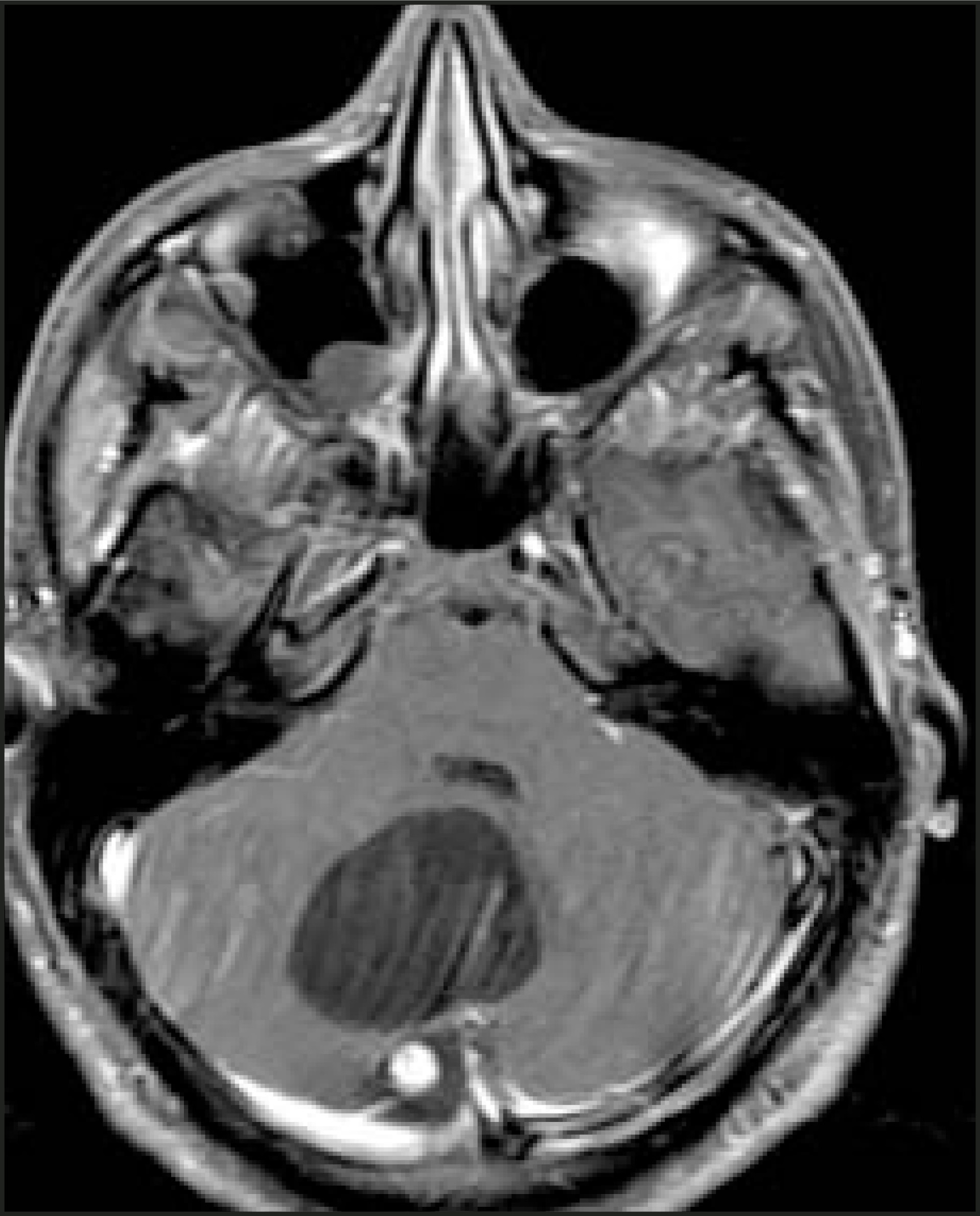
Pediatric neoplasms
Among children, infratentorial neoplasms account for 48% of all intracranial masses. These tumors, which are especially common between the ages of 2 and 10, are essentially all intra-axial. Any structure within the posterior fossa may be involved. The most common lesions are pilocytic astrocytoma, medulloblastoma, ependymoma, and brainstem glioma. Once again, as extra-axial pediatric neoplasms are exceedingly rare, only intra-axial lesions will be discussed.
Intra-axial neoplasms
Pilocytic astrocytoma—Pilocytic astrocytoma is the most common infratentorial neoplasm in the pediatric age group. This tumor is cystic in 60% to 80% of cases. Being the least aggressive form of glioma (WHO Grade I), the overall prognosis for pilocytic astrocytoma is very good, with a 5-year survival rate >90%. In a minority of cases, especially in older children, it may be solid or infiltrating. Such cases are associated with a much poorer prognosis. Pilocytic astrocytoma may occur in the hypothalamic/chiasmatic region, where it is associated with neurofibromatosis type 1 (NF-1).
Classically, pilocytic astrocytoma is a cystic lesion, which is isointense to CSF on T1- and T2-weighted imaging with an avidly enhancing mural nodule. It is typically located off-midline in the cerebellar hemisphere, with mild enhancement of the thin cyst wall (Figure 15). Calcification is uncommon.1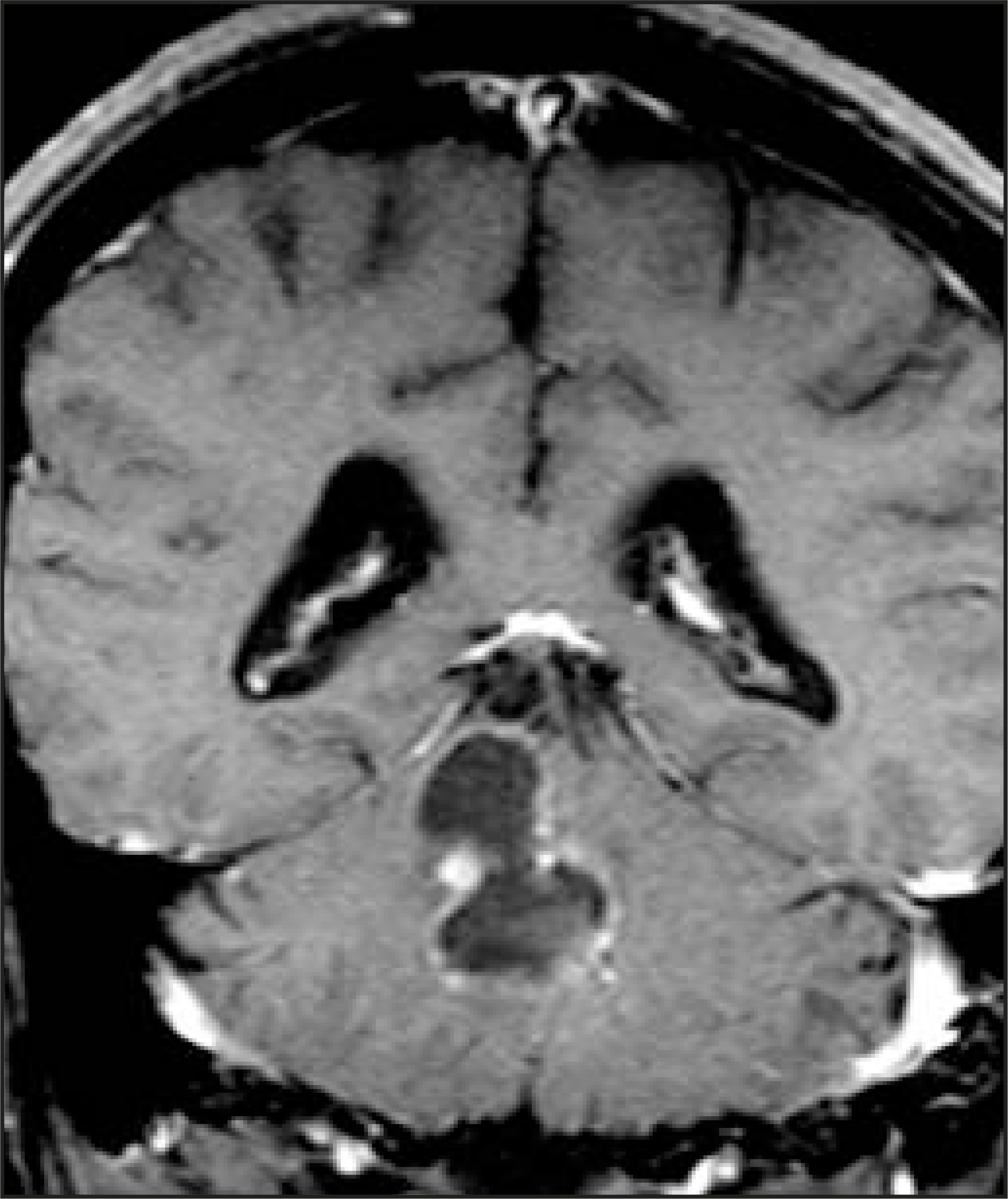
Medulloblastoma, PNET—Medulloblastoma comprises approximately 30% of all pediatric posterior fossa tumors. It is a midline mass associated with the cerebellar vermis. It arises from the superior medullary velum and distorts and compresses the fourth ventricle, often presenting with hydrocephalus. Subarachnoid spread along the leptomeninges is common, occurring in 15% to 40% of cases. As a result, screening MR examination of the spine should be performed to search for metastases. Since the primitive “small blue” cells that comprise the lesions have a high nuclear-to-cytoplas-mic ratio, medulloblastoma is typically hyperdense on noncontrast CT and demonstrates intermediate to low signal intensity on T2-weighted MR imaging. Moderate, heterogeneous enhancement is seen with contrast administration. Calcification is uncommon.
While usually seen in the pediatric population, medulloblastoma can also occur rarely in young adults. Unlike its pediatric counterpart, it is typically cystic rather than solid. It is most often found within the cerebellar hemispheres rather than the midline; in this circumstance, it usually demonstrates a more aggressive course.15
Ependymoma—Ependymoma arises most commonly from within the fourth ventricle, expanding the fourth ventricle as it grows, allowing distinction from medulloblastoma. Unlike medulloblastoma, calcification is common, occurring in 40% to 50% of cases. Ependymoma tends to be more poorly defined and commonly spreads via the foramina of Magendie and Luschka, further differentiating the two neoplasms (Figure 16). Like medulloblastoma, however, ependymoma may present with hydrocephalus. Imaging characteristics are not distinctive compared with most neoplasms, being hypointense on T1- and hyperintense on T2-weighted MR imaging, with avid enhancement. Again, however, calcification is commonly seen on CT.16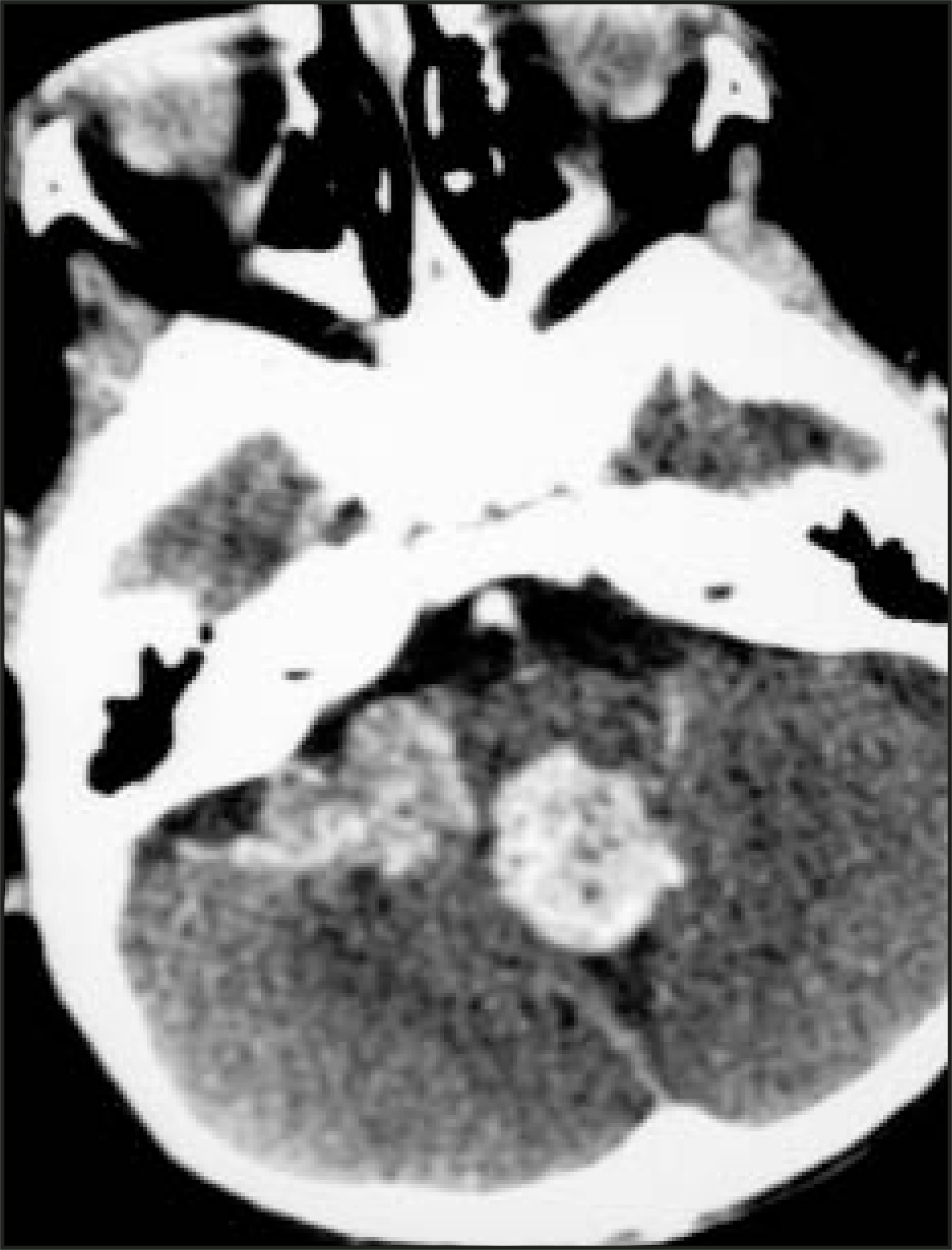
Brainstem glioma—Brainstem glioma is a slow-growing neoplasm that accounts for 20% of posterior fossa masses in children. It is an infiltrative lesion that presents later in childhood than do most pediatric posterior fossa neoplasms. It may be isolated to the brainstem, notably affecting the pons, or it may demonstrate an exophytic pattern. The enhancement pattern of brainstem glioma is variable, often demonstrating only subtle, if any, enhancement. It demonstrates high signal on T2-weighted or FLAIR MR images, which are the best imaging sequences for visualizing this lesion (Figure 17). Due to the infiltrative nature of brainstem glioma, surgical treatment is typically precluded; therefore, although usually noncurative, radiation therapy is the recommended treatment of choice.1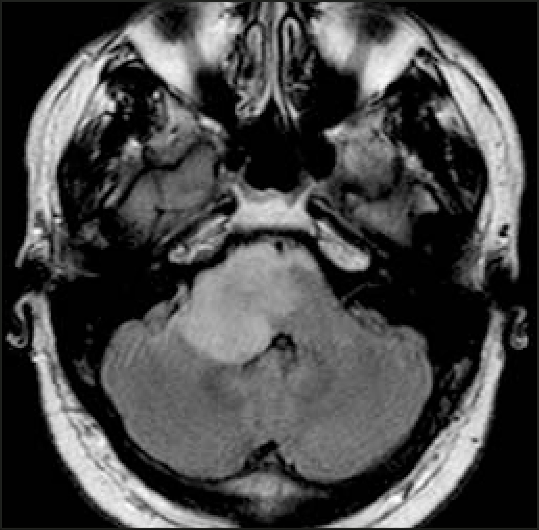
Conclusion
Many neoplastic lesions can occur in the intracranial compartment. In order to help the referring clinician make an accurate diagnosis, radiologists must anwer several essential questions: 1) adult or pediatric patient, 2) supratentorial or infratentorial compartment, and 3) intra-axial or extra-axial location. With a logical approach and the answers to these basic questions, the differential diagnostic possibilities can be limited to generate a complete, yet not overwhelming, list for the referring clinician.
References
- Grossman R, Yousem D. Neuroradiology: The Requisites. 2003:97-172.
- Del Carpio-O’Donovan R, Korah I, Salazar A, Melancon D. Gliomatosis cerebri. Radiology. 1996;198:831-835.
- Altman N, Purser R, Post M. Tuberous sclerosis: Characteristics at CT and MR imaging. Radiology. 1998;167:527-532.
- Dwyer K, Naul L, Hise J. Gliosarcoma: MR features. J Comput Assist Tomogr. 1996;20:719-723.
- Schwaighofer B, Hesselink J, Press G. Primary intracranial CNS lymphoma: MR manifestations. AJNR Am J Neuroradiol. 1989;10:725-729.
- Sheporaitis L, Osborn A, Smirniotopoulos J. Intracranial meningioma. AJNR Am J Neuroradiol. 1992;13:29-37.
- Shin J, Lee H, Khang S. Neuronal tumors of the central nervous system: Radiologic findings and pathologic correlation. RadioGraphics. 2002;22:1177-1189.
- Davis P, Wichman R, Takei Y, Hoffman J. Primary cerebral neuroblastoma: CT and MR findings in 12 cases. AJNR Am J Neuroradiol. 1990;11:115-120.
- Lipper M, Eberhard D, Phillips C. Pleomorphic xanthoastrocytoma, a distinctive astroglial tumor: Neuroradiologic and pathologic features. AJNR Am J Neuroradiol. 1993;14:1397-1404.
- Ostertun B, Wolf H, Campos M. Dysembryoplastic neuroepithelial tumors: MR and CT evaluation. AJNR Am J Neuroradiol. 1996;17:419-430.
- Chang T, Teng M, Guo W, Sheng W. CT of pineal tumors and intracranial germ-cell tumors. AJNR Am J Neuroradiol. 1989;10:1039-1044.
- Zagardo M, Castellani R, Rees J. Radiologic and pathologic findings of intracerebral schwannoma. AJNR Am J Neuroradiol. 1998;19:1290-1293.
- Horowitz B, Chari M, James R, Bryan R. MR of intracranial epidermoid tumors: Correlation of in vivo imaging with in vitro 13C spectroscopy. AJNR Am J Neuroradiol. 1990;11:299-302.
- Lee S, Sanches J, Mark A. Posterior fossa hemangioblastomas: MR imaging. Radiology. 1989;171:463-468.
- Koeller K, Rushing E. From the archives of the AFIP: Medulloblastoma: A comprehensive review with radiologic-pathologic correlation. RadioGraphics. 2003;23:1613-1637.
- Spoto G, Press G, Hesselink J, Solomon M. Intracranial ependymoma and subependymoma: MR manifestations. AJNR Am J Neuroradiol. 1990;11:83-91.
Citation
. Supratentorial and infratentorial brain neoplasms. Applied Radiology. 2004;33(11):23-33. doi:10.37549/AR1294.