Technology of computer-aided detection
Applied Radiology — Vol. 33 , Issue 9 , pp. 30 -37
DOI: 10.37549/AR1277
Published: September 1, 2004
Categories
Researchers began to experiment with using computers to assist in the diagnosis of disease, particularly cancer, in the early 1980s. Unfortunately, the computers in use at that time were not powerful enough to perform such complex tasks in a clinically feasible manner. By the late 1990s, however, computer technology had improved, and computer-aided detection (CAD) became a reality. In June 1998, the United States Food and Drug Administration (FDA) officially opened the doors to CAD for breast cancer when it granted marketing approval to R2 Technology, Inc. (Los Altos, CA) for the ImageChecker M1000. Initially approved for use only in film-based screening mammography, the use of CAD expanded in May 2001 with its approval for use in diagnostic mammography. In January 2002, two additional CAD systems were cleared for marketing: Second Look (CADx Medical Systems, Inc., Northborough, MA) and MammoReader (Intelligent Systems Software, Inc., Clearwater, FL). In July 2002, Intelligent Systems Software merged with film digitizer vendor Howtek (Hudson, NH) to become iCAD, Inc. (Nashua, NH). In December 2003, iCAD acquired CADx, leaving two major players in the current American CAD mammography market.
In April 2002, GE Medical Systems (now GE Healthcare, Waukesha, WI) received FDA approval to modify their Senographe 2000D Full-Field Digital Mammography System (FFDM) to allow the integration of a CAD component, thereby bringing CAD to digital mammography and film-based systems.
The biggest boost to the use of CAD, however, came in April 2001 when the U.S. Centers for Medicare and Medicaid Services (CMS) began to offer reimbursement for this procedure. The current rate of reimbursement for the use of CAD in screening mammography, established in March 2003, is $19.13 for global CAD mammography (CPT code 76085). The professional component (modifier 26) is $3.31 and the technical component (modifier TC) is $15.82. For CAD use in diagnostic mammography, the rate is $28.31 ($3.31 professional and $25.00 technical).1 On April 1, 2003, CMS began reimbursing for CAD with digital mammography at the same rates as film mammography.
“Medicare has been very accepting of this technology, and that really has been a big help in getting the other insurance companies to follow suit, and I think it’s justifiable,” said Michael N. Linver, MD, Director of Mammography for X-Ray Associates of New Mexico, Albuquerque, NM. “The literature shows overwhelmingly that there is a benefit in using CAD, even for the most advanced readers of mammograms.”
CAD for mammography
According to the FDA, CAD is “intended to identify and mark regions of interest on standard mammographic views to bring them to the attention of the radiologist after the initial reading has been completed. Thus, the system assists the radiologist in minimizing observational oversights by identifying areas on the original mammogram that may warrant a second review.”2
Each CAD system uses proprietary computer image-analysis algorithms to search for characteristics of the microcalcifications and spiculations commonly seen in breast cancer. The system then ranks the findings and marks those areas that meet the programmed threshold of possible abnormalities. Each system is programmed differently for exactly which features it marks and how those areas are marked. Mammographic findings that might result in a CAD “mark” include clusters of bright spots that may indicate microcalcifications, possible masses, and patterns of dense regions with radiating lines.
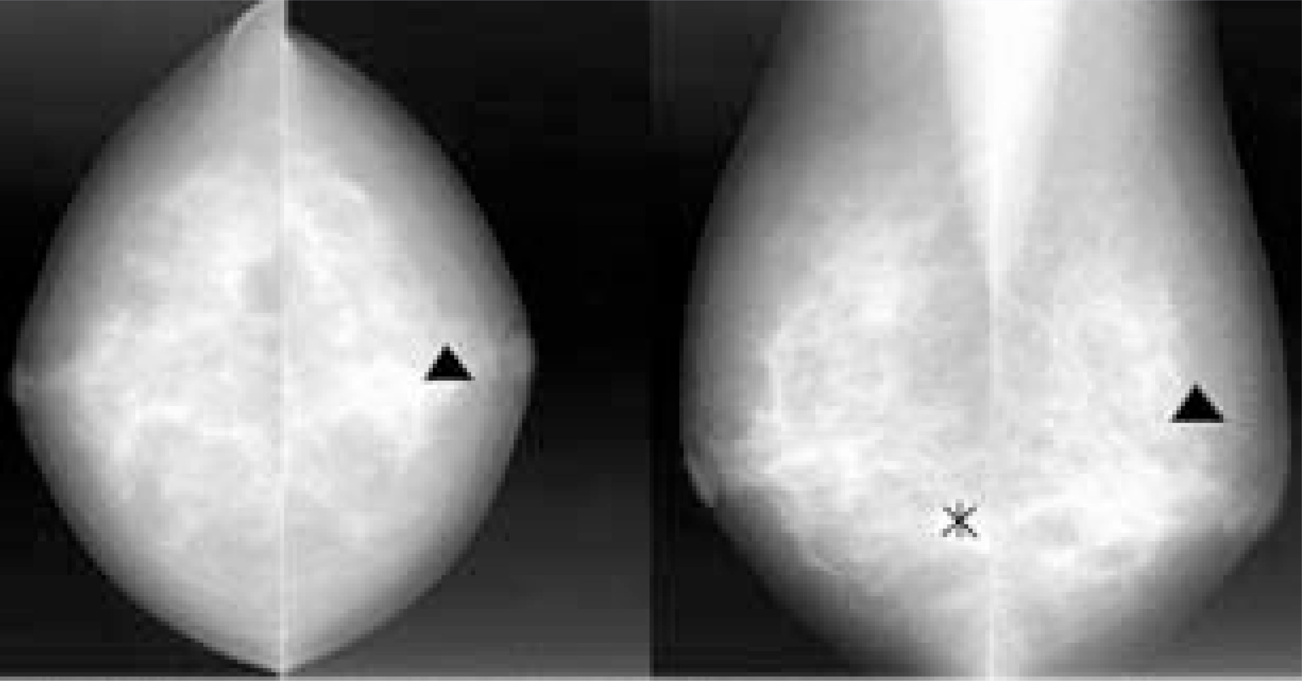
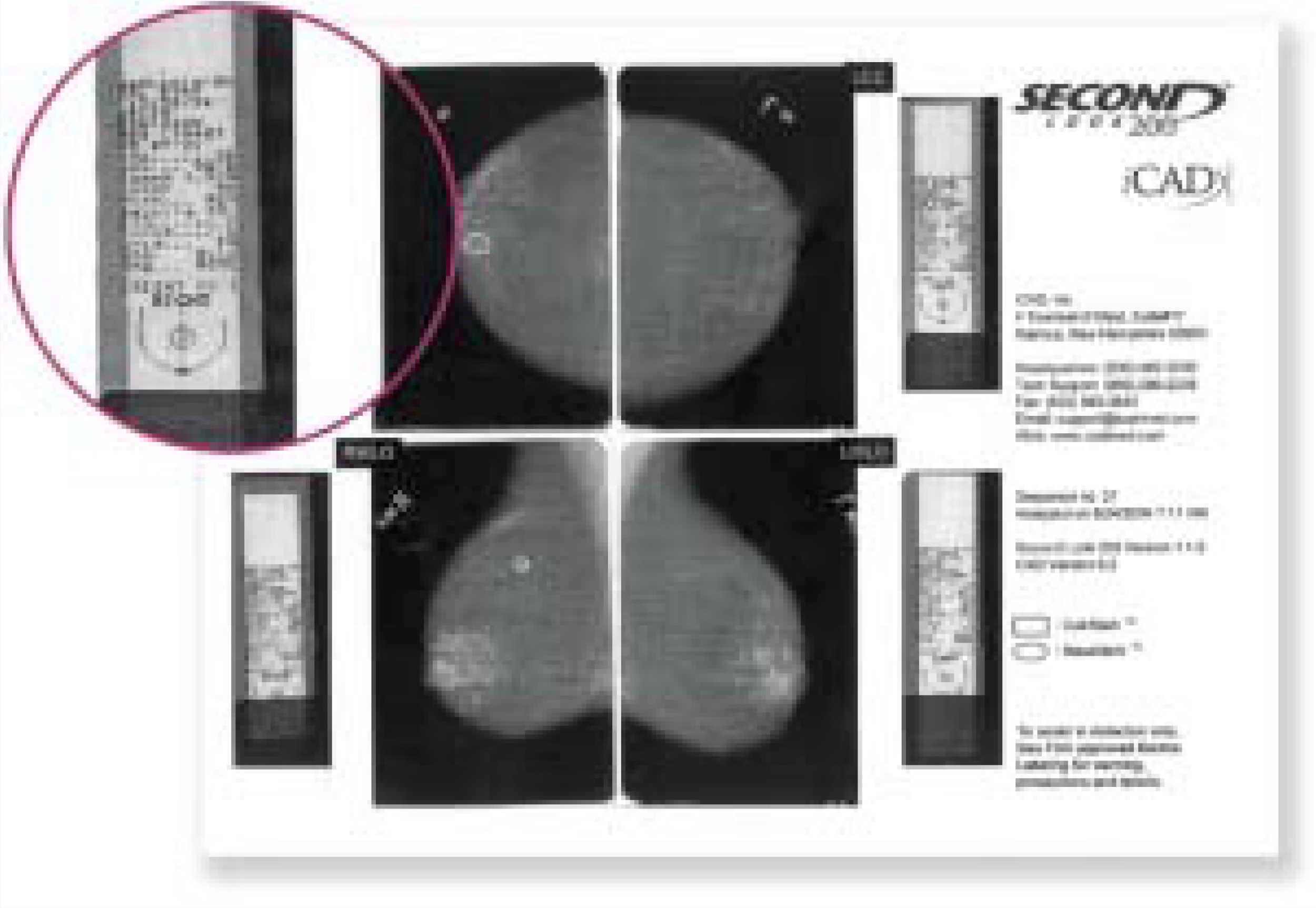
The CAD marks indicate specific areas that may need close attention by the radiologist. The CAD review can be displayed on a computer screen or printed out on a hard copy (Figure 1). Each system uses its own patented algorithms and symbols, such as triangles, squares, asterisks, or arrows, to indicate specific regions of interest (ROIs) (Figure 2). However, CAD review and marks do not alter the original mammographic images in any way.
Incorporating CAD into the radiology workflow
When using CAD with film-screen mammography, the images are acquired in the standard manner. Once finished, the films are fed into the processing unit to be digitized and processed by the algorithms. The digitized films are then sent to the viewer. The radiologist then reads the mammogram without any CAD markings and makes an interpretation. Once the reading is complete, the radiologist presses the button to activate the CAD display. This shows the search results on a monitor without marking the original film. The radiologist then reviews the ROIs indicated on the CAD review and updates the interpretation if necessary. Alternatively, the system can print out a hard-copy version of its findings, and the radiologist can refer to that rather than viewing the CAD results on screen.
With digital mammography, there are no hard-copy films to be digitized, so the initial soft-copy review is conducted as usual and then the CAD review is initiated. In all cases, however, it is essential that the radiologist fully read and interpret the findings before activating the CAD option or referring to the CAD findings.
The use of CAD in film-screen mammography has been shown to add 17 seconds of time for the radiologist and 1 minute, 21 seconds for the technologist. The added radiologist time is similar to the 16 seconds typically required to examine the mammogram with a magnifying glass.3
“We do not read mammograms online,” said Linver. “We batch-read our screening mammograms. We have the assistant hang the films and write down the result. This allows the reading radiologist to concentrate fully on the films, which increases accuracy and efficiency. We use the paper printout because by doing so we can read on all the multiviewers and are not tied to the multiviewer that has the CAD attached to it. The paper printouts then become part of the patient record,” he continued. “We read at the multiviewers, make up our minds, then look at the paper, and review the marks made by the CAD. Then we decide whether or not to change our initial im-pression. If we do change it, we do so right at that time. We always make our de-cisions first before we look at the CAD. I think if you try to do it any other way, your thinking is going to be overly colored, and you may overlook things that are not marked by the CAD.”
The value of CAD for mammography
A 2001 study of more than 12,800 screening mammography patients compared reading results with and without the use of CAD.4 The study found that the use of CAD increased the recall rate from 6.5% to 7.7% but did not change the positive predictive value for biopsy (38%). Overall, there was a 19.5% increase in the number of cancers detected and the proportion of early-stage malignancy detection (defined as stages 0 and I) rose from 73% to 78%. The authors concluded that “the use of CAD in the interpretation of screening mammograms can increase the detection of early-stage malignancies without undue effect on the recall rate or positive predictive value for biopsy.”4 This figure of a 20% increase in detected cancers is now widely accepted.
Interestingly, this increased rate of detection is higher than that found with double reading, which has been shown to increase sensitivity by approximately 10% when compared with single reading.5
“The sensitivities for CAD’s ability to find early breast cancers have increased dramatically,” said Linver. “Its overall sensitivity is roughly 90% now, with highest sensitivities being for calcifications. It almost never misses calcifications on the film, but it still has some way to go in finding small, early invasive cancers—those that either consist of a mass or an area of architectural distortion. It finds appproximately 84% to 86% of known masses when they are presented to the computer. Overall, it is doing remarkably well as an adjunctive tool to the radiologist.”
“I think it has the most trouble finding architectural distortions without a true mass,” Linver continued. “But then again, so do we as radiologists. CAD is very much a reflection of what we do. In fact, that is how CAD was taught to find what it finds: by feeding it images with specific objects that we as radiologists believed were abnormal. So that’s how it learns: from us. Therefore, the things that are most difficult for us to find are going to be the most difficult for CAD to find as well.”
Another advantage of CAD is that it is always available and always at peak performance, unlike human readers. “The nice thing about CAD is that it is available and ready for action 24/7, whereas radiologists are subject to the frailties of the human condition,” said Linver. “We are sharp sometimes and we’re not so good at other times. The machine is going to be rock solid anytime it’s used. So in that sense, it has the potential to be much better than we are.”
Drawbacks and pitfalls
As with many new technologies, the greatest barrier to widespread utilization lies in its cost. A CAD unit, depending on the model, can cost upwards of $200,000. Practices that perform a small volume of mammograms—and most mammograms are performed at facilities that do only a small volume of such studies each day—often will not be able to amortize that cost, even with the reimbursement that is currently available.
In addition, there is a learning curve with the use of this technology. “At first we are inclined to say, ‘If the computer finds it, then it must be significant,’” explained Linver. “But you have to remember that the computer is looking at these images in a vacuum and we are not. When we look at a mammogram, we have the advantage of being able to look at both views of each breast and make a decision based on both views. The computer has to make its decision on the basis of one view at a time. Not only that, it does not have the advantage of being able to review all the previous mammograms. So again, we have a tremendous advantage over the computer in our ability to sort out whether or not a marking is real, imaginary, or of no significance.” For this reason, the FDA-approved indications clearly state that the radiologist must interpret all images before reviewing the CAD data and the CAD information should be used only as an adjunct once the radiologist has formed his or her own clinical opinion.
Digital mammography and CAD
The use of CAD with digital mammography is very similar in its use with film-based systems, but there are a few key differences. With FFDM, the need to digitize films is eliminated because the original image is acquired in a digital format. This may increase accuracy by avoiding any artifacts that may arise during the film digitization process.
“I think it’s an ideal marriage to use digital CAD with FFDM because there is no chance of any lost information,” said Linver, “Every single time the CAD evaluates the digital image, it will get exactly the same answer. There is a much greater consistency in the CAD interpretation. That is one of the problems with film-based CAD. If you run the films through the digitizer and run the CAD algorithms on a few of them, you are not going to get exactly the same answer each time. It’s a little unnerving when that happens and you say, ‘Which is the right one?’ That is never going to happen with digital mammography.”
CAD for X-ray lung cancer detection
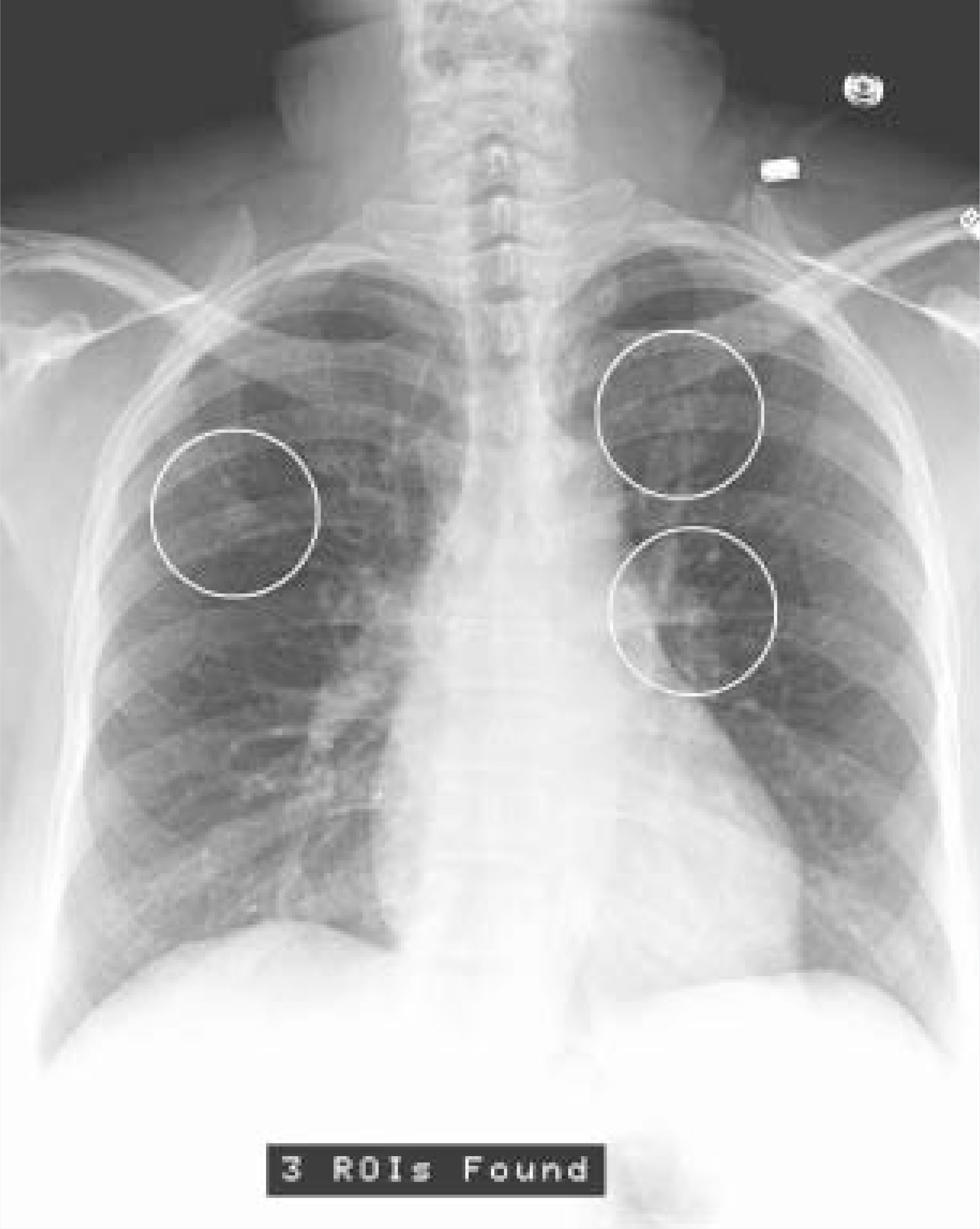
While mammography is clearly the dominant modality associated with CAD, this technology is also being used for the detection of lung cancer, both with analog and digital X-ray systems, as well as computed tomography (CT). In July 2001, the FDA approved the first CAD system for lung cancer detection, the RapidScreen RS-2000 from Deus Technologies (Rockville, MD). This stand-alone analog system digitizes existing chest X-rays and uses proprietary algorithms to mark circles to indicate ROIs that may
contain nodules indicating early-stage lung cancer. The results are shown on the display unit and a printout of the image is generated for inclusion with the original film. For the radiologist, the review process is similar to that used with mammography. The original films are read and, once a clinical opinion is formed, the CAD markings are consulted and the marked ROIs are re-examined by the radiologist.
Two years after the analog system was approved, the FDA granted Deus marketing approval for their Digital Imaging and Communications in Medicine (DICOM)-compatible digital X-ray CAD system, the RapidScreen RS-2000D. With this system, the CAD processing is performed in the background on a DICOM-formatted chest image. The CAD markings are either “burned in” to generate a new image or they can be set to become part of the original DICOM image. The images are sent to the picture archiving and communication system (PACS) server both before and after CAD processing. Once completed, the system sends the detection results to the PACS server where they are associated with the original study and available for viewing though the networked PACS workstation.
The digital system marks the ROIs (Figure 3) in the same manner as the analog system and adds a note at the bottom indicating the number of ROIs found as a backup to help ensure that the clinician does not miss any of the marked areas due to screen glare or poor contrast.
CAD for CT lung cancer detection
In July 2004, the FDA opened a new modality to CAD technology when it granted approval to R2 Technology to begin marketing the ImageChecker CT CAD Software system. This system was designed to provide CAD for solid lung nodules during review of multidetector CT chest exams.
The use of CAD with CT is also similar to the process established for digital mammography. “The current methodology for CAD, as suggested by R2 and agreed to by the FDA,” explained Pablo Delgado, MD, Associate Professor of Radiology at the University of Missouri, Kansas City, MO, “is that radiologists should look at the CT studies first and make up their mind or at least look for abnormalities without CAD. Subsequently, radiologists can activate the CAD or query the information of the CAD program that has been processed by the software and feed that information into a CAD report or CAD finding. Radiologists can then review those CAD findings and decide if they wish to either include CAD information that might add to their original findings or, possibly, refute the CAD findings by saying that they don’t agree with it.”
The value of CAD in CT lung scans
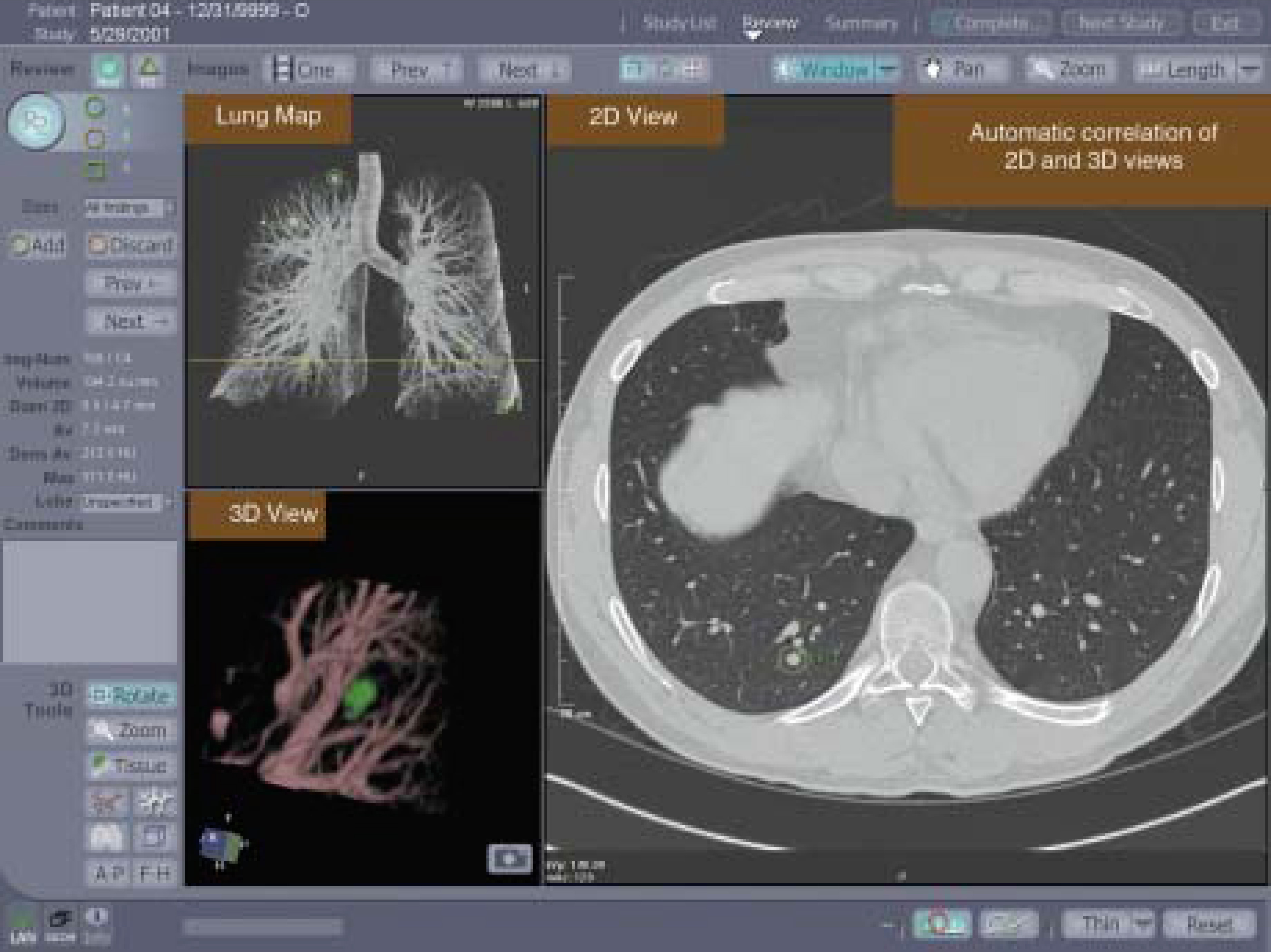
Designed to assist the radiologist in detecting lung nodules, the system’s proprietary algorithms use automatic measurement and characterization information to examine the study in three dimensions and mark potential ROIs for additional review by the radiologist (Figure 4).
Two additional software packages were also recently approved for use with this system. One provides automatic three-dimensional registration and the ability to track lung nodule progression or regression over time. The other was designed to help radiologists see and evaluate pulmonary emoboli and other filling defects in pulmonary arteries.
Dr. Delgado has been using the CAD CT system for lung screening as part of the beta testing process. “The CAD primarily locates solid nodules of a small-to-medium range (anywhere from 4 to 30 mm), nodules that are of approximately fluid or soft-tissue density, those with calcium density, and nodules that are spherical or ovoid, or some derivation thereof,” continued Delgado. “The algorithm is not currently de-signed to pick up ‘groundglass’ opacities, just solid nodules.”
“This technology is particularly useful in patients who are being worked up for a suspicious nodule,” continued Delgado, “and in those being followed up from a chest X-ray. Also, in patients with a known and sustained nodule that is being monitored—which is currently accepted for certain nodules due to their size, morphology, or any associated history or findings in the patient—CAD can be used to look for potential changes over time. I think that is where there is a lot of use with CAD—that it can give you some information that the radiologist’s eye may have more difficulty in seeing, such as the volume, density, and radius of the nodule. Just the fact that there are nodules in the lungs may not truly be all the information available. We want to know more about each individual nodule and, specifically, how it changes related to therapy, such as chemotherapy, radiation, and other treatments.”
Learning curve
As with CAD for mammography, there is a learning curve associated with CAD for CT lung scanning. “Radiologists’ acceptance of CAD, I think, hinges on their ability to change, just as they did with positron-emission tomography (PET) or MR imaging or any new modality,” explained Delgado. “CAD is basically just an extension of what we have already been doing. Radiologists need to take a small leap-of-faith to delve into the new world of CAD—a bunch of digital information from a software program—and somehow incorporate that into their clinical practices. With CT systems going from 4-slice to 8-slice to 16-slice to 64-slice, radiologists do not have time to scrutinize every single image to such extreme detail. They need to still look at the case diligently, but they need to accept that they might need a little bit of assistance, that they might overlook certain things, such as potential early lung cancers.”
As in the use of CAD with mammography, it is essential that radiologists not use CAD as a crutch. All images must be read and clinical opinions must be formed before the CAD data is considered. “Radiologists still need to be in control of their ultimate interpretations and their conclusions based on their own observations of the CAD information,” said Delgado. “I think the biggest caveat that people need to consider is that the CAD does not become this all-knowing computer brain that knows everything. CAD does have false-positives. There are certain clinical settings in which the radiologist may completely disagree with the CAD or override the CAD findings; and that’s perfectly legitimate. Radiologists should not become afraid or always follow CAD findings as if they were gospel.”
Other CAD applications
As CAD technology continues to evolve, additional applications will be developed. Currently, the use of CAD for CT colonography is in an advanced stage of investigation.
CT or “virtual” colonoscopy is an extremely data-intensive process, and the addition of CAD would help with the diagnostic review of CT images. CT CAD for the colon is being designed to use two- and three-dimensional volume-rendered data to locate and mark three-dimensional objects within the colon. These marks will then be displayed on the reconstructed colon landscape. Similar to other CAD applications, this system will use pre-programmed proprietary algorithms based on engineering measurements of the physical characteristics of interest to clinicians. Located objects will be compared with a library of similarly characterized objects. In addition, the system may be designed to provide subsequent registration cueing with an optical colonoscope to help guide the physician to the target for removal.
The future of CAD
The use of computers in the diagnosis of cancer has come a long way in the last 20 years; from theory in the 1980s, to clinical practice for mammography in the late 1990s, to use with CT in lung screening in 2004. As technology continues to evolve, the possibilities for future uses seem nearly endless.
Linver summed up this vision for the future of CAD by quoting his late friend and colleague, Carl Vyborny, MD, of the University of Chicago. According to Linver, Vyborny, while moderating a Plenary Session on CAD mammography at the Annual Meeting of the Radiological Society of North America on December 1, 1999, said, “We now call CAD computer-aided human detection. But I assure you, within 20 years, it’s going to be called human-aided computer detection. And in 40 years, there’s a good chance it’s going to be called computer-aided computer detection. No human beings involved at all.”
“I like that quote very much,” concluded Linver, “because I think it puts everything in perspective in terms of where this technology is going. There’s no question; it’s just going to continue to get better and better.”
References
- Medicare Reimbursement for Mammography Services in Calendar Year 2003. 2004.
- PMA Final Decisions Rendered for January 2002. 2004.
- Shile P. Changes in workload with the use of computer-aided detection in mammography. Appl Radiol. 2001;30(1):33-34.
- Freer T, Ulissey M. Screening mammography with computer-aided detection: Prospective study of 12,860 patients in a community breast center. Radiology. 2001;220:781-786.
- Anderson E, Muir B, Walsh J, Kirkpatrick A. The efficacy of double reading mammograms in breast screening. Clin Radiol. 1994;49:248-251.
Citation
. Technology of computer-aided detection. Applied Radiology. 2004;33(9):30-37. doi:10.37549/AR1277.