Testicular carcinoma and a horseshoe kidney in 2 patients
Applied Radiology — Vol. 37 , Issue 7 , pp. 40A -40B
DOI: 10.37549/AR1632
Published: July 1, 2008
Categories
CASE SUMMARY
Case 1
A 43-year-old man presented to his family doctor with an achy sensation in his right groin. Other than this complaint, he was in his usual state of health. He was very active and worked as a carpenter. He had examined himself and felt a hard mass, which was confirmed on a routine physical examination. The remainder of his social and family history and a review of systems were normal. The physical examination also revealed a well-developed, well-nourished man who had no supraclavicular or cervical masses and had no gynecomastia. An examination of the genitals revealed a hard, irregular mass in the right testicle, which was confirmed on scrotal ultrasound.
Case 2
A 29-year-old man presented to his primary physician complaining of 3 days of left testicular pain and swelling. Other than this complaint, he was in his usual state of health. At presentation, he had no fever or voiding symptoms and had a normal urinalysis. An examination of his left scrotum showed a 4× 6-cm inflamed mass. He was placed on antibiotics with anti-inflammatory medication and was told to return for a follow-up visit. To evaluate for possible testicular torsion, the patient was sent for a nuclear flow scan, the results of which were negative. The patient returned in 6 weeks, at which time his examination was unchanged. His preoperative alpha fetal protein (AFP) level was normal, and hisbeta–human chorionic gonadotrophin (HCG) level was 203.
IMAGING FINDINGS
Case 1
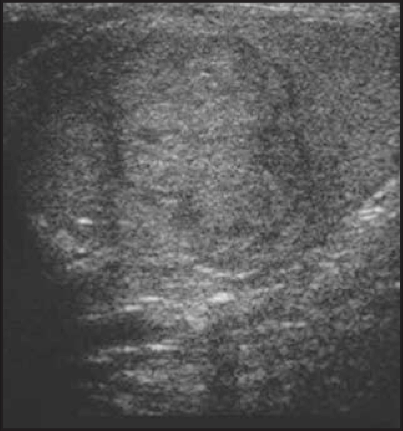
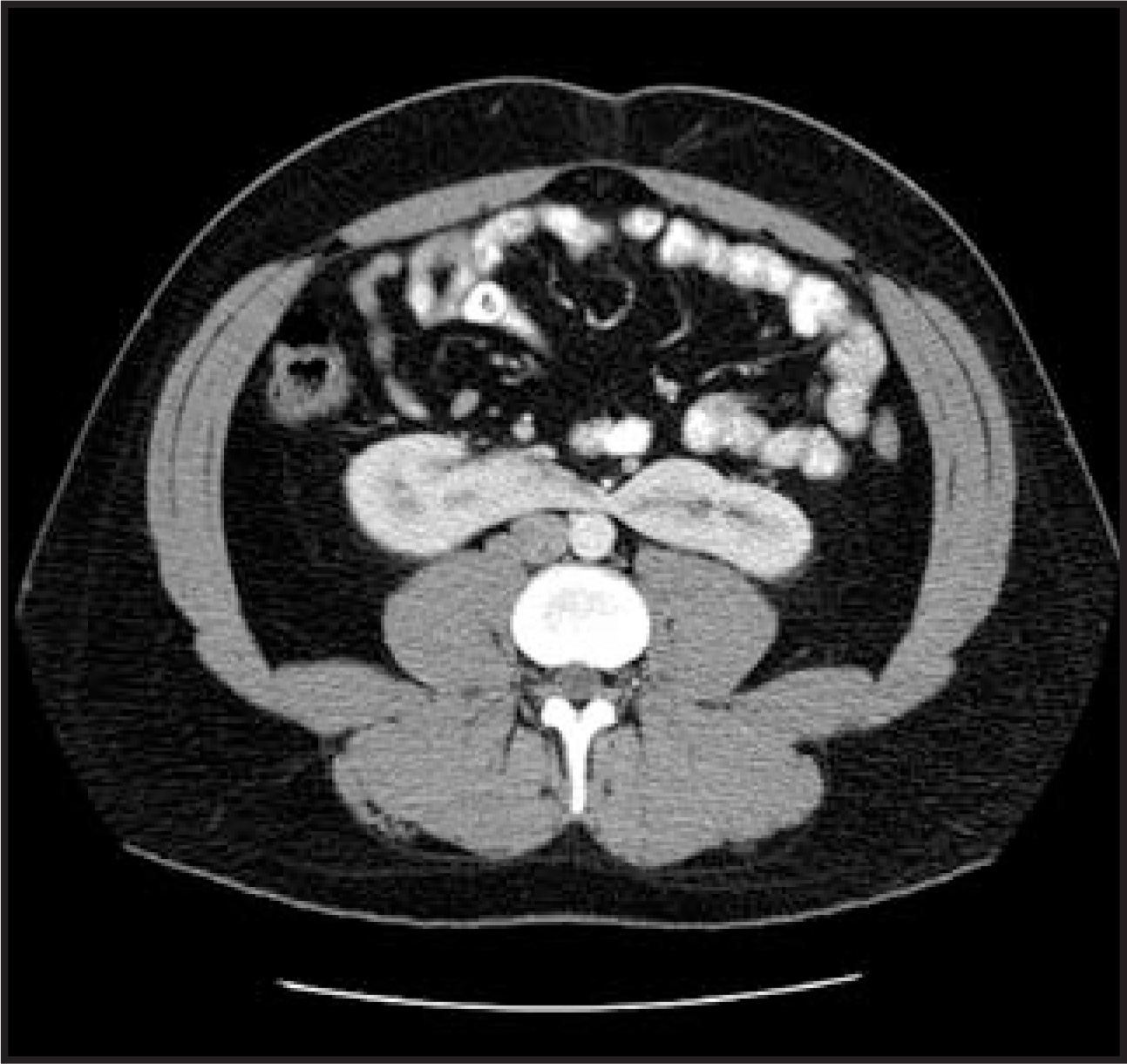
Sagittal ultrasound was performed with a 13-MHz probe of the right testicle and revealed a 2× 2-cm isoechoic, intratesticular mass (Figure 1). A contrast-enhanced axial CT image at the level of the lower pole of the kidneys (Figure 2) shows a bridging isthmus between the right and left kidneys, which is indicative of a horseshoe kidney.
Case 2
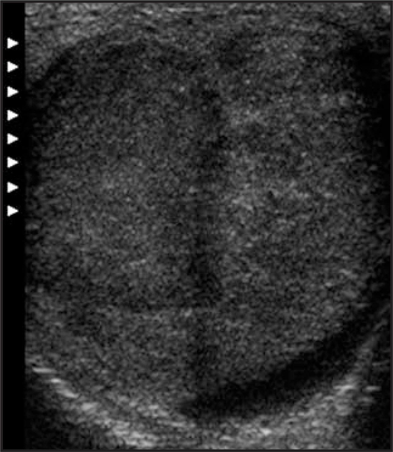
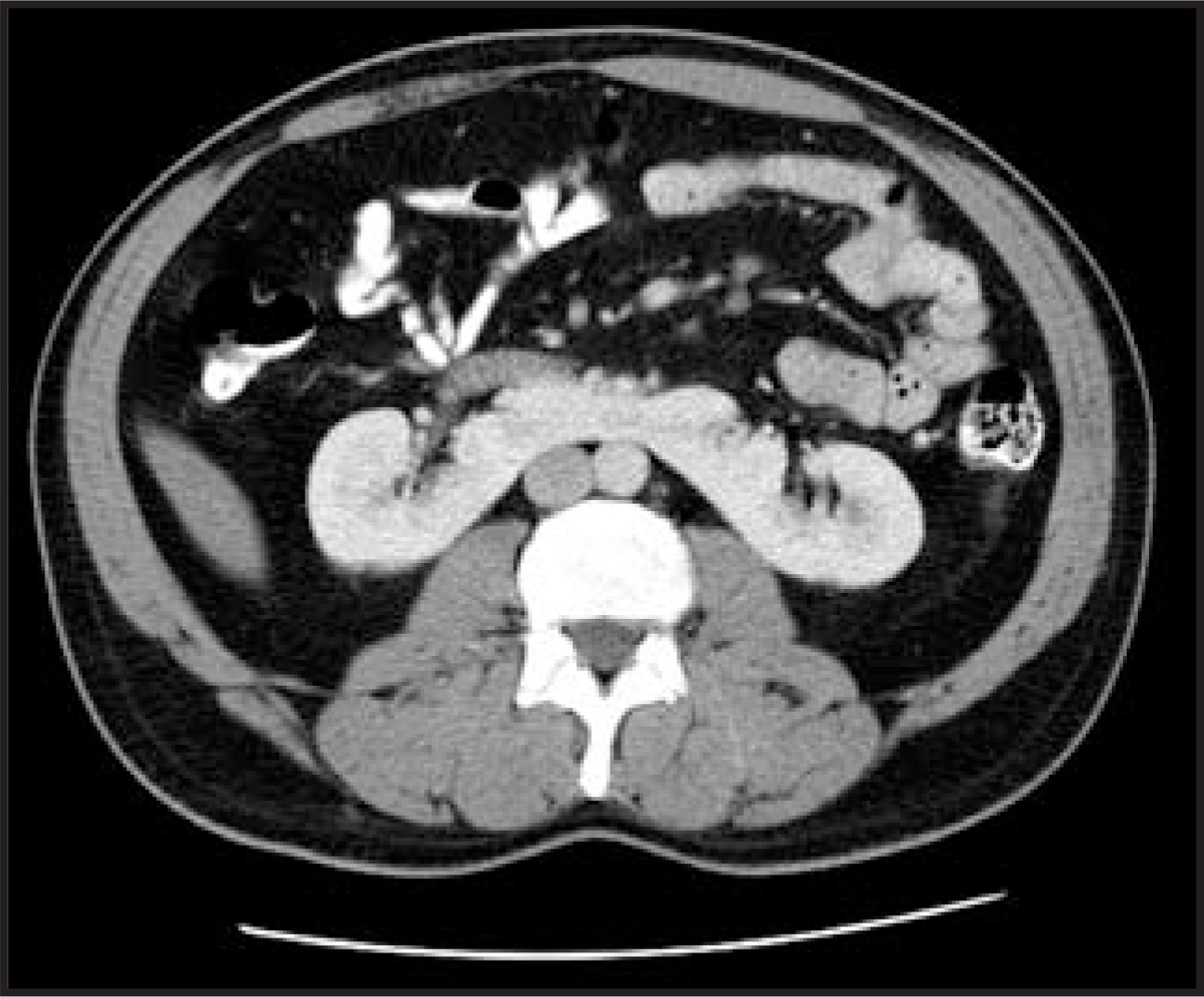
A transverse ultrasound image shows a solid, hypoechoic, intratesticular mass within the left testicle, measuring approximately 5 cm(Figure 3). A contrast-enhanced axial CT image shows a horseshoe kidney in the same patient (Figure 4).
DIAGNOSIS
Testicular carcinoma in patients with a horseshoe kidney
CASE FOLLOW-UPS
Case 1
The patient underwent a radical orchiectomy. Histological examination revealed it to be a mixed germ cell tumor that was composed of embryonal (60%) and seminomal (40%) cells. Local and lymphatic invasion was noted, but there were clear margins. His beta-HCG was elevated at 20 mIU/mL; his AFP level was 2.1 mIU/mL. He was subsequently referred to a tertiary care center and advised on his treatment options, which included 1) retroperitoneal lymph node dissection (RPLND), 2) chemotherapy, or 3) rigid surveillance.
Case 2
The patient underwent radical orchiectomy that revealed a pure seminomatous tumor of the left testicle measuring 7.5 cm in its maximum dimension. The patient subsequently received postoperative radiation therapy to the periaortic and left pelvic nodes, with 2550 cGy administered over a 3-week time period.
DISCUSSION
Horseshoe kidney is said to occur in approximately 1:400 births, with an increased incidence in males of approximately 2–3:1.Embryologically, it is thought to be due to failure of the cleavage of the metanephrons as they ascend out of the pelvis, shifting their blood supply from the iliac branches to segmental aortic branches, to their definitive lumbar position. This occurs at roughly 4 to 8 weeks of embryogenesis. In 95% of cases, the lower poles are fused by a non-functioning isthmus at approximately the L3–L4 level. The fused lower poles usually lie anterior to the aorta. What causes this fusion is not definitively known. Carleton and Segura1,2 postulated that a horseshoe kidney is the result of local conditions within the developing pelvis, such as medial compression of the umbilical arteries that run retroperitoneally from the umbilicus along the bladder, anterior to the ureters and the hypogastric arteries.
As with other congenital defects of the genitourinary system, horseshoe kidney seems to be related to a higher incidence of other anomalies in both the genitourinary system as well as other organ systems. Boatman3 reviewed the record of all patients with horseshoe kidney seen between 1939 and 1969 and found 96 cases. Associated congenital anomalies were found in 32 cases, with many patients having multiple defects. Some of the more common associations with horseshoe kidney include cryptorchidism, renal calculi, uretopelvic junction obstruction, and frequent urinary tract infections secondary to stasis. Hildebrand first reported carcinoma in a horseshoe kidney in 1895.4 Buntley5 reported111 malignant tumors associated with horseshoe kidney, mostly adenocarcinoma and carcinomas of the renal pelvis, between 1895 and 1976.In their series, Blackard and Mellinger6 described a similar finding of an increased incidence of renal pelvic tumors associated with horseshoe kidney.
Extrarenal anomalies associated with horseshoe kidney include congenital heart defects (such as ventricular septal defect and coarctation), central nervous system disorders (such as hydrocephalus and meningomyelocele), and various other gastrointestinal anomalies (such as omphalocele, tracheoesophageal fistula, and duodenal stenosis). Although there is a well known association between cryptorchidism and testicular carcinoma, and (as just mentioned) horseshoe kidney and cryptorchidism, an exhaustive search of the literature could find no direct relationship between horseshoe kidney and testicular carcinoma. The only closely related reference found in the literature was by Sogani et al,7 who reported on 310 patients who underwent RPLND for germ cell tumors of the testis, 4 of whom were subsequently found to have horseshoe kidney. This author believes that the 2 cases presented here represent a unique and purely coincidental occurrence during a 1-year period.
CONCLUSION
After an extensive search of the literature, this author could find no other report of a testicular carcinoma arising in a patient with horseshoe kidney, and it is, therefore, my belief that there is neither increased incidence nor direct relationship between neoplasms of the testicle and horseshoe kidney.
References
- Carleton A. Crossed ectopia of the kidney and its possible cause. J Anat. 1937;71:292-308.
- Segura J, Kelalis P, Burke E. Horseshoe kidney in children. J Urol. 1972;108:333-337.
- Boatman D, Kölln C, Flocks R. Congenital anomalies associated with horseshoe kidney. J Urol. 1972;107:205-207.
- Nagar R, Sanwal B. J Indian Med Assoc. 1989;87(1):14-16.
- Buntley D. Malignancies associated with horseshoe kidney. Urology. 1976;8:146-148.
- Blackard C, Mellinger G. Cancer in a horseshoe kidney. A report of 2 cases. Arch Surg. 1968;97:616-627.
- Sogani P, Whitmore W. Retroperitoneal lymphadenectomy for germ cell tumor of testis in association with horseshoe kidney. Urology. 1981;18(5):446-452.
Citation
. Testicular carcinoma and a horseshoe kidney in 2 patients. Applied Radiology. 2008;37(7):40A-40B. doi:10.37549/AR1632.