The acute pediatric chest
Applied Radiology — Vol. 33 , Issue 10 , pp. 26 -36
DOI: 10.37549/AR1285
Published: October 1, 2004
Categories
This article will systematically review the imaging aspects of respiratory distress in the newborn infant. First, the discussion will address vascular access lines, endotracheal/gastric tube placement, and possible complications, such as pneumothorax and pneumomediastinum. Next, the differential diagnosis of respiratory distress is adapted using gestational age at birth, lung volume, and the time course of opacities. Clinical information, such as the presence of meconium aspiration and cesarean section, are essential in order to determine an accurate diagnosis.
Medical disease causing respiratory distress in the newborn can be logically separated into disease affecting the preterm infant (such as hyaline membrane disease) and disease in the term infant (such as transient tachypnea, meconium aspiration syndrome, and neonatal pneumonia). Surgical disease presenting with respiratory distress includes congenital lung disease, such as congenital diaphragmatic hernia (CDH) and congenital cystic adenomatoid malformation (CCAM) that presents with respiratory distress at birth, and a group that presents later, such as congenital lobar emphysema (CLE) and sequestration.
Lines and tubes
The proper position of tubes and lines on a neonatal chest radiograph is very important. The endotracheal tube tip (ETT) should be located beneath the thoracic inlet and 1.5 vertebral bodies above the carina. Malposition of the ETT, often due to head movement when placed too low, in either of the main bronchi (most commonly, the right) causes resorption atelectasis of the contralateral lung. The nasogastric tube tip (NGT) should be within the stomach. The desired tip position of the umbilical venous catheter (UVC) is at the junction of the inferior vena cava and the right atrium.1 The UVC generally projects over the right side of the spine on the supine radiograph and has a “bend” along its course through the falciform ligament. Most complications are caused by malposition of the UVC in the portal veins, with the concomitant risk of thrombosis, which can be identified with sonography2 or, if chronic, on radiographs as a calcification. 3 Complications caused by malpositioning of the UVC in the lungs or heart include pulmonary infarction, hydrothorax, arrhythmias, nonbacterial thrombotic endocarditis, atrial wall perforation, and cardiac tamponade. 4
Optimum placement of the umbilical artery catheter (UAC) tip is either above or below visceral branches of the aorta. High lines have the tip positioned between T8-T12, whereas low lines have their tips between L3-L5. Umbilical artery catheterization with the tip at the 8th thoracic segment may be associated with fewer complications than at lower positions.5,6 The UAC generally projects over the left side of the spine on the anteroposterior film and initially courses caudally through the internal iliac artery (hypogastric artery).
The tips of the extracorporeal membrane oxygenation (ECMO) arterial and venous catheters are often nonopaque, and their exact positions are often difficult to ascertain. 7 In arterial-venous ECMO, the tip of the arterial catheter should be within the aortic arch, and the tip of the venous catheter should be within the right atrium. In venous-venous ECMO, the tip of the sole venous catheter should be within the right atrium.
Medical disease
The premature infant
Hyaline membrane disease—Hyaline membrane disease (HMD), also known as idiopathic respiratory distress syndrome or surfactant deficiency syndrome, is the most common cause of respiratory distress in the neonatal period. It almost always occurs in premature infants and is recognizable, aside from demographic information, by lack of subcutaneous fat, no humeral ossification center, and an ETT. The incidence of HMD varies according to gestational age at birth (50% at 27 weeks, 16% at 31 weeks, and 5% at 34 weeks). Hyaline membrane disease in a term neonate is unlikely.
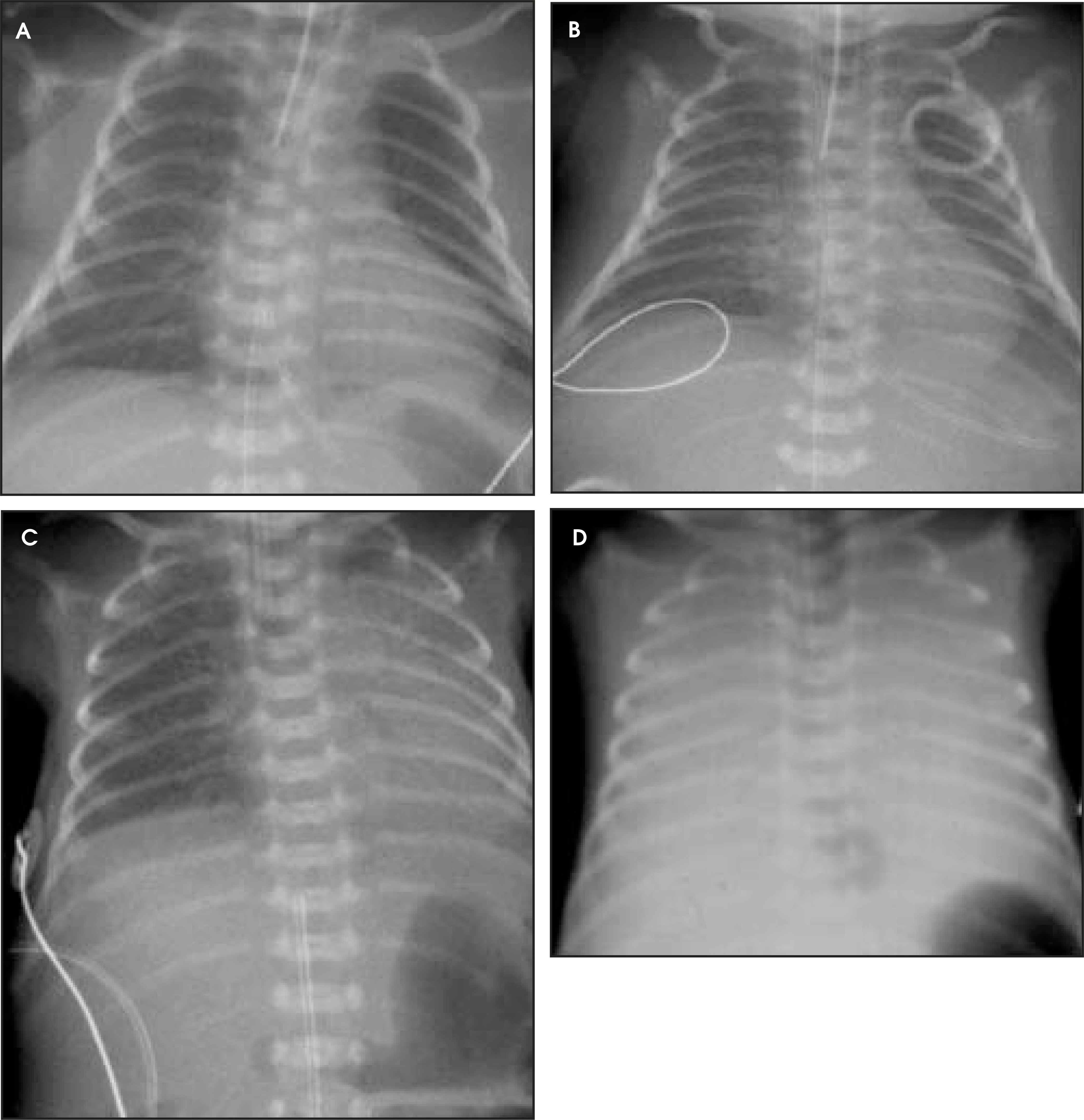
Lack of surfactant causes microatelectasis, which can be seen as a reticulogranular (ground-glass) appearance and hypovolemic lungs, which are recognizable as a bell-shaped thorax (Figure 1). Hyperinflation (if the infant is not intubated) excludes HMD. Pleural effusions are seldom seen with HMD. The spectrum of HMD appearances on the chest radiograph can be divided into 4 disease stages (Figure 1): Stage I-reticulogranular; stage II-additional air brochograms; stage Ill-hazy or indistinct cardiac contour; and stage IV-completely opacified lungs. 8 The major role of the radiologist is to assess complications of assisted ventilator therapy.
Complications of HMD include leaky lung syndrome (LLS), patent ductus arteriosus, pulmonary interstitial emphysema (PIE), pneumothorax, and bronchopulmonary dysplasia (BPD). Pulmonary edema is a difficult problem in premature infants on positive pressure ventilation. It results from damage to the capillaries secondary to hypoxia and toxic effects of high concentrations of oxygen with leaking of fluid into the pulmonary intersitium9 and, as such, constitutes LLS. Leaky lung syndrome can be diagnosed and distinguished from HMD by the characteristic time-span of development. It develops after initial clearing of the lungs following surfactant therapy, when the lungs become hazy again in the presence of slight enlargement of the cardiac silhouette. Pulmonary edema may be distinguished from HMD by the predominant pattern of haziness (HMD shows a predominantly reticulogranular pattern) and the presence of pleural effusions. Pulmonary edema in these infants may also result from a left-to-right shunt through a reopened patent ductus arteriosus and, in such cases, is also associated with slight cardiomegaly.
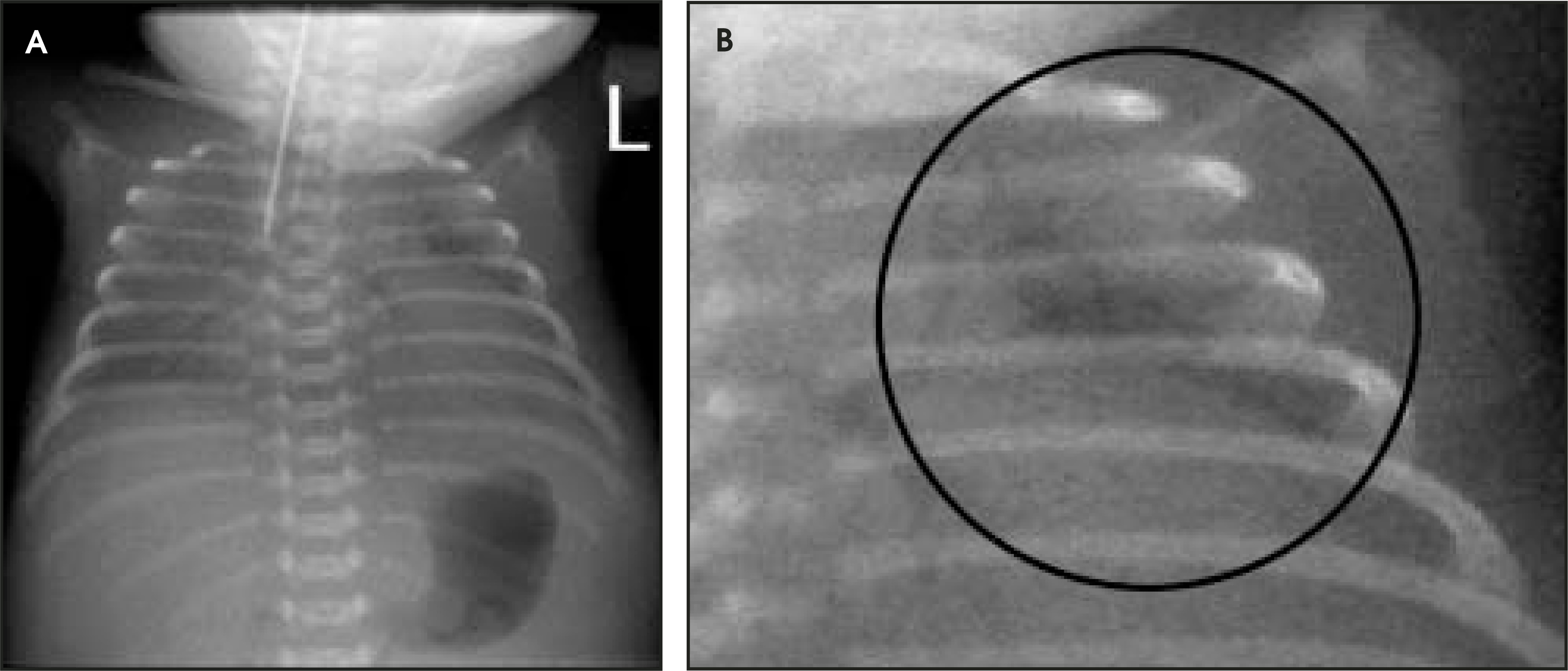
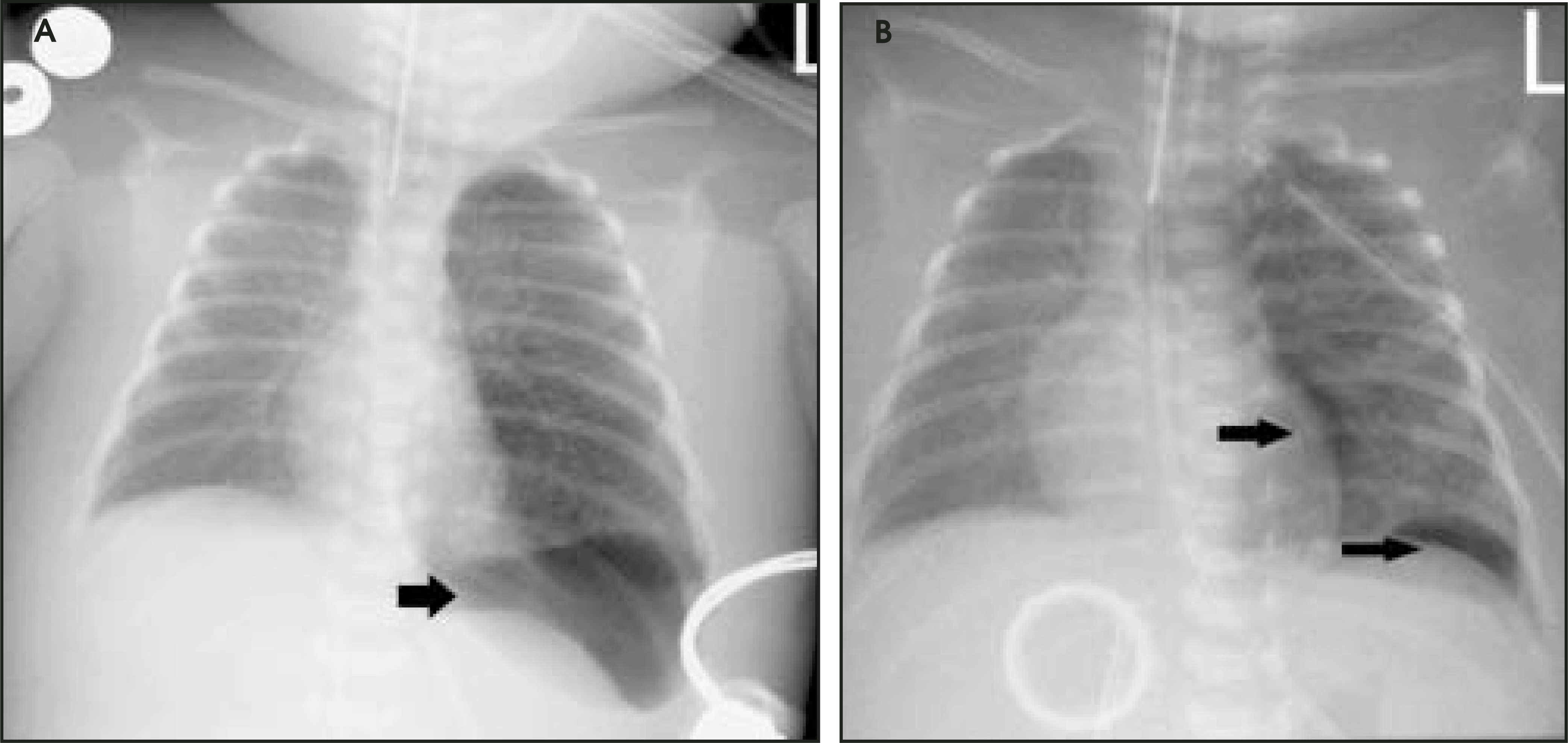
Pulmonary interstitial emphysema refers to accumulation of interstitial air in peribronchial and perivascular spaces and is characterized by tortuous linear lucencies, which radiate outward from the hilar regions extending all the way to the periphery of the lung (Figure 2). Complications of PIE include pneumo-thorax, pneumomediastinum, and pneumopericardium (Figure 3).
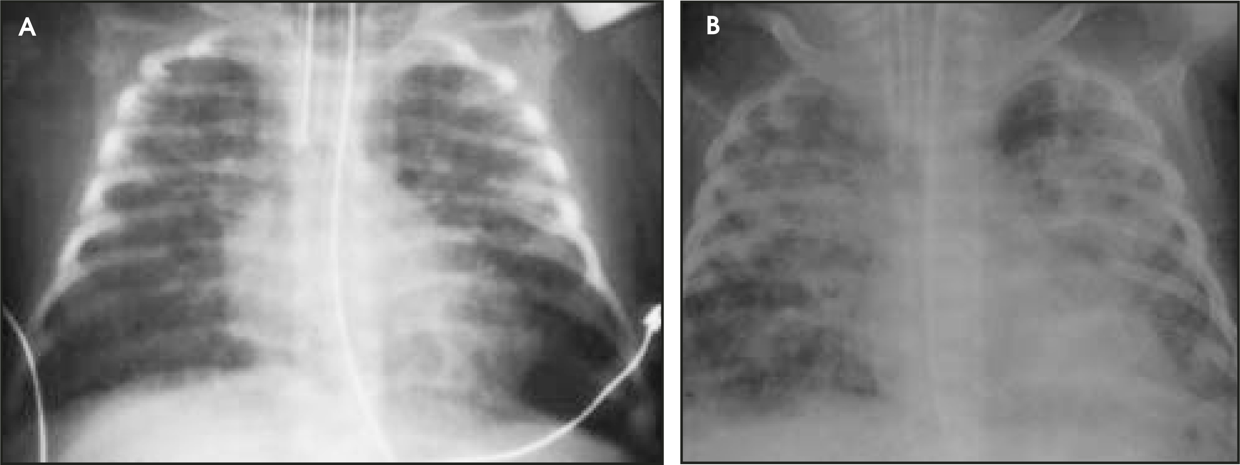
Bronchopulmonary dysplasia is a disease entity defined by agreement of the American Association of Pediatricians as one that appears after more than 28 days of high-pressure, high-rate ventilatory assistance and includes a radiographic picture of interstitial fibrosis, cyst-like emphysematous changes, and increased lung volume. 10 However, recently, a more extensive definition has been proposed. 11 The most common radiographic appearance of BPD is that of diffuse interstitial thickening with mild to moderate hyperinflation (Figure 4). 12 In infants who survive the neonatal intensive care unit, the chest radiograph will have normalized in the majority of patients. However, their pulmonary function tests will remain restrictive until much later.
The term infant
Transient tachypnea of the newborn—Transient tachypnea of the newborn (TTN, or wet lung) is a clinical diagnosis and comprises a physiologic phenomenon of retained fetal pulmonary amnion fluid, which is physiologically cleared in the newborn by means of the birth canal “squeeze,” lymphatics, and capillaries. The absence of one of these mechanisms, such as in case of cesarean section and prolonged (meaning in the presence of medications) labor will exacerbate the wet lung; therefore, accurate clinical information is essential for diagnosis. Typically, the infant will present with respiratory distress at 2 to 4 hours of age or earlier, with normalization after 48 hours. The most common characteristic changes on the chest radiograph consist of hyperinflation, prominent interstitium shadows, and effusions (Figure 5). Transient tachypnea of the newborn can be differentiated from HMD by assessment of gestational age at birth and the typical disappearance within the 48-hour follow-up period.
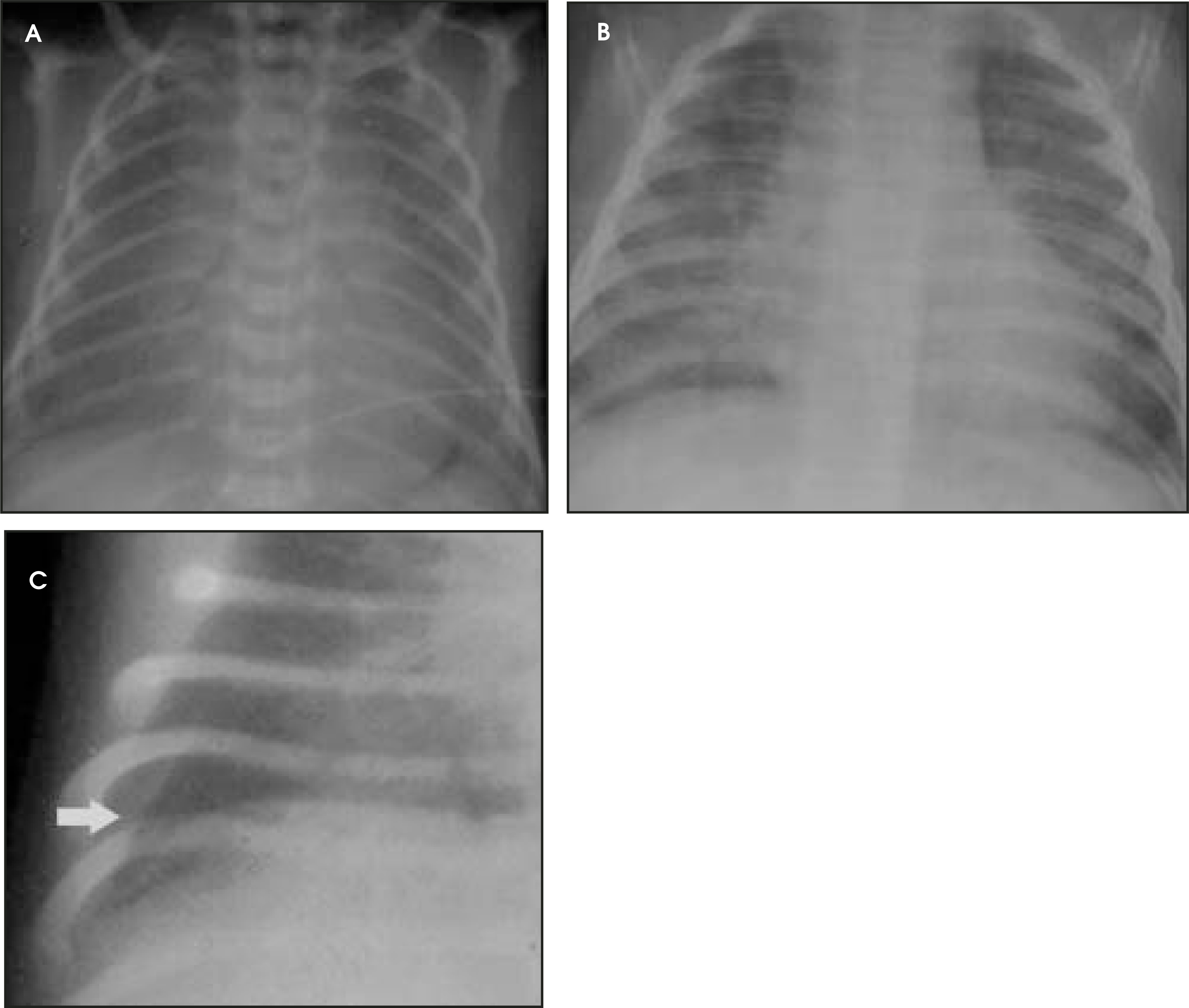
Meconium aspiration syndrome—Meconium aspiration syndrome (MAS) is related to perinatal stress (such as hypoxia or prolonged labor), with a vagal response postulated as the triggering factor for intrauterine evacuation of meconium by the fetus. In 10% of normal births, there is meconium in the amniotic fluid; in 10% of those cases, the infant may aspirate this meconium-laden fluid during the first breath. Aspiration of meconium particles, lodged in the small peripheral bronchi, induces bronchial obstruction and air trapping due to a check-valve mechanism, which causes both atelectasis and overaeration. The irritative effect of meconium aspirate, a chemical pneumonitis, may cause focal alveolar atelectasis. Additionally, meconium may also cause surfactant dysfunction, resulting in diffuse atelectasis (secondary HMD). 13 This complicating chemical pneumonitis has been shown to occur after approximately 48 hours. 14
Conventional radiographs of the chest typically reveal hyperinflated nodular lungs, lack of effusions, and an ETT (Figure 6). Complications of MAS include pneumothorax and pneumomediastinum (in up to 25% of cases). Since the intrapulmonary pressure may remain high, MAS is often complicated by persistent fetal circulation caused by a persistent ductus arteriosus and open foramen ovale. Treatment options include oxygen and antibiotics and, in severe cases, ECMO or high-flow oxygenation.
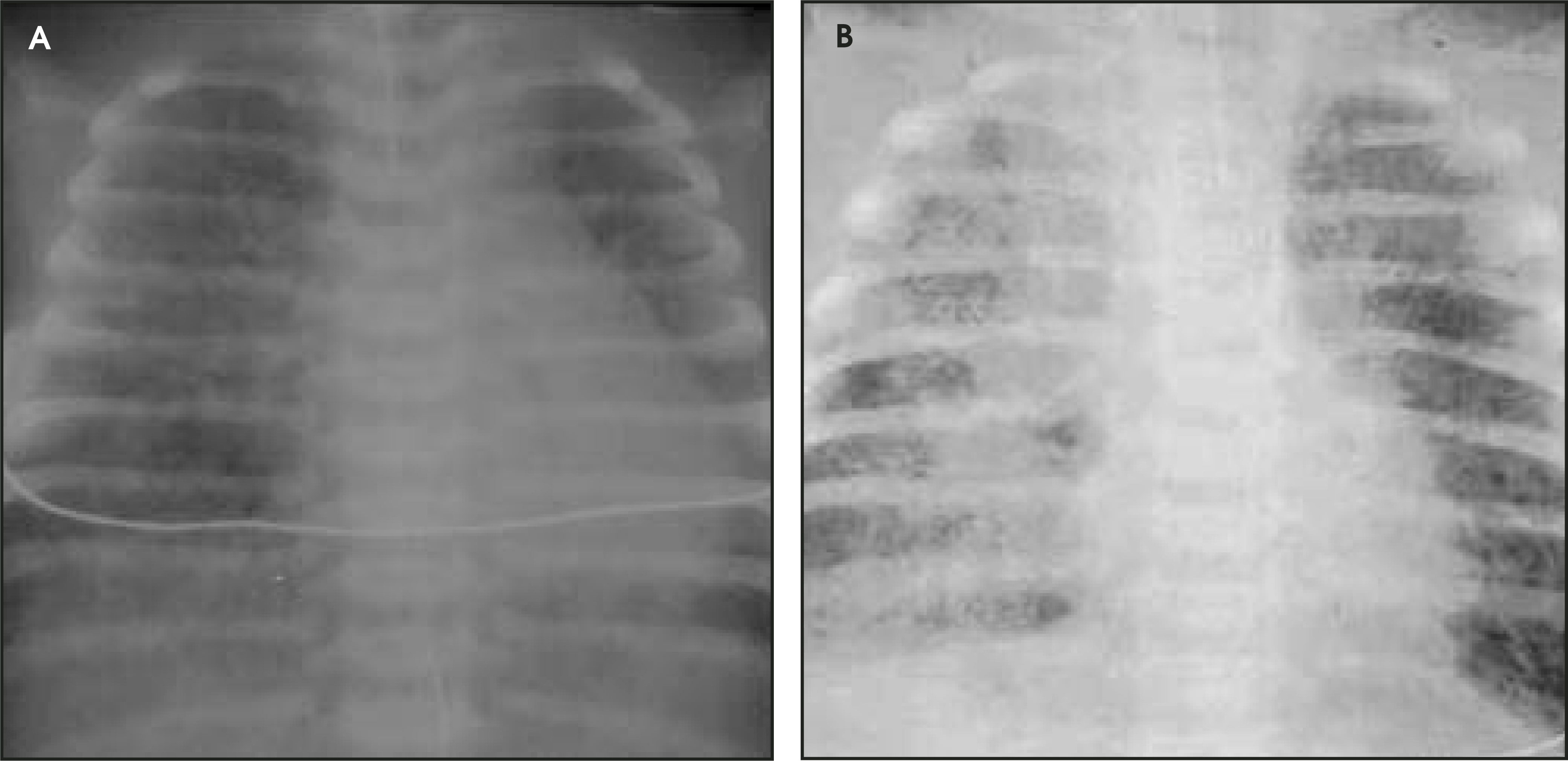
Neonatal pneumonia—Most early pneumonias are the result of fetal inhalation of an infection picked up in the birth canal (G-hemolytic streptococci, Escherichia coli, viral). Prolonged labor, premature rupture of membranes, placental infection, and ascending infection from the perineum are predisposing factors. The radiologic appearance of neonatal pneumonia can mimic that of transient tachypnea of the newborn, early HMD or MAS. A patchy, occasionally asymmetric, radiating, bilateral interstitial opacity is commonly seen in a hyperinflated chest (Figure 7). Important clues suggesting pneumonia are the presence of pleural effusions (excluding HMD) and the absence of an endotracheal tube, as patients with neonatal pneumonia are rarely intubated. Furthermore, diffuse hazy opacification suggestive of HMD in a term or near-term infant should always raise the suspicion of neonatal pneumonia. In addition, meconium aspiration syndrome, retained fluid, and HMD may co-exist with pneumonia.
Surgical disease
Disease present at birth
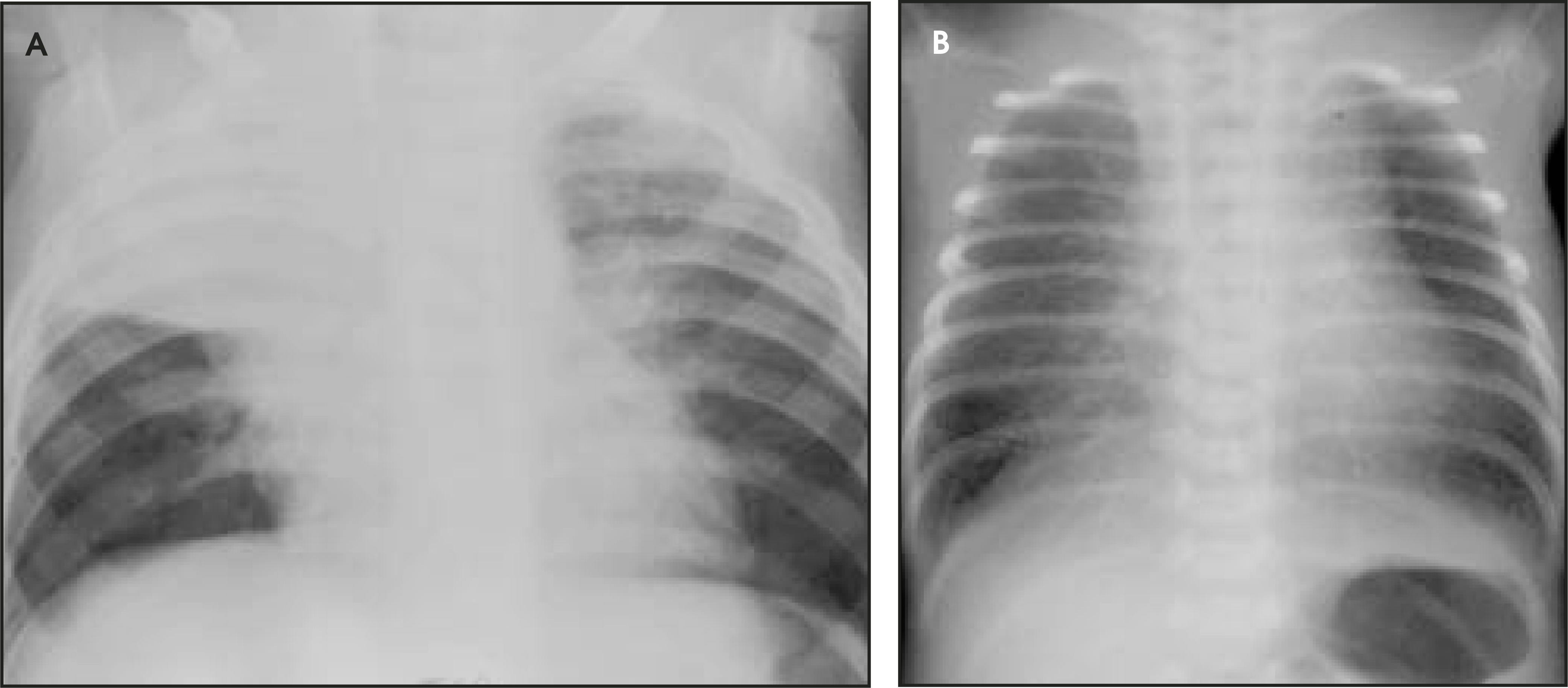
Congenital diaphragmatic hernia—Congenital diaphragmatic hernia (CDH) is caused by bad timing of the closure of the pleuroperitoneal canal and occurs lateral to the spine, most frequently on the left side (90% of cases). The Bochdalek hernia is the most frequently (85% of all CDH) encountered form, which herniates through the posterior foramen of Bochdalek (a helpful mneumonic: Bochdalek-Back). The other type of herniation is rare (3% to 5%) and occurs through the middle foramen of Morgagni (Morgagni-Middle). The Morgagni hernia occurs mostly on the right side, as the heart protects the left side. Congenital diaphragmatic hernia can constitute a major surgical emergency, due to respiratory distress caused by compression of the contralateral lung by the herniation of a large amount of bowel herniated into the chest. The classic radiologic appearance is one in which the trachea is deviated to the right, and the left hemithorax is either opaque or filled with “cystic” structures (bowel loops), which may be seen after air has been swallowed (Figure 8). An abdomen that is relatively devoid of intraluminal air also often accompanies these findings. The stomach may be an important clue in differentiating a CDH from a CCAM. In a CDH, the stomach is either not identified or is located in an abnormal position, whereas in CCAM, the stomach is in a normal position. The main complications of CDH include persistent fetal circulation and pulmonary hypoplasia. Despite surgical treatment and the advent of ECMO, mortality remains high (40% to 50%). A congenital diaphragmatic hernia is associated with extradiaphragmatic anomalies in 53% to 64% of all cases.15,16 Most anomalies affect the cardiovascular system (22% to 39%) (without a typical heart defect),15,17,18 the genitourinary system (15% to 18%),15,19 neural tube defects (16% to 28%),15,18,20 and malformations of the musculoskeletal system (18%).15
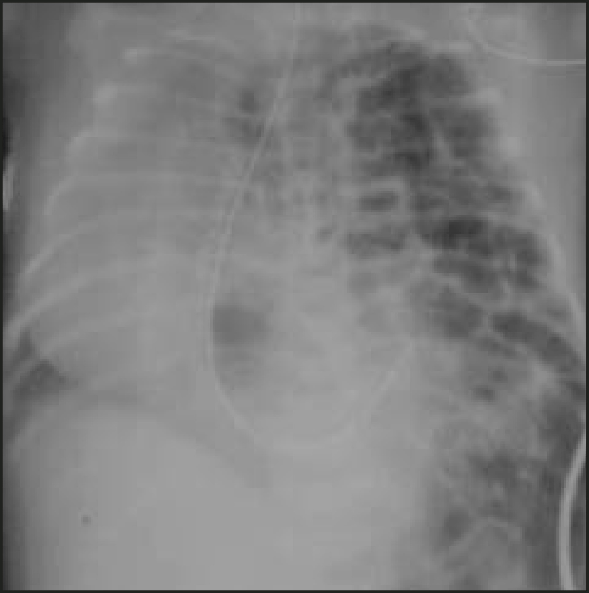
Congenital cystic adenomatoid malformation—Congenital cystic adenomatoid malformation is a relatively rare congenital hamartomatous lesion of the lung, which lacks normal bronchial communication and often presents in the first month of life (80% of cases present <6 months of age) as a cause of respiratory distress secondary to a space-occupying effect that comprises lung tissue. Typically, there is no respiratory distress at birth. There are three types: Type I (50%) is characterized by single or multiple large cysts of varying size, each measuring >2 cm in diameter (Figure 9). Type II (40%) consists of multiple small cysts of more uniform size, none >2 cm in diameter. Type III (10%) consists of large, bulky, solid-appearing lesions that contain multiple microscopic-sized cysts. 21
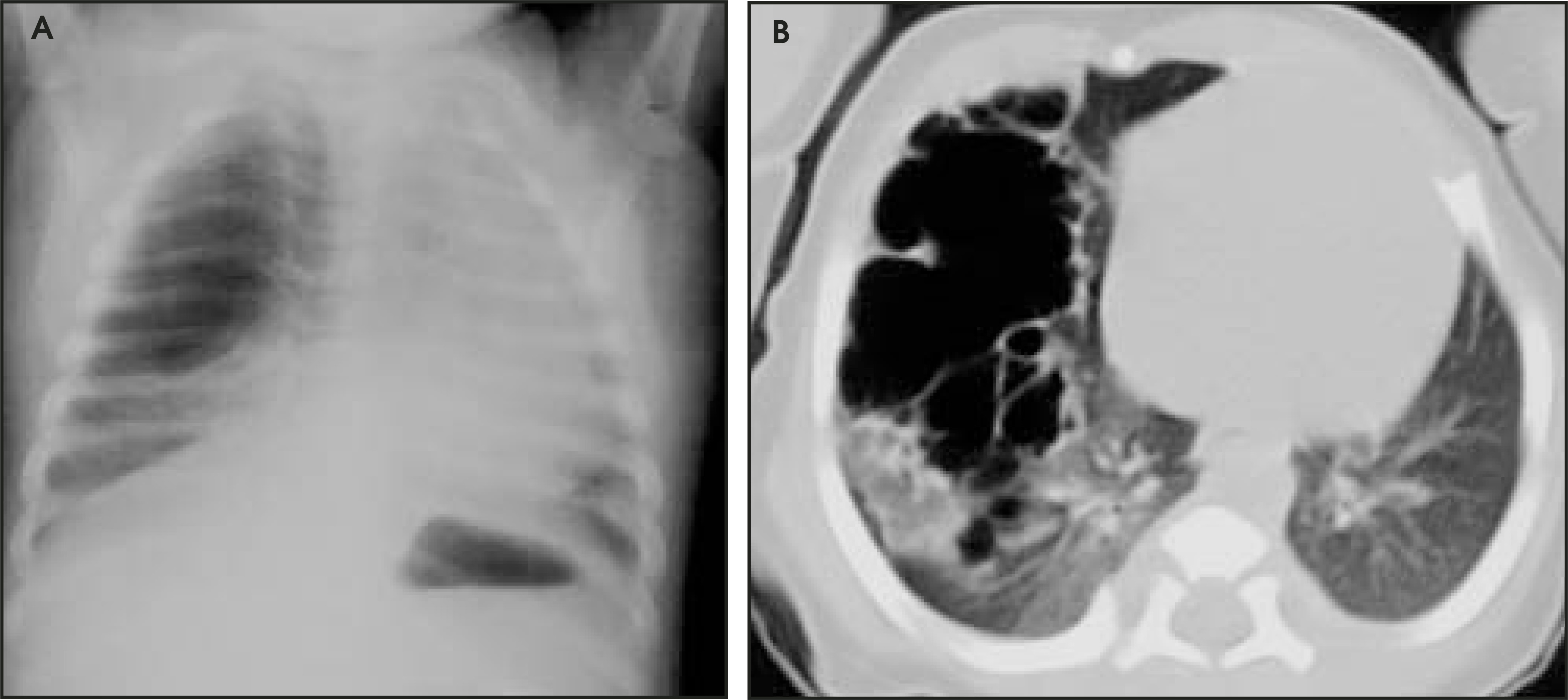
The radiographic findings are variable and correlate with the type of lesion; however, the classification has no clinical relevance except as a reminder that CCAM can have a variable appearance. A more generalized classification is used by others that incorporates ultrasound findings. 22 According to this classification, microcystic lesions (<5 mm) are usually associated with fetal hydrops and a poor prognosis, whereas macrocystic lesions (>5 mm) are not usually associated with hydrops and have a favorable prognosis. Chest radiographs of type I lesions typically show unilateral single or multiple air-filled cysts in the thorax of a neonate with respiratory distress. A CCAM is most often unilobar, with no lobar predilection, contrary to congenital lobar emphysema. A CCAM often enlarges after birth and shows tracheal deviation and a variable cystic-solid component, which may mimic staphylococcal pneumonia and diaphragmatic hernia. However, the former is unusual directly postpartum and the latter is often combined with an abdomen devoid of gas. In a CCAM, air-fluid levels may be present in cysts and there is a variable thickness of the cyst wall. Therapy of CCAM is surgical.
Congenital lobar emphysema—Congenital lobar emphysema (CLE) is one of the leading causes of respiratory distress in the neonate and is often diagnosed in utero or shortly after birth. It is caused by narrowing of bronchi secondary either to bronchial cartilage underdevelopment or extrinsic compression.
The narrowing of bronchi leads to a check-valve mechanism, with overdistention of the affected lobe. A preferred term for CLE is congenital lobar overexpansion, because it more accurately describes the basic pathologic abnormality, which is overdistention of otherwise normal alveoli without destruction of alveolar walls. 23 Congenital lobar emphysema presents within 6 months of birth and affects boys 3 times more often than girls. Congenital lobar emphysema usually involves one lobe, with the following distribution: left upper lobe (41%), middle lobe (34%), and the right upper lobe (21%). Involvement of the lower lobes and/or multilobar disease is extremely rare. Congenital lobar emphysema is associated with a ventricle septal defect and persistent ductus arteriosus in 15% of cases.
Radiographic hallmarks of CLE are hyperaerated lobe(s) (this may mimic pneumothorax), with shift of the mediastinum to the contralateral side, flattened hemidiaphragm, and widened intercostal spaces. In cases of upper-lobe overdistention, there may be compression of the ipsi-lateral lower lobe, or in the case of middle lobe overdistention, there may be compression of both the ipsilateral lower and upper lobes. During the first few days of life, there may be alveolar opacification, because there is no clearance of lung fluid through bronchi. Later on, as fluid is cleared, the classic appearance of CLE is seen (Figure 10). The differential diagnosis includes CCAM, bronchogenic cyst and cystic hygroma. Computed tomography is helpful in selected cases to differentiate between CLE, a large type I CCAM, and contralateral pulmonary hypoplasia.24
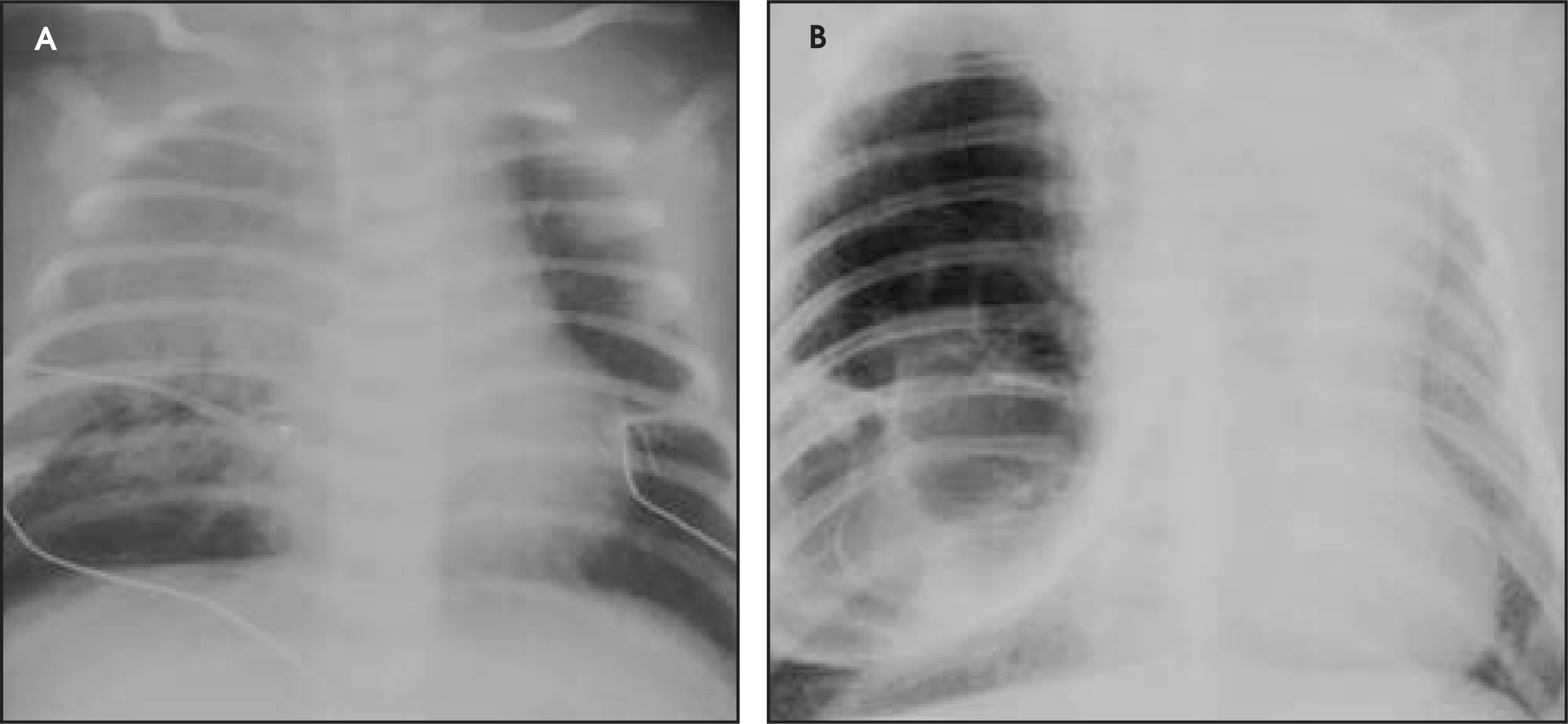
Most often, treatment consists of emergency lobectomy. But if the infant is not in serious respiratory difficulty, no immediate treatment is necessary and treatment consists of maintaining ventilatory settings as low as possible in order to avoid ventilator-related hyperexpansion.
Sequestration—Pulmonary sequestration, accessory lung, and brochopulmonary foregut malformation probably all describe the same entity. Many also count CCAM, CLE, and bronchogenic cyst as part of this spectrum. 25 A sequestration is defined as a congenital mass of aberrant pulmonary tissue that has no normal vascular or bronchial connection. The intralobar and extralobar variants can be differentiated most reliably on the basis of venous drainage. Intralobar drainage occurs primarily via the left atrium or pulmonary veins; extralobar drainage is primarily via the systemic venous plexus, including the inferior vena cava or azygos system. Both intra- and extralobar sequestrations have a vascular supply consisting of systemic arteries arising from the aorta or its branches. The majority of sequestrations are intralobar, and relatively few are extralobar. Commonly, an intralobar sequestration is supplied by a large systemic vessel, is contained within the lung without a separate pleural covering, occurs on the left side 60% of the time, and presents in teenagers. An extralobar sequestration is often supplied by a small systemic vessel, has its own pleural covering, occurs on the left side 90%, and presents in boys 60% of the time. The extralobar variety presents in the first month of life, and 61% of them present before 6 months.
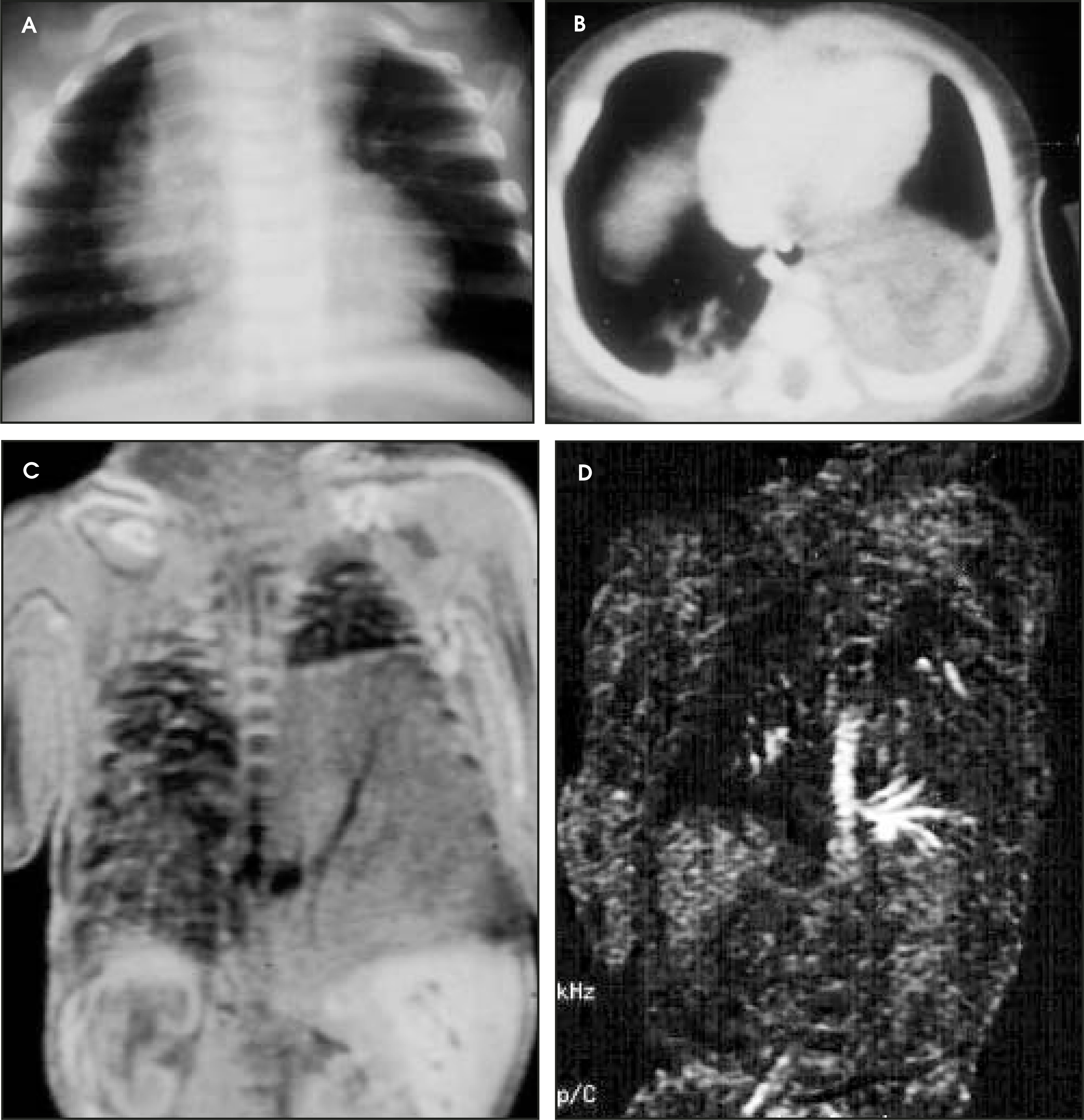
Most sequestrations are located in the posterior basilar segments of the lower lobes and are only occasionally bilateral. Conventional radiographs may be normal or may show a (recurrent) lower lobe consolidation or a relatively lucent cystic lower lobe component (Figure 11). The differentiation between intralobar and extralobar sequestration cannot be made at imaging and does not affect surgical management. Complete surgical removal to treat recurrent infection is the therapy of choice; if the sequestration is an incidental finding, removal is not necessary. Visualization of the supplying systemic artery is the characteristic finding and the documentation that surgeons look for before removing the lesion. Magnetic resonance angiography will often delineate the vascular supply (Figure 11).
Conclusion
Causes of respiratory distress in the newborn can be diagnosed accurately by the chest radiograph. Surfactant is needed to hyperinflate; therefore, hyperinflation of the lungs excludes hyaline membrane disease (if the infant is not intubated). Conversely, underinflation in a nonintubated infant usually indicates prematurity and, thus, surfactant deficiency. In the event one sees an endotracheal tube, its position can also be an important clue: deviation is likely to predict surgical intervention, such as in congenital diaphragmatic hernia, cystic adenomatoid malformation, and congenital lobar emphysema.
The main task of the radiologist in HMD is detection of treatment complications, such as the leaky lung syndrome, pulmonary interstitial emphysema, persistent ductus arteriosus, pneumothorax, and bronchopulmonary dysplasia. Neonatal congenital lesions can also be classified by location, appearance and presentation.
References
- Narla D, Hom M, Lofland G. Evaluation of umbilical catheter and tube placement in premature infants. RadioGraphics. 1991;11:849-863.
- Kim J, Lee Y, Kim S. Does umbilical vein catheterization lead to portal venous thrombosis? Prospective U.S. evaluation in 100 neonates. Radiology. 2001;219:645-650.
- Rizzo A, Haller J, Mulvihill D. Calcification of the ductus venosus: A cause of right upper quadrant calcification in the newborn. Radiology. 1989;173:89-90.
- Bjorklund L, Malmgren N, Lindroth M. Pulmonary complications of umbilical venous catheters. Pediatr Radiol. 1995;25:149-152.
- Barrington K. Umbilical artery catheters in the newborn: Effects of position of the catheter tip. Cochrane Database Syst Rev. 2000.
- Mokrohisky S, Levine R, Blumhagen J. Low positioning of umbilical-artery catheters increases associated complication in newborn infants. N Engl J Med. 1978;299:561-564.
- Irish M, OToole S, Kapur P. Cervical ECMO cannula placement in infants and children: Recommendations for assessment of adequate positioning and function. J Pediatr Surg. 1998;33:929-931.
- Northway W, Rosan R. Radiographic features of pulmonary oxygen toxicity in the newborn: Bronchopulmonary dysplasia. Radiology. 1968;91:49-58.
- Brasch R, Berthezene Y, Vexler V. Pulmonary oxygen toxicity: Demonstration of abnormal capillary permeability using contrast-enhanced MRI. Pediatr Radiol. 1993;23:495-500.
- Bancalari E, Abdenour G, Feller R, Gannon J. Bronchopulmonary dysplasia: Clinical presentation. J Pediatr. 1979;95(5 Pt 2):819-823.
- Jobe A, Bancalari E. Bronchopulmonary dysplasia. Am J Respir Crit Care Med. 2001;163:1723-1729.
- Northway W. Bronchopulmonary dysplasia: Twenty five years later. Pediatrics. 1992;89:969-973.
- Clark D, Nieman G, Thompson J. Surfactant displacement by meconium free fatty acids: An alternative explanation for atelectasis in meconium aspiration syndrome. J Pediatr. 1987;110:765-770.
- Swaminathan S, Quinn J, Stabile M. Long-term pulmonary sequelae of meconium aspiration syndrome. J Pediatr. 1989;114:356-361.
- Bollmann R, Kalache K, Mau H. Associated malformations and chromosomal defects in congenital diaphragmatic hernia. Fetal Diagn Ther. 1995;10:52-59.
- Benacerraf B, Adzick N. Fetal diaphragmatic hernia: Ultrasound diagnosis and clinical outcome in 19 cases. Am J Obstet Gynecol. 1987;156:573-576.
- Greenwood R, Rosenthal A, Nadas A. Cardiovascular abnormalities associated with congenital diaphragmatic hernia. Pediatrics. 1976;57:92-97.
- Puri P, Gorman F. Lethal nonpulmonary anomalies associated with congenital diaphragmatic hernia: Implications for early intrauterine surgery. J Pediatr Surg. 1984;19:29-32.
- Knight P, Clatworthy R. Screening for latent malformations: Cost effectiveness in neonates with correctable anomalies. J Pediatr Surg. 1982;17:123-129.
- David T, Illingworth C. Diaphragmatic hernia in the Southwest of England. J Med Gen. 1976;13:253-262.
- Stocker J, Madewell J, Drake R. Congenital cystic adenomatoid malformation of the lung. Classification and morphologic spectrum. Human Pathology. 1977;8:155-171.
- Adzick N, Harrison M, Glick P. Fetal cystic adenomatoid malformation: Prenatal diagnosis and natural history. J Pediatr Surg. 1985;20:483-488.
- Kuhn C, West W, Craighead J, Damjanov I, Linder J. Anderson’s Pathology. 1996:1471.
- Dee P. Imaging of Diseases of the Chest. 2000:689-725.
- Panicek D, Heitzman E, Randall P. The continuum of pulmonary developmental anomalies. RadioGraphics. 1987;7:747-772.
Citation
. The acute pediatric chest. Applied Radiology. 2004;33(10):26-36. doi:10.37549/AR1285.