The purpose of this article is to summarize pitfalls in the interpretation of magnetic resonance (MR) imaging due to aeration of the anterior or posterior clinoid processes.
Advertisement
Advertisement
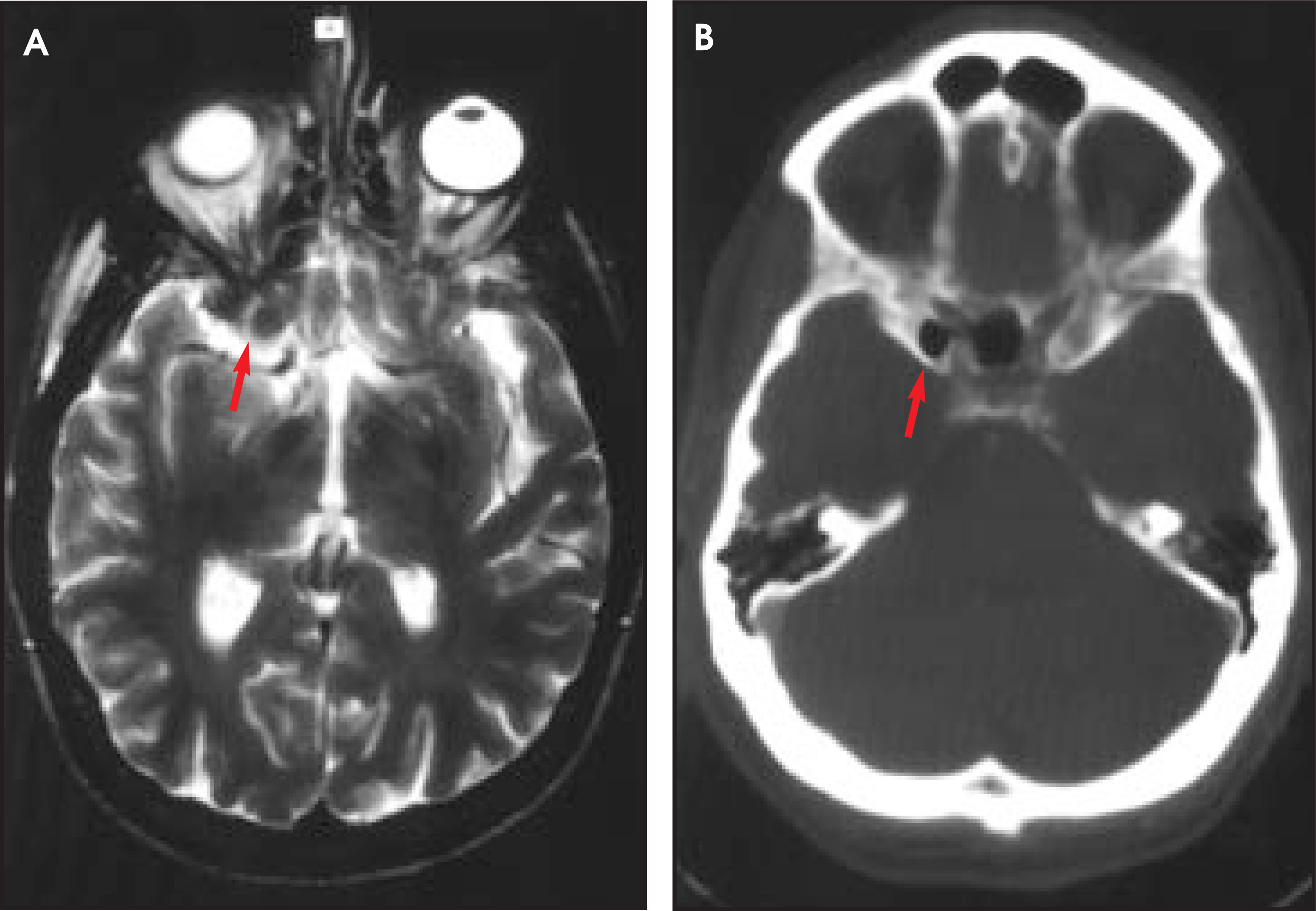
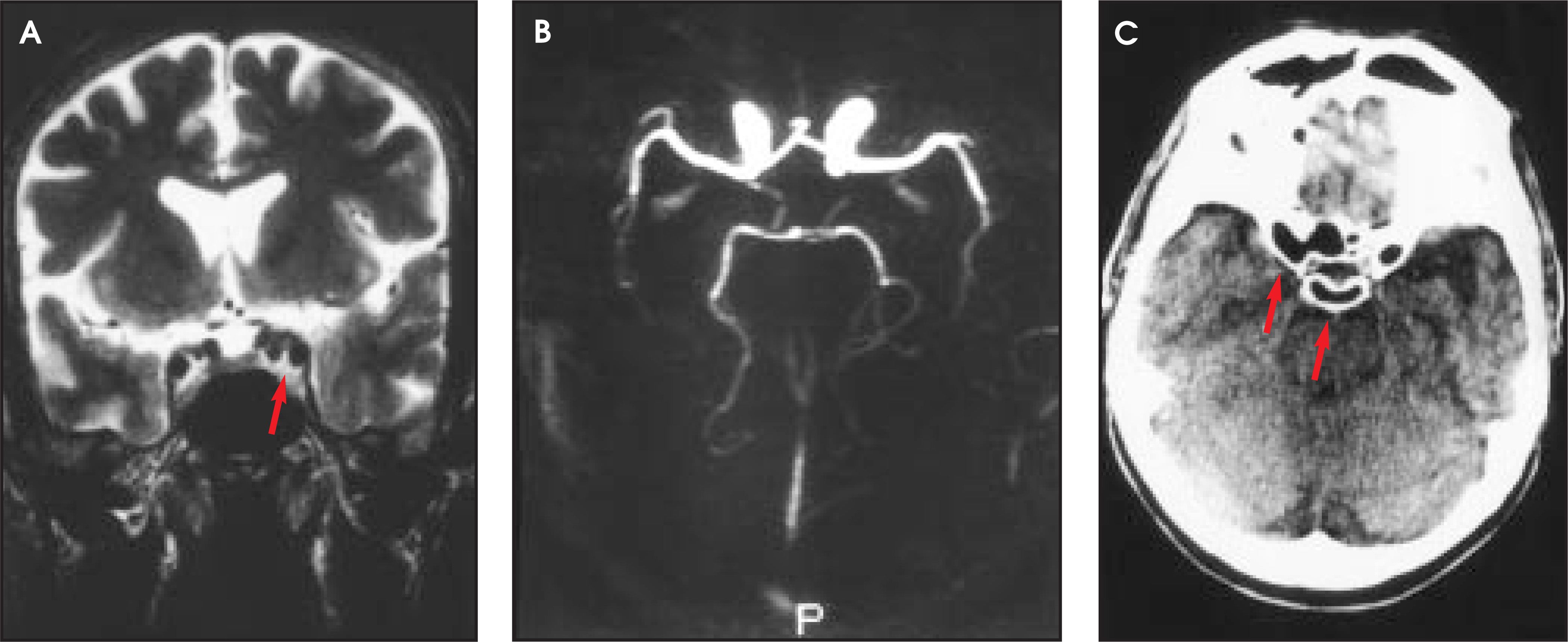
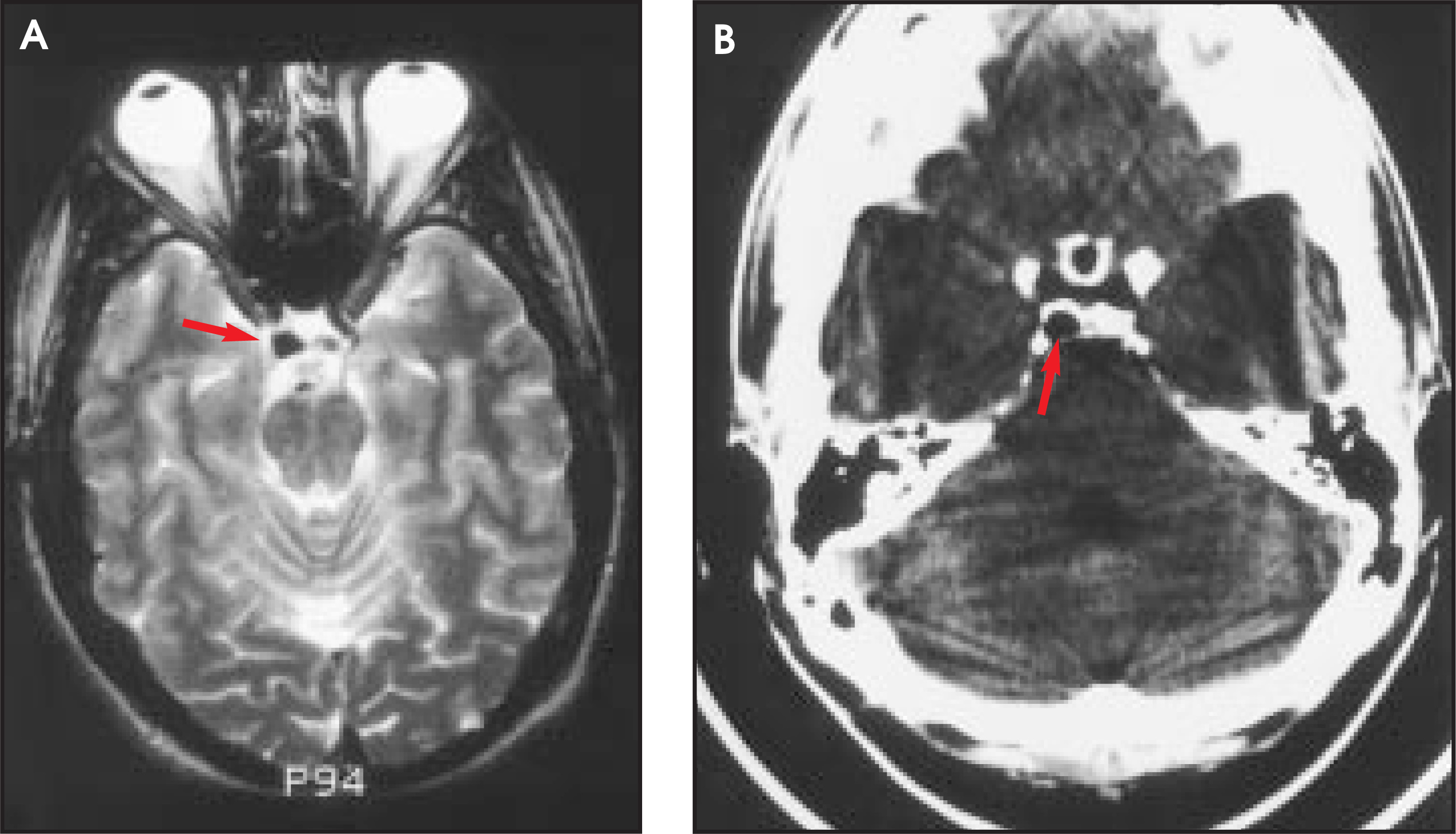
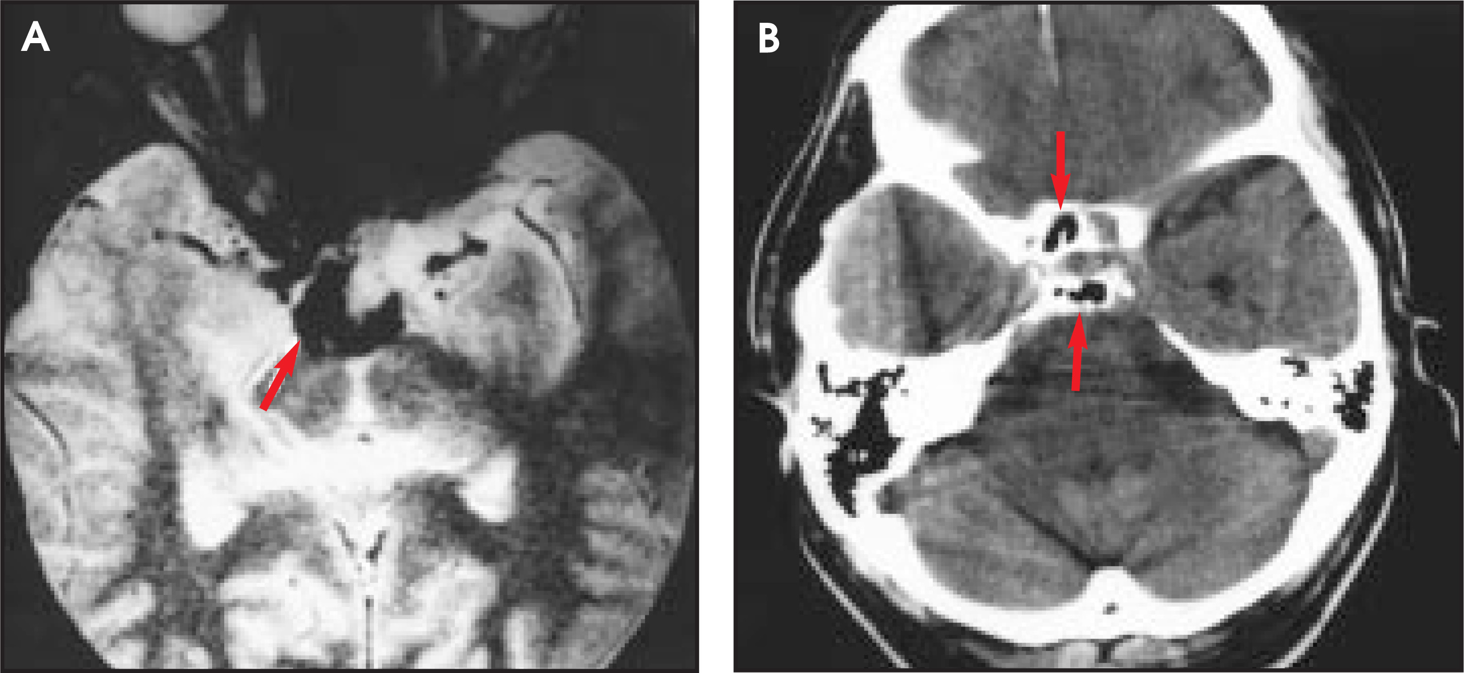
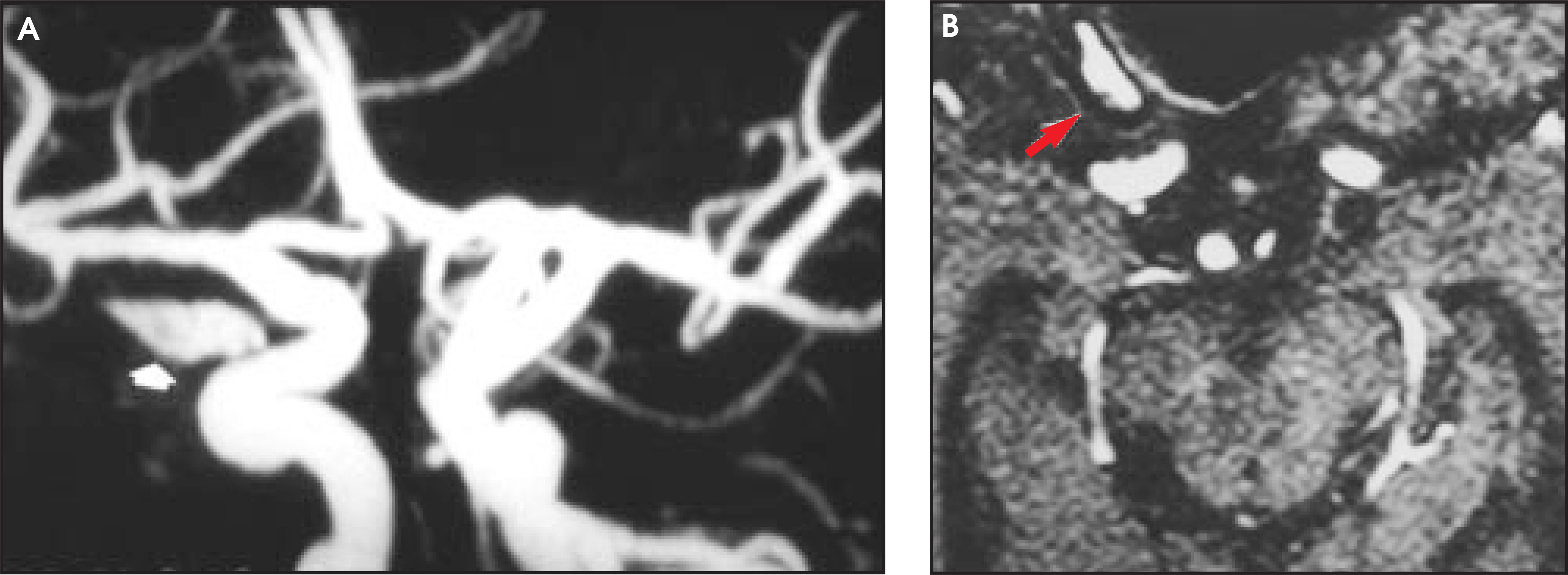
In the bygone days of plain-film neuroradiology, careful examinations of the anterior clinoid processes were of diagnostic importance. Knowledge of anatomy in this region is still important, as an aerated anterior clinoid process may be a confusing pitfall in modern MR imaging; it may mimic a tumor (Figure 1), an aneurysm (Figures 2 and 3), blood (Figure 4), calcium, or an abnormal vessel. Less frequently, an aerated dorsum sella or a posterior clinoid process may cause similar diagnostic confusion (Figures 3 and 4). Figure 5 illustrates a potential pitfall in the interpretation of a maximum intensity algorithm cerebral angiogram caused by normal fat in a nonaerated anterior clinoid process.FIGURE 1. (A) T2 axial image of a patient with right-eye visual disturbances. A pseudolesion (arrow) is seen near the optic canal. This lesion, which is isointense to the brain, was suspected initially to be meningioma. (B) CT scan reveals extensive sphenoid sinus aeration of the right anterior clinoid process, which corresponds to the pseudolesion seen on MR.FIGURE 2. (A) T2 coronal image shows apparent third (extra) flow void in the area of the left cavernous carotid artery (arrow), suggesting the possibility of aneurysm. Upon closer inspection, a small apparent third flow void is also seen in the area of the right cavernous carotid artery. (B) MR angiogram shows no evidence of a distal carotid aneurysm. (C) CT scan shows extension of sphenoid sinus aeration into both anterior clinoid processes (arrows)—explaining the pseudoflow voids.FIGURE 3. (A) In a T2 axial view, the question of a possible extra flow void (arrow) consistent with carotid aneurysm was raised. (B) CT scan revealed that asymmetrical aeration of the dorsum sella (arrow) was the reason for the low-density pseudoflow void.FIGURE 4. (A) Gradient-echo image revealed findings consistent with a subarachnoid hemorrhage in the suprasella cistern (arrow) in this patient with a severe headache. (B) CT scan reveals air in the tuberculum sella and dorsum sella (arrows) as the source of the magnetic susceptibility artifact superimposed over the subarachnoid space on the gradient-echo MR sequence.FIGURE 5. (A) Maximum intensity projection (MIP) image of an MR angiogram illustrates an apparent vascular structure (arrow) near the expected location of the right ophthalmic artery. (B) Source image reveals cortical low signal line around the fatty marrow-filled anterior clinoid process (arrow) accounting for the structure seen on the MIP MR angiogram.
Advertisement
Advertisement
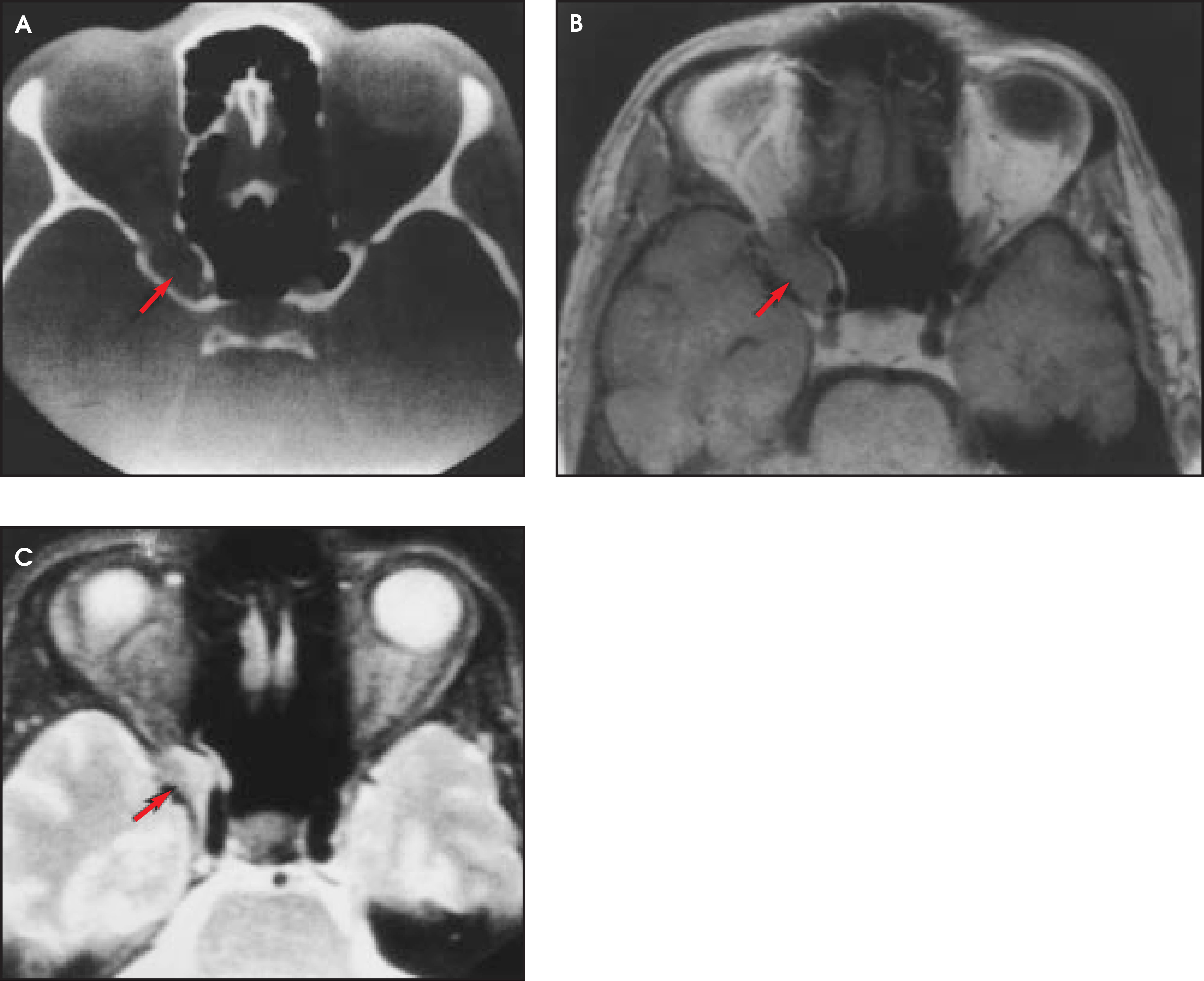
An aerated clinoid process may also be subject to diseases that typically occur in other paranasal sinuses, such as mucocoele (Figure 6), that may cause marked visual loss due to proximity to the optic nerve and ophthalmic artery.FIGURE 6. (A) Mucocele of the anterior clinoid process. Contrast-enhanced CT demonstrates expansion of the bony right anterior clinoid process by a nonenhancing soft-tissue lesion. The bony margin of the right anterior clinoid process is thinned, when compared with the normal bony margin of the left anterior clinoid process. (B) T1-weighted MR imaging confirms the CT finding of a soft-tissue mass (arrow) in the right anterior clinoid. (C) T2-weighted image illustrates inhomogeneity of the lesion (arrow). (Images reprinted with permission from Schwaighofer BW, Sobel DF, Klein MV, et al. Mucocele of the anterior clinoid process: CT and MR findings. J Comput Assist Tomgr. 1989;13:501-503.)
Endoscopic surgeons need to be aware of the anterior clinoid process because surgical instruments in an aerated clinoid process are quite close to the optic nerve and the carotid and ophthalmic arteries. AR
Citation
Healy JF. The aerated anterior and posterior clinoid processes: A pictorial review of potential pitfalls in MR imaging. Applied Radiology. 2003;32(1):17-19. doi:10.37549/AR1154.