The child with a neck mass
Applied Radiology — Vol. 34 , Issue 8 , pp. 8 -22
DOI: 10.37549/AR1359
Published: August 1, 2005
Categories
Evaluation of neck masses is a common indication for imaging children. Fortunately, most lesions are either congenital or inflammatory in origin, and only 5% of all childhood neoplasms occur in the head and neck. The primary goal of imaging is to determine the extent of the disease and suggest a differential diagnosis based on the location and imaging characteristics. A reasonable differential diagnosis can usually be made based on location (midline or lateral), clinical history (congenital or acquired, presence or absence of fever and tenderness), and imaging characteristics (cystic versus solid). The remainder of this manuscript will focus on the more common cystic and solid masses that occur in the pediatric neck.
Imaging modalities
Potential imaging modalities include conventional radiographs, ultrasound (US), computed tomography (CT), magnetic resonance imaging (MRI) and nuclear medicine. Conventional radiographs are used for evaluation of patients with stridor, suspected retropharyngeal abscess or adenoid hypertrophy. Ultrasound is ideal for optimally determining whether a mass is cystic or solid, as well as for assessing whether a node is suppurative and for guiding abscess drainage. In addition, it is the imaging modality of choice in children with suspected thyroglossal duct cyst, not only to prove the cystic nature of the midline neck mass but also to confirm the presence of a normal- appearing bi-lobed thyroid gland in the lower neck.1 Ultrasound is also ideal for evaluating patients with suspected fibromatosis colli.2,3 Ultrasound, CT, or MRI can be used for evaluating jugular vein patency in patients with suspected Lemierre syndrome (internal jugular vein thrombophlebitis and septic emboli secondary to pharyngotonsillitis).4,5 CT and/ or MRI are frequently used to evaluate the total extent of more diffuse diseases, including inflammatory, congenital, and neoplastic processes. Since imaging with MRI frequently requires sedation in children less than 6 or 7 years of age, CT of the neck is more frequently performed on these children. CT of the neck is also frequently performed in combination with CT of the chest/abdomen/pelvis in children with neoplasms such as lymphoma. CT is ideal for evaluating osseous erosion in children with suspected rhabdomyosarcoma, with MRI frequently performed as an adjunct in patients with suspected intracranial or intraspinal extension. MRI is the preferred method of imaging in children with suspected hemangioma of infancy, congenital vascular malformations, cervical neuroblastoma, and neurofibroma. Nuclear medicine imaging is frequently used in combination with CT and/or MRI for evaluation of children with neuroblastoma (I-123), lymphoma (gallium-67 citrate or fluorodeoxyglucose positron emission tomography [FDG-PET]), osteomyelitis (technetium [Tc]-99m-methylene diphosphonate [MDP], gallium-67 citrate) and other neoplasms.
Cystic neck masses
The majority of cystic neck masses in children are congenital malformations and include thyroglossal duct cysts, branchial apparatus cysts, dermoid cysts, and lymphatic malformations.
Thyroglossal duct cysts
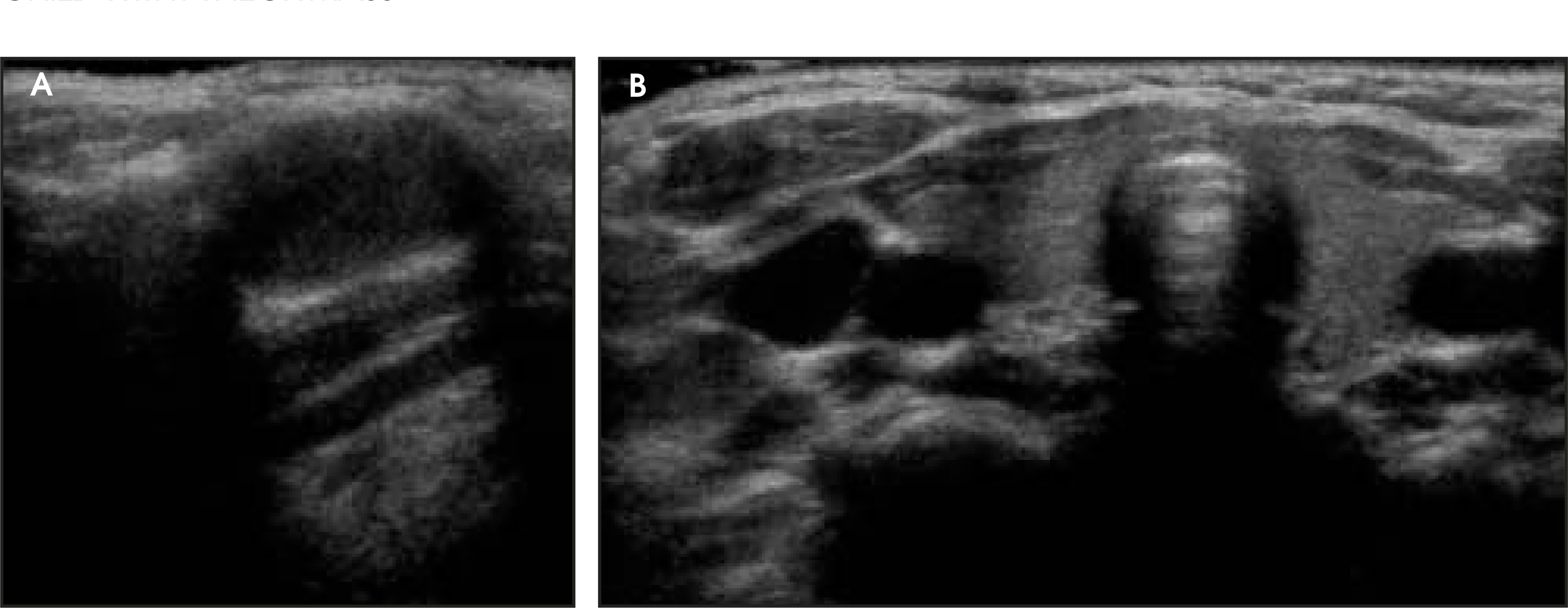
The thyroglossal duct (TGD) is a normal fetal structure that extends from the foramen cecum at the posterior aspect of the tongue to the lower neck in the region of the thyroid bed. The TGD normally involutes during the 5th or 6th week of fetal life. If any portion of the duct does not involute, remnant secretory epithelium may form a cyst.6,7 Most TGD cysts occur in the midline. Twenty percent to 25% are in the suprahyoid neck, 15% to 59% are at the hyoid bone, and 25% to 65% are in the infrahyoid neck.7,8 Patients typically present with a midline neck mass that elevates with swallowing or protrusion of the tongue. If infected, they might present with warmth, erythema, and fever. In the past, nuclear medicine imaging with Tc-99m-pertechnetate was used to assess for the presence of a bi-lobed thyroid gland in the lower neck and rule out ectopic thyroid in the midline mass. More recently, US has been used to evaluate for a normal-appearing bi-lobed thyroid gland in the lower neck and to confirm the cystic nature of the midline neck mass.1 Uncomplicated thyroglossal duct cysts may be anechoic, hyperechoic, or heterogeneous on US. The presence of intralesional echoes does not imply superimposed infection or hemorrhage (Figure 1).9 On CT and MRI, thyroglossal duct cysts will appear as fluid-filled cysts of variable attenuation and signal intensity, depending on the protein content of the cyst (Figure 2). If there is superimposed infection, there may be edema in the adjacent soft tissues and peripheral contrast enhancement. Treatment of thyroglossal duct cysts is surgical excision (Sistrunk procedure) that includes excision of the cyst, the entire remnant tract, and a central portion of the hyoid bone.10
Branchial apparatus anomalies
Normal branchial apparatus embryology is quite complex and beyond the scope of this manuscript. However, in general, the branchial apparatus structures develop between the 4th and 6th week of gestation and consist of 6 pairs of mesodermal branchial arches separated by 5 paired endodermal pharyngeal pouches internally and 5 paired ectodermal bran-chial clefts externally. Anomalies of the branchial apparatus may be in the form of cysts, sinus tracts, or fistulae. Branchial apparatus cysts are the most common branchial apparatus anomalies that require imaging.
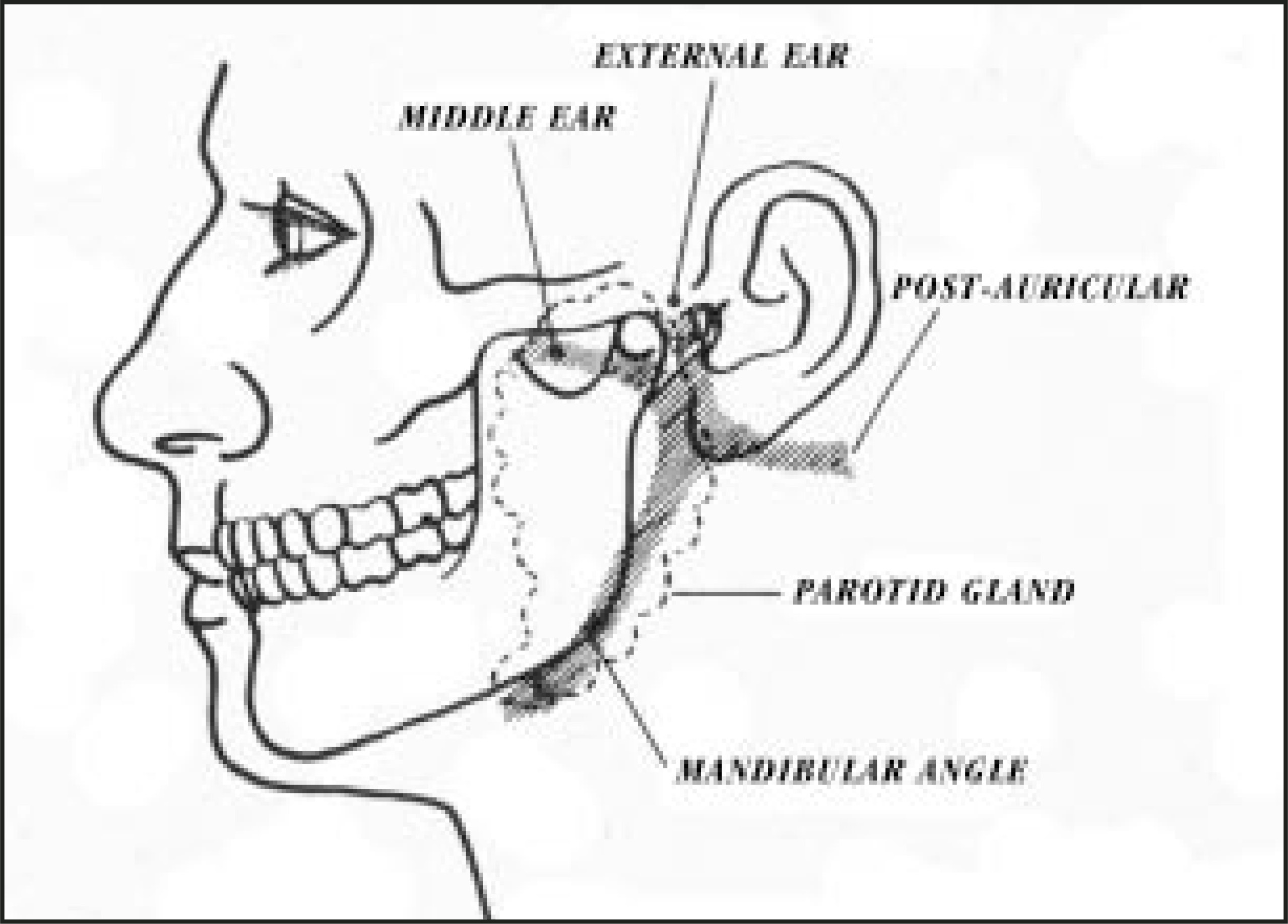
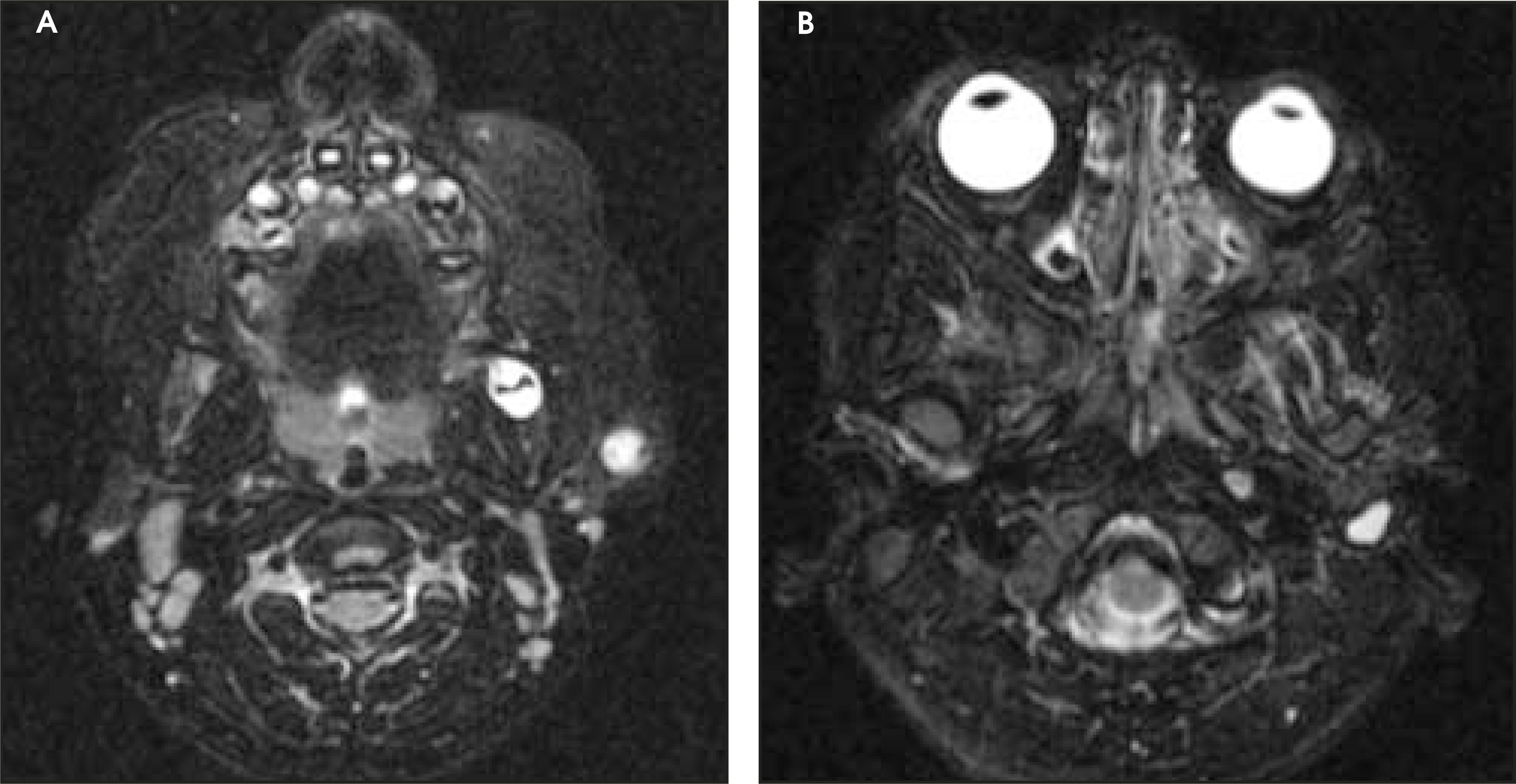
First branchial anomalies account for only 8% of all branchial anomalies and are usually cysts or sinuses near the external auditory canal, the pinnae, or the region of the parotid gland (Figure 3).11-13 Patients typically present with masses or sinus tracts, with or without recurrent infection. The cysts may be imaged with CT, US, or MRI. MR imaging is ideal in evaluating the fluid-filled tract, which may extend from the more superficial cyst to the external auditory canal (Figure 4).
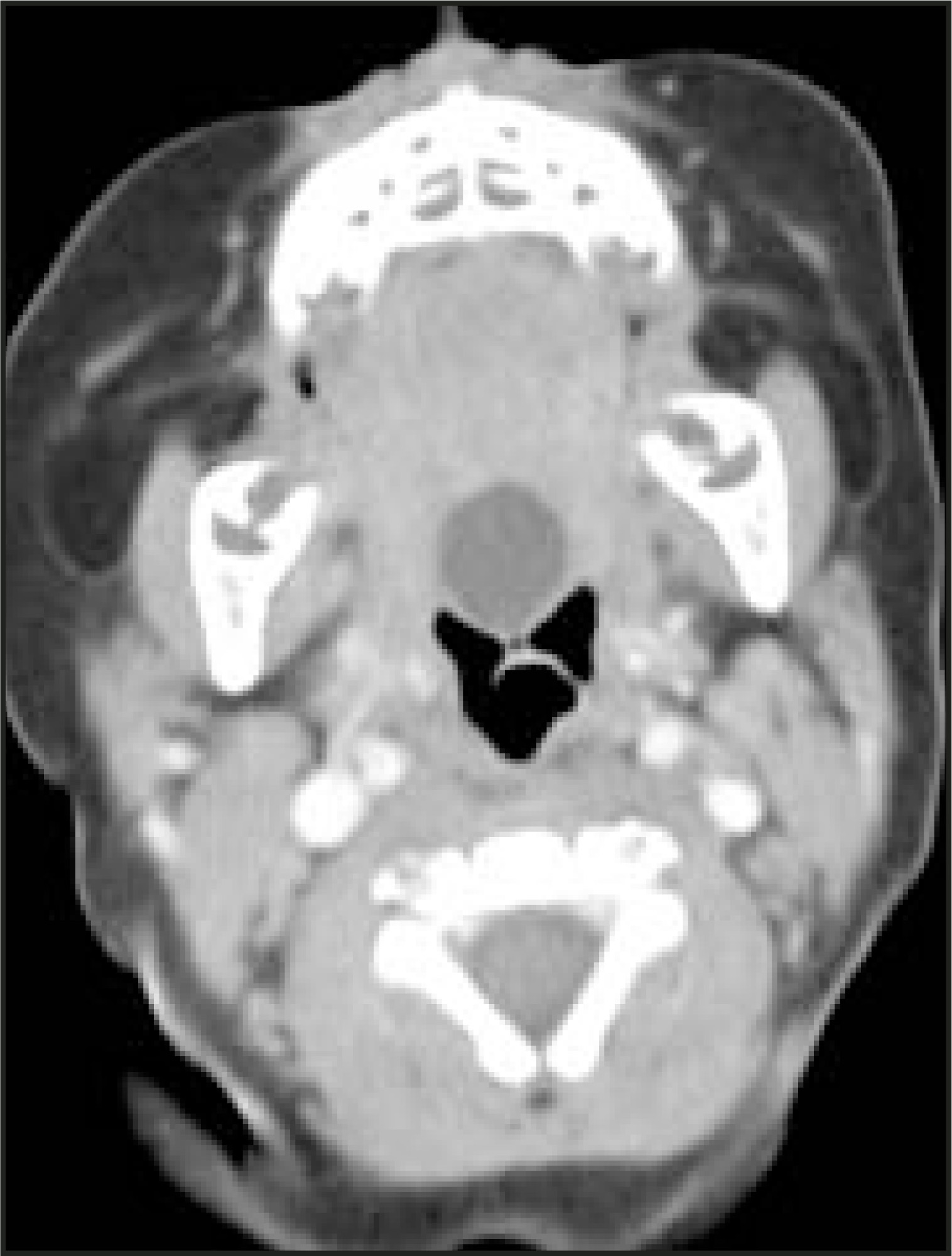
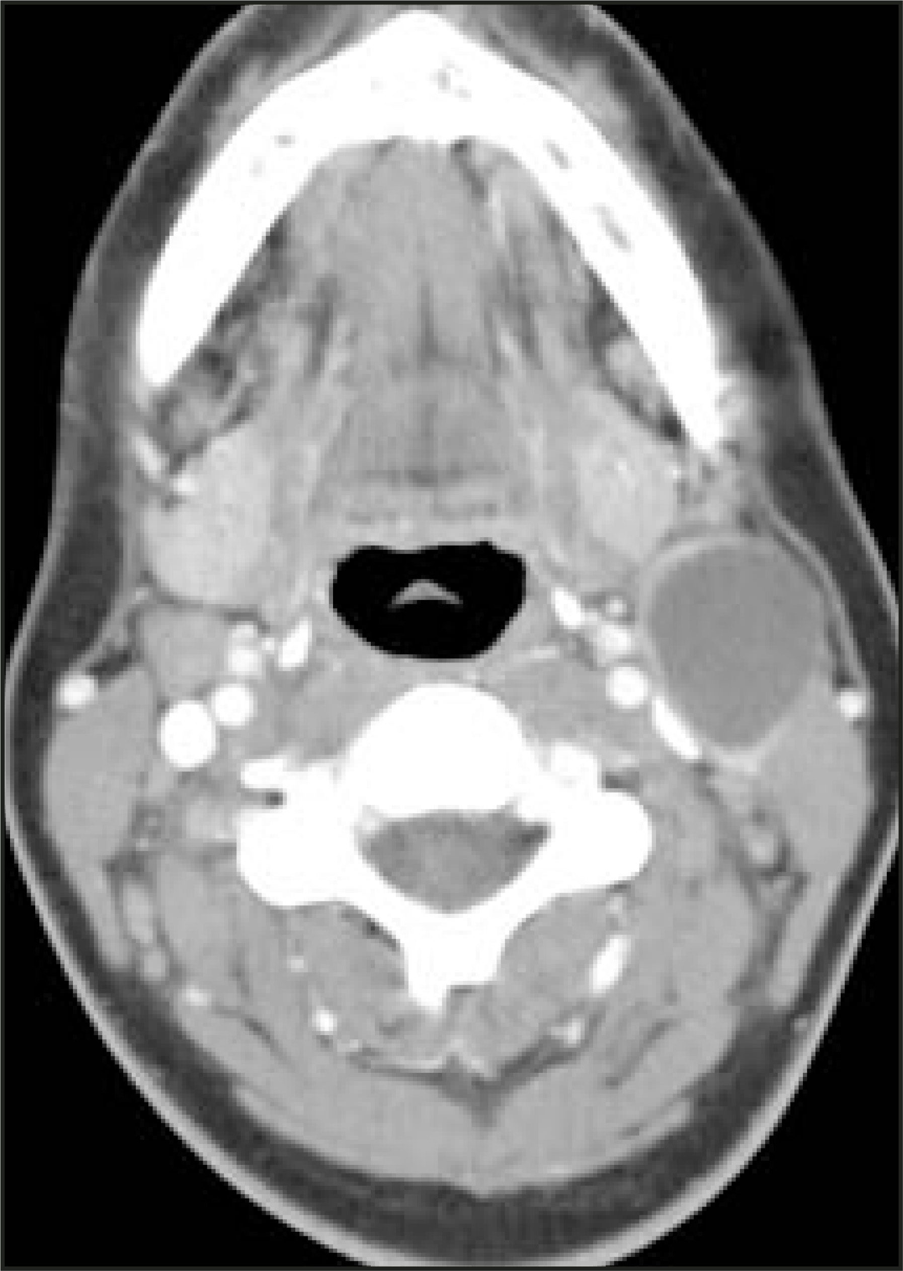
Second branchial apparatus cysts are the most common and account for up to 95% of all branchial apparatus anomalies. Most are located anterior to the sternocleidomastoid muscle, posterior to the submandibular gland and lateral to the carotid sheath (Figure 5). Occasionally, cysts may protrude between the internal and external carotid arteries; rarely, they are deep to the platysma muscle and anterior to the sternocleidomastoid muscle or located directly adjacent to the pharyngeal wall.14 As with other cysts, these lesions will be hyperechoic or anechoic on US, low in attenuation on CT, and fluid in signal intensity on MRI unless there is associated superimposed infection, which will change the imaging characteristics on all modalities. The key to making this diagnosis is the location of the lesion.
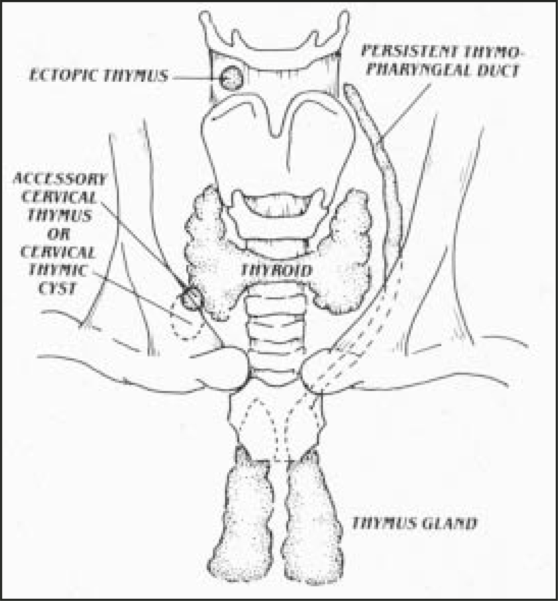
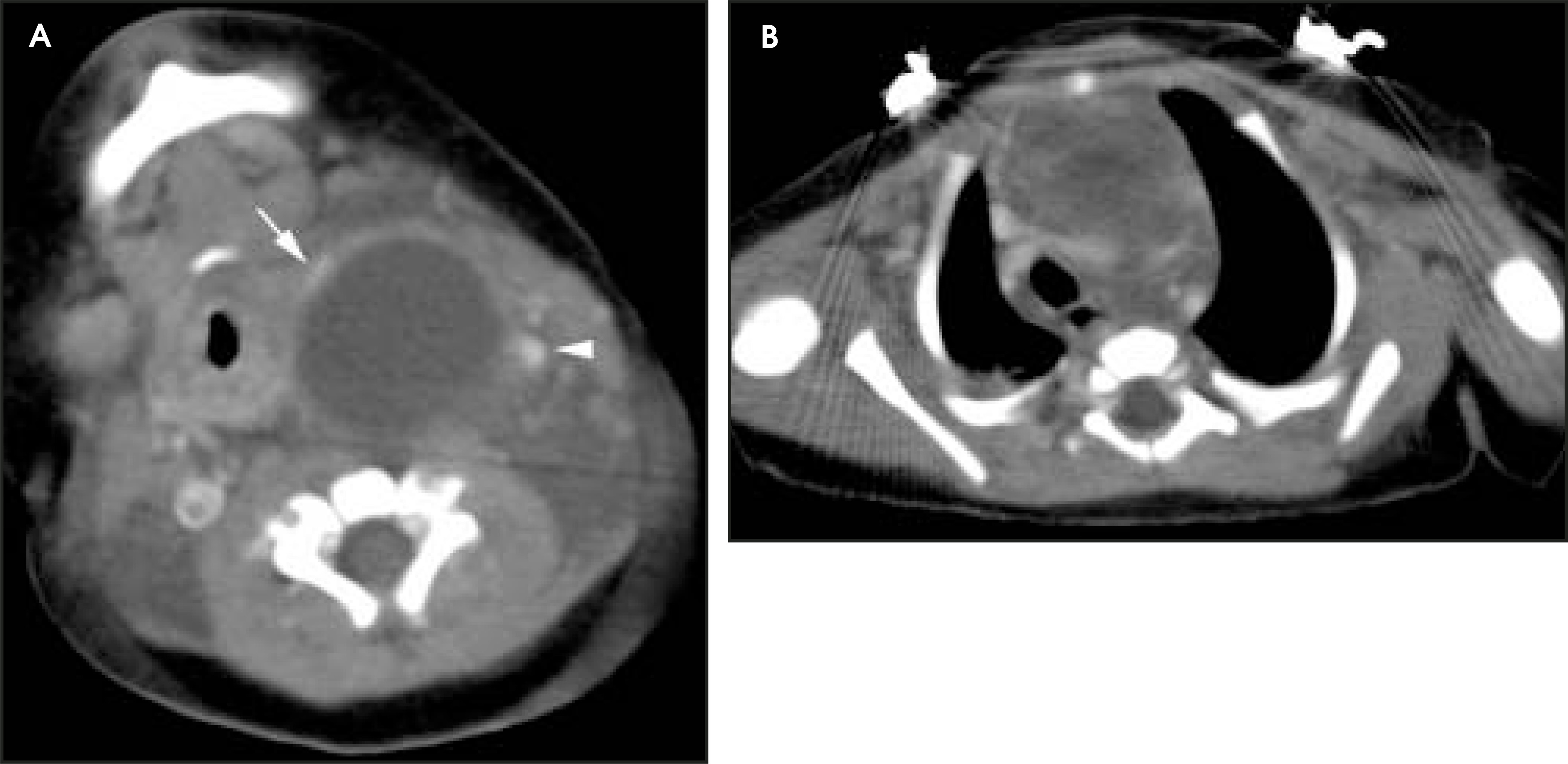
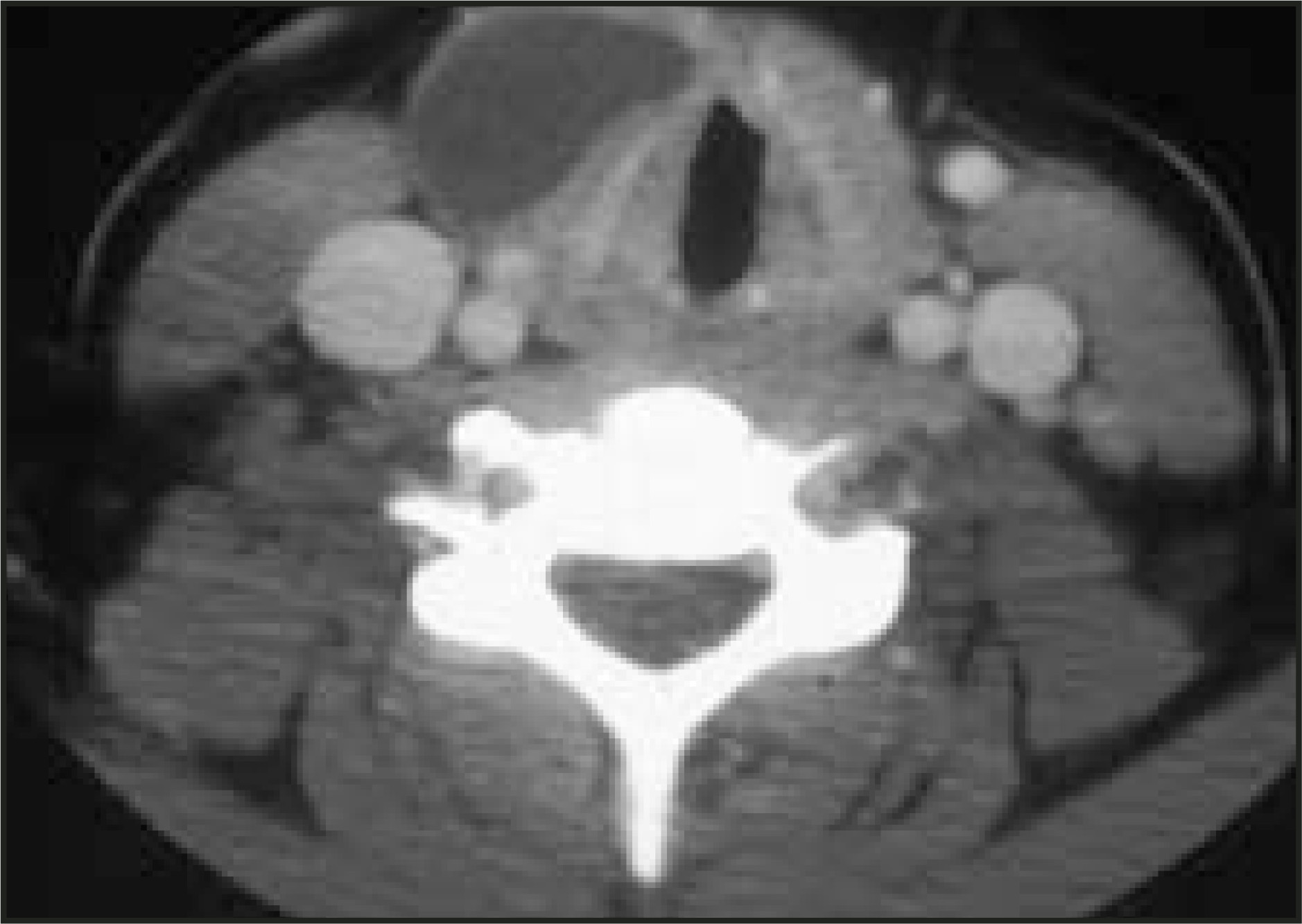
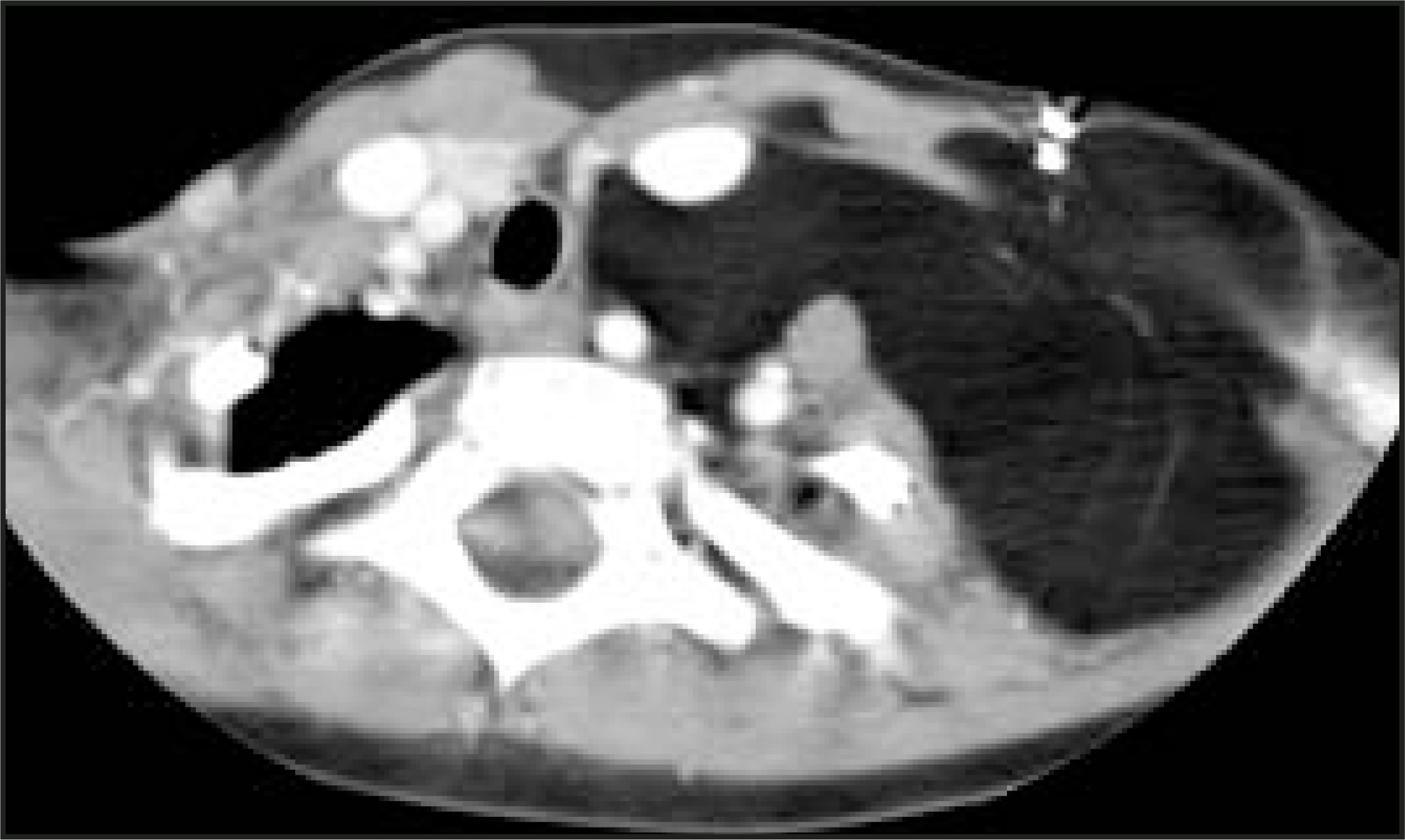
The most common third branchial apparatus anomaly is the thymic cyst. These are rare remnants of the third branchial pouch. In normal embryologic development, the thymic primordia migrates from the pharynx caudally and fuses in the anterior mediastinum along the course of the thymopharyngeal duct. Similar to the thyroglossal duct cysts, when the duct fails to involute, a cyst may occur anywhere along the thymopharyngeal duct from the angle of the mandible to the upper mediastinum (Figure 6).12,15There may be intralesional echoes on US with or without superimposed hemorrhage or infection. Up to 50% of cervical thymic cysts will be continuous with the mediastinal thymus (Figure 7).16 Cervical thymic cysts are intimately associated with the carotid sheath; they frequently splay the carotid artery and jugular vein, particularly when they involve the suprahyoid neck. As with other branchial apparatus anomalies, the location is the key to suggesting the diagnosis. In a child, any cystic mass that involves the neck and also extends to the anterior mediastinum should certainly suggest a cervical thymic cyst.
Dermoid cysts and epidermoid cysts
Dermoid cysts may contain squamous epithelium and skin appendages such as hair follicles and sebaceous glands. Epidermoid cysts contain only squamous epithelium. Both may present as midline neck masses with variable attenuation, echogenicity, and signal intensity, depending on internal contents (Figure 8). If fat globules are identifiable on CT or MRI, then a dermoid cyst is the most likely diagnosis. If there are associated calcifications, then the lesion is more likely a teratoma or venous malformation. If the lesion is entirely composed of fat, then simple lipoma is the most likely diagnosis (Figure 9).
Lymphatic malformations
Lymphatic malformations (LMs) are vascular malformations composed of primative embryonic lymph sacs of varying sizes. Lymphatic malformations typically increase in size as the child grows, and they may show rapid increase in size in association with upper respiratory tract infection or intralesional hemorrhage. Although they may be seen in genetic syndromes such as Turner’s syndrome, Noonan’s syndrome, and trisomy 21 syndrome, most LMs occur in children with normal karyotype.
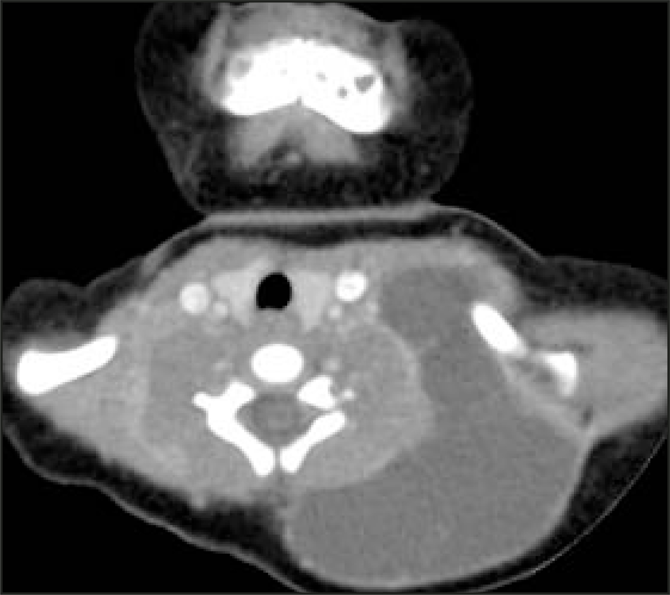
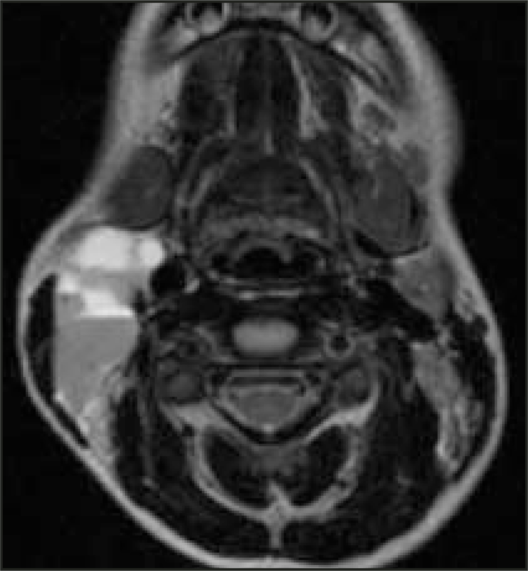
Lymphatic malformations can be divided into microcystic, macrocystic, or mixed lesions. They may be unilocular, multilocular, focal, or diffuse/infiltrative. The fluid-filled spaces are usually anechoic or hypoechoic on US and usually have low attenuation on CT and fluid signal intensity on MRI. There may be minimal enhancement of internal septations.17-20 They are the primary cystic malformation to present with transspatial involvement (Figure 10). Of all cystic lesions in the neck, LMs are the most likely to hemorrhage, frequently resulting in intralesional fluid-fluid levels (Figure 11). As with other cystic lesions, if there is superimposed infection, the imaging characteristics will change accordingly.
Lymphatic malformations are frequently part of mixed vascular malformations, with the most common additional component being a venous malformation. The venous malformation component may appear as a cystic malformation on precontrast images but will show postcontrast enhancement and may contain phleboliths (Figure 12). When LMs are complex and transspatial or when they involve multiple structures of the neck (eg, airway, tongue, floor of mouth) or the parotid gland, and in patients with combined vascular malformations, MR imaging is the preferred modality.18-20
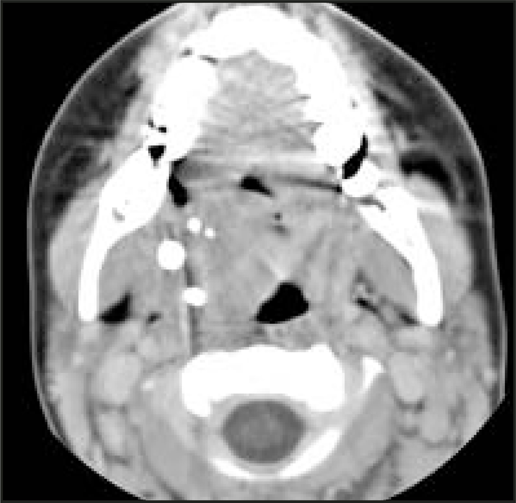
The preferred treatment of LMs is surgical excision. However, complete surgical excision may be difficult when the lesions are microcystic and infiltrative. Macrocystic lesions may also be treated with percutaneous sclerotherapy, including alcohol solution, cyclophosphamide, bleomycin and doxycycline.21-23
Solid neck masses
The remainder of this manuscript will focus on benign and malignant extra thyroid solid neck masses in children, including fibromatosis colli, inflammatory adenopathy, hemangioma, neurofibroma, teratoma, lymphoma, rhabdomyosarcoma, neuroblastoma, and metastatic adenopathy.
Fibromatosis colli
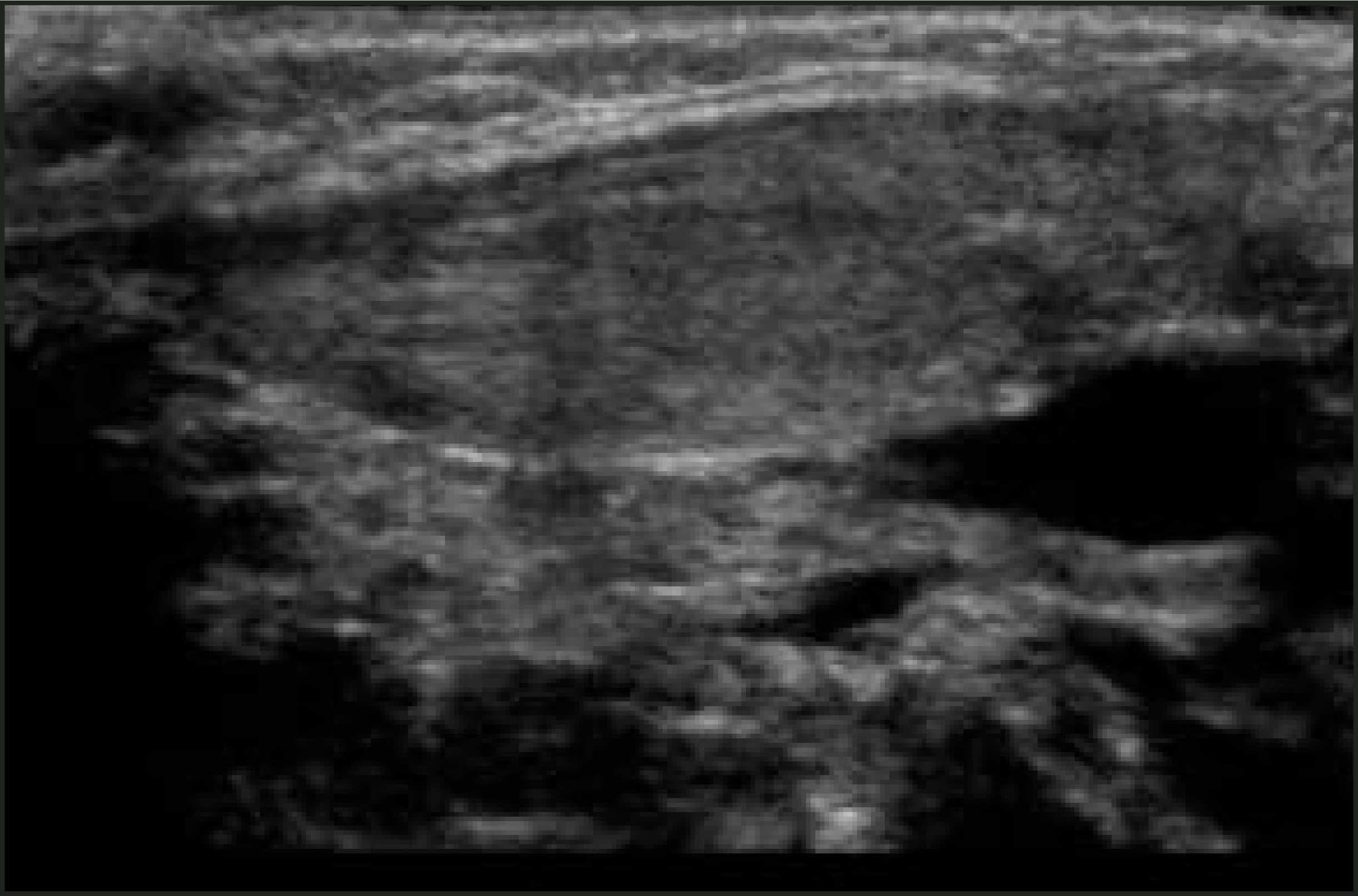
Fibromatosis colli or sternocleidomastoid tumor of infancy occurs in neonates. The etiology is uncertain but may be related to intramuscular hemorrhage or fibrosis. There is an increased risk in neonates with a history of breech presentation and/or difficult delivery. Patients typically present at 2 to 4 weeks of age with a unilateral neck mass, with or without torticollis. Ultrasound is the imaging modality of choice, showing focal or diffuse enlargement of the sternocleidomastoid muscle (SCM), which may be homogeneous or heterogeneous in echotexture (Figure 13). There may be a rim of decreased echoes thought to represent compressed normal adjacent sternocleidomastoid muscle.2,3,24
Inflammatory adenopathy
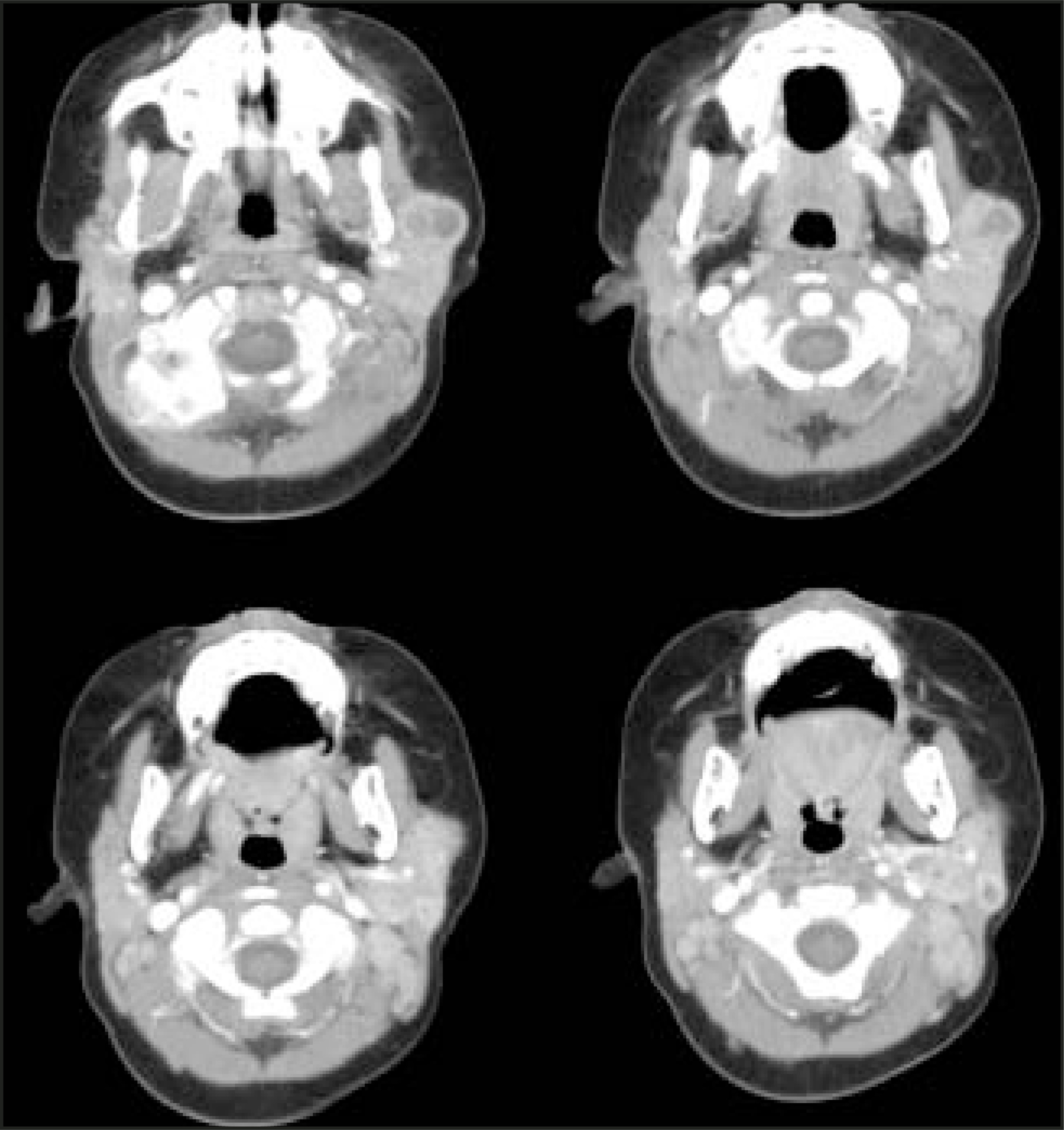
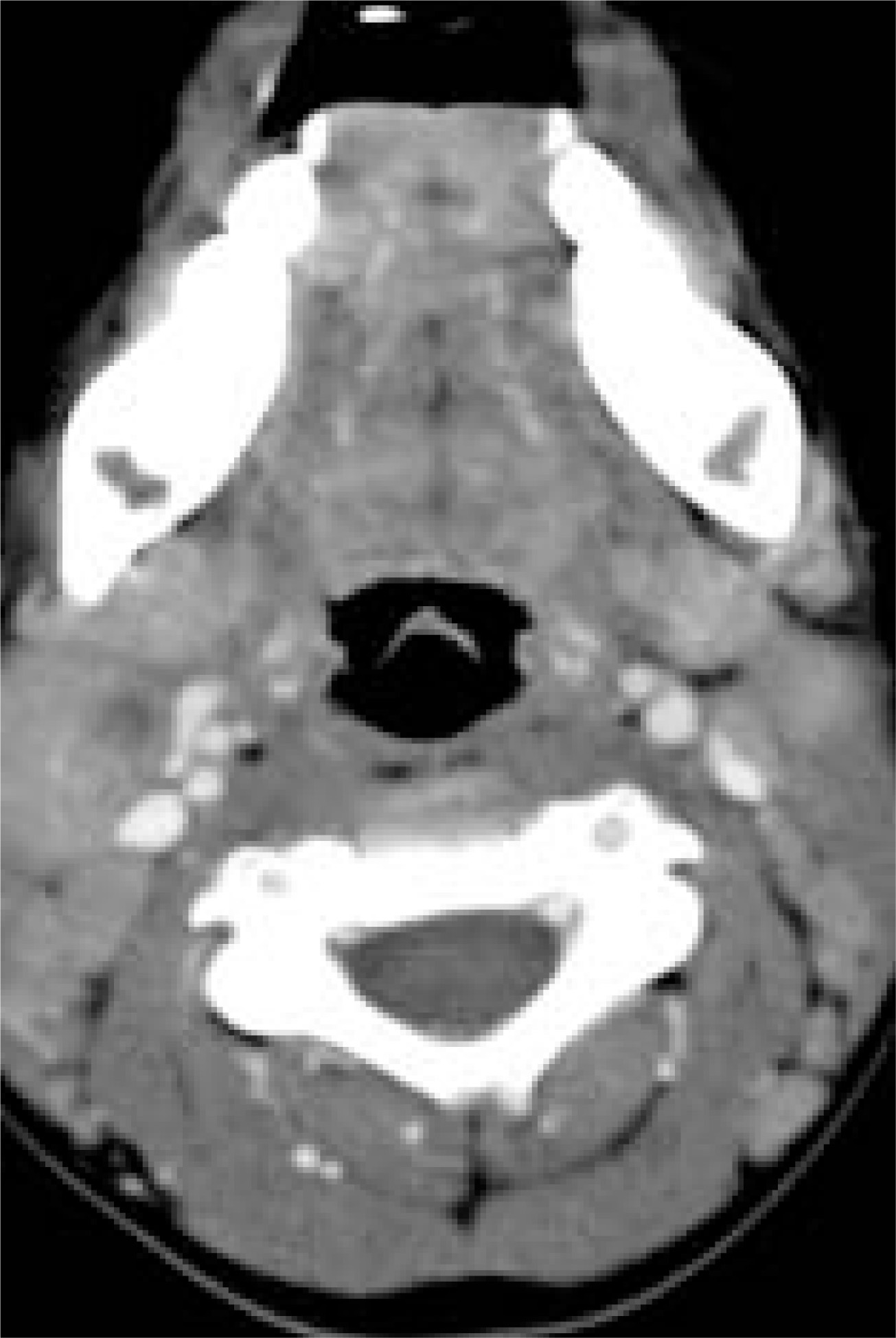
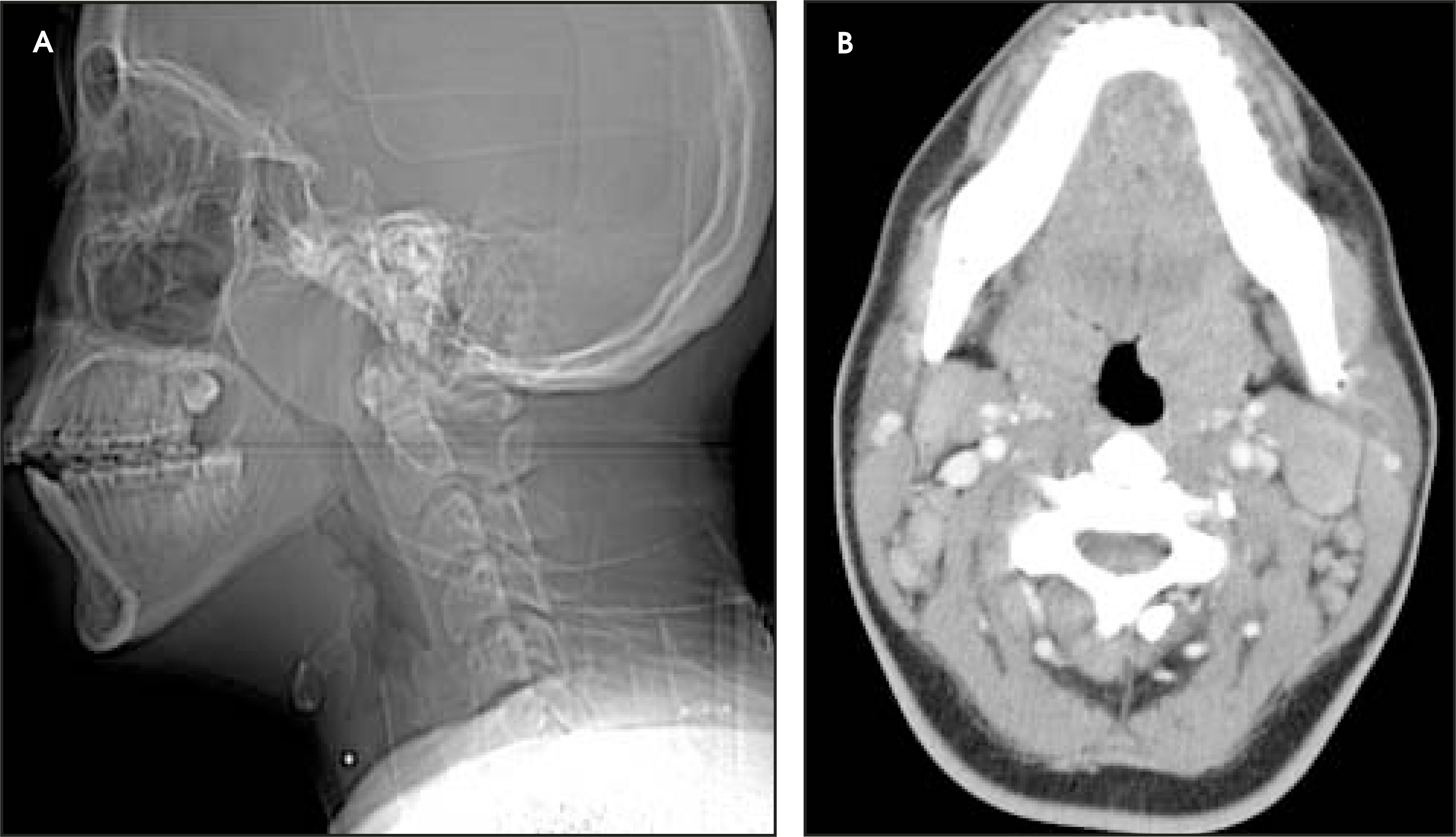
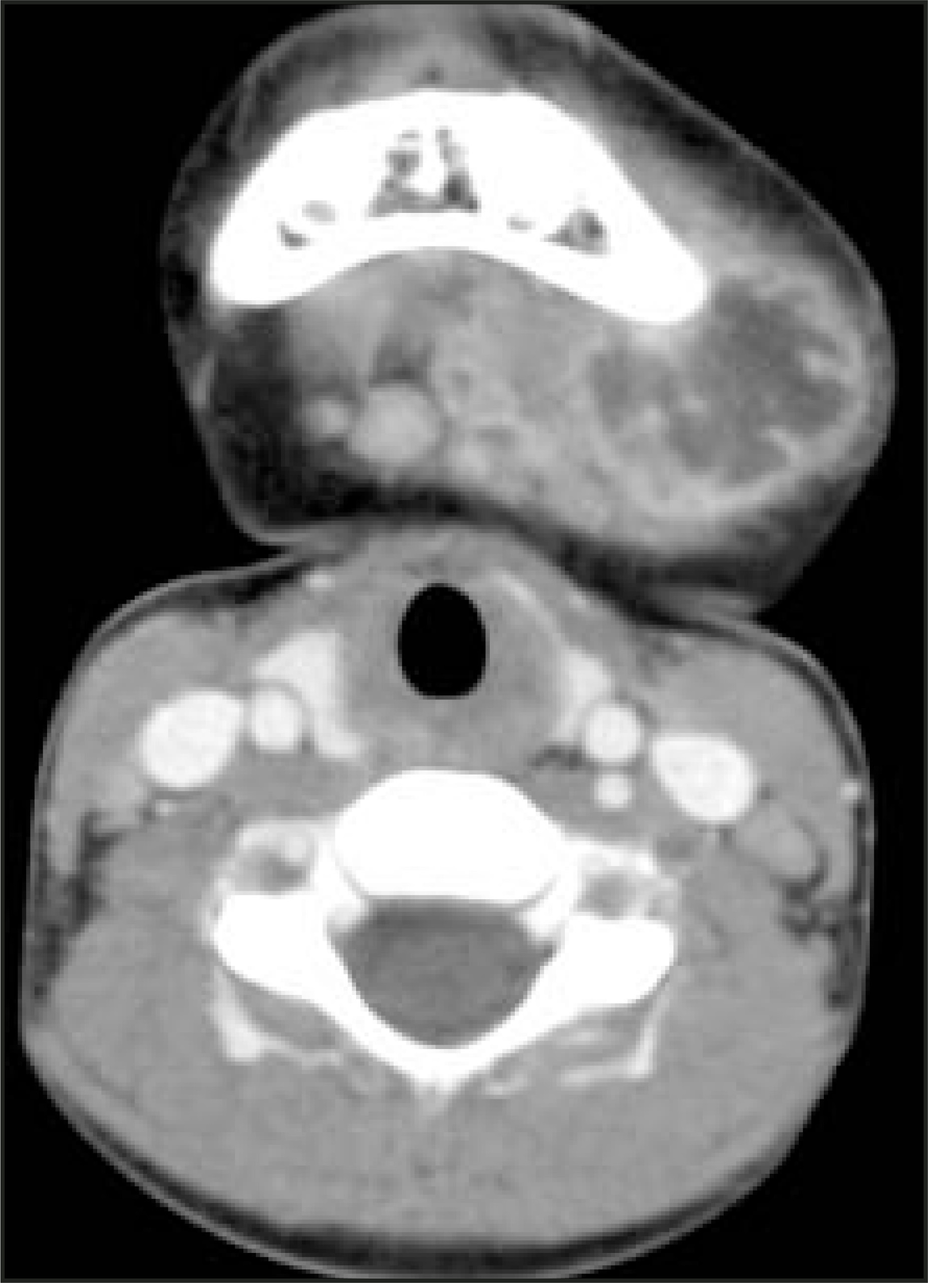
Nonsuppurative adenitis is the most common non-neoplastic solid neck mass in children. The majority of children with uncomplicated cervical adenitis are treated medically and do not require imaging. If they are toxic-appearing or have symptoms that increase despite administration of antibiotic therapy, postcontrast CT imaging is frequently performed to assess the extent of the inflammatory process and rule out a focal abscess. When imaging children with cervical adenitis, the key questions to be answered are location of abscess, extent of cellulitis and myositis, and evaluation of the vascular structures of the neck to exclude jugular vein compression or thrombosis as well as narrowing of the internal carotid artery.25 Lymphadenitis may be secondary to viral disease, bacterial disease, mycobacterial disease, or fungal disease. Nonsuppurative inflammatory adenitis has a typical appearance on all imaging modalities showing multiple (sometimes conglomerate) nodes. Hilar flow is demonstrated on US. Nontuberculous mycobacterial disease may lack evidence of surrounding cellulitis and myositis and may contain small calcifications or low-attenuation centers (Figure 14).26-28 Cat-scratch disease is not uncommon in children and may also present with cervical adenopathy, without significant associated cellulitis (Figure 15). This diagnosis should be considered in any child with a history of exposure to cats. If a child without evidence of cellulitis presents with significant cervical adenopathy and associated enlargement of the palatine and adenoid tonsils, mononucleosis should be considered (Figure 16). A well-defined or irregularly shaped fluid collection with an enhancing rim is certainly suggestive of an abscess (Figure 17). However, these imaging characteristics are not 100% specific, and the appearance of phlegmon without true drainable pus can be similar.29
Hemangioma of infancy
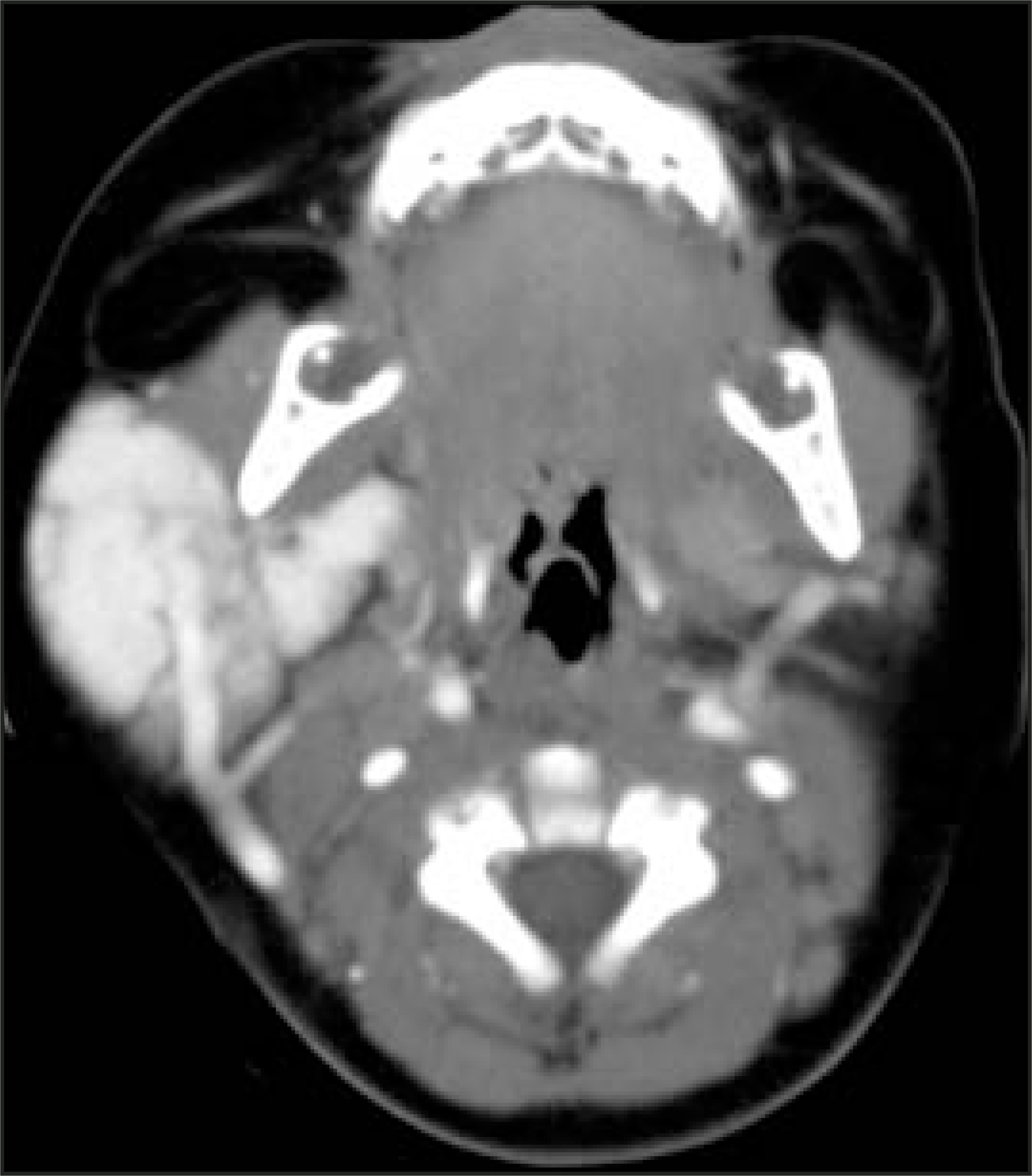
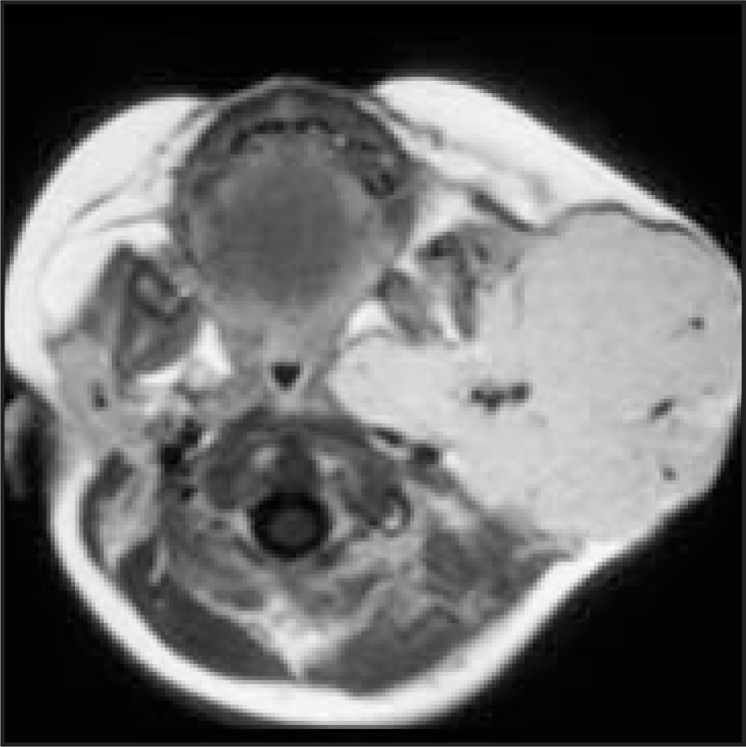
Hemangioma of infancy is a benign neoplasm resulting from endothelial proliferation, which usually presents in children less than 6 months of age, gradually increases over the next 2 years, and spontaneously involutes over the next 5 to 7 years. In the proliferative phase, they demonstrate diffuse intense contrast enhancement (Figure 18). High-flow intralesional flow voids will be demonstrated on MRI (Figure 19). During the involutional phase, they show evidence of fatty infiltration and decrease in size.30,31 Attention to the age of the patient with a significantly enhancing neck mass is extremely important. In an older child without a prior history of a neck mass in infancy, similar imaging characteristics should raise the question of sarcoma rather than of benign hemangioma. Treatment of hemangioma occurring in an infant is usually expectant waiting. Steroids, interferon, and, rarely, surgery are reserved for lesions that compromise the airway or are associated with significant loss of skin integrity.
Neurofibromas
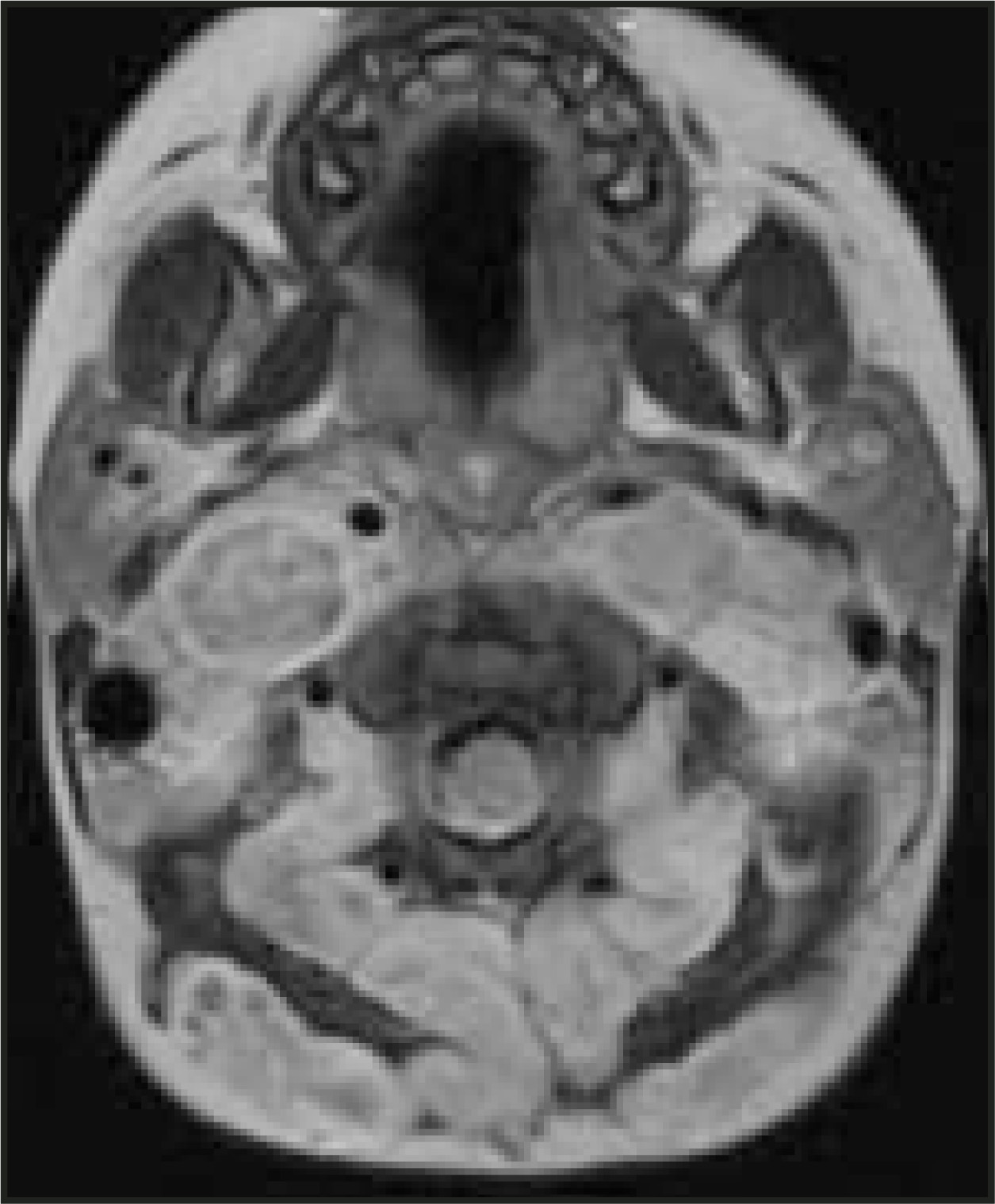
Neurofibromas are unencapsulated benign nerve sheath tumors of Schwann cell origin. They may be single or multiple; when multiple or plexiform, they almost always occur in children with neurofibromatosis Type 1. Solitary lesions may be very well defined; plexiform lesions are ill defined and infiltrative. Frequently, neurofibromas will show variable contrast enhancement, with or without a “target sign” composed of a central area of decreased T2 signal intensity and decreased contrast enhancement (Figure 20).32
Teratomas
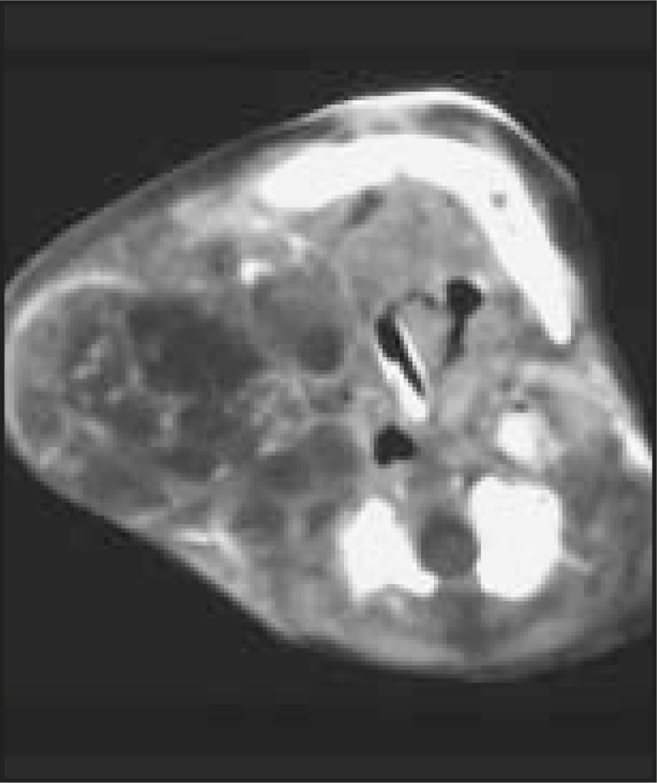
Teratomas are neoplasms composed of multiple tissues that are foreign to the part of the body in which the lesion arises.33 Teratomas in children are most commonly sacrococcygeal, and 5% to 14% occur in the head and neck region; most of these children are less than 1 year of age at the time of diagnosis. These lesions contain all 3 germ layers, originate from pleuripotential cells, and may be composed of mature or immature elements. In the neonate, the presence of immature elements does not correlate with malignant potential, and most teratomas are benign. Most are very large and complex lesions containing solid and cystic components, frequently with fat and calcification (Figure 21). Treatment is surgical excision.
Malignant solid masses
Lymphoma
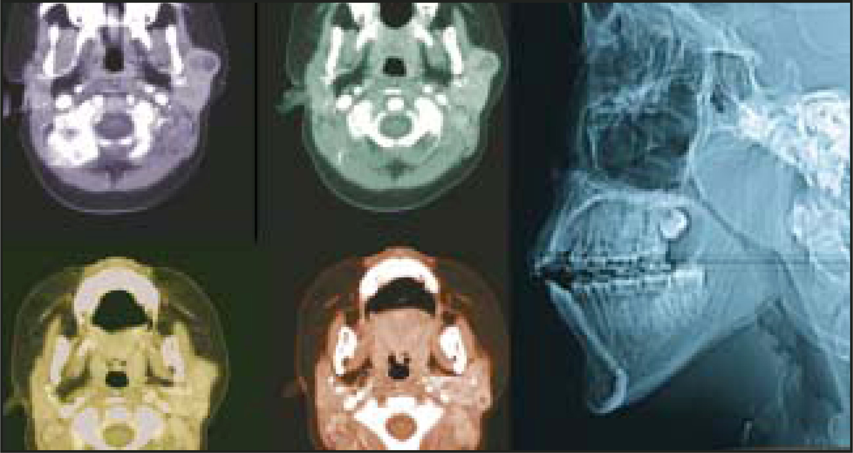
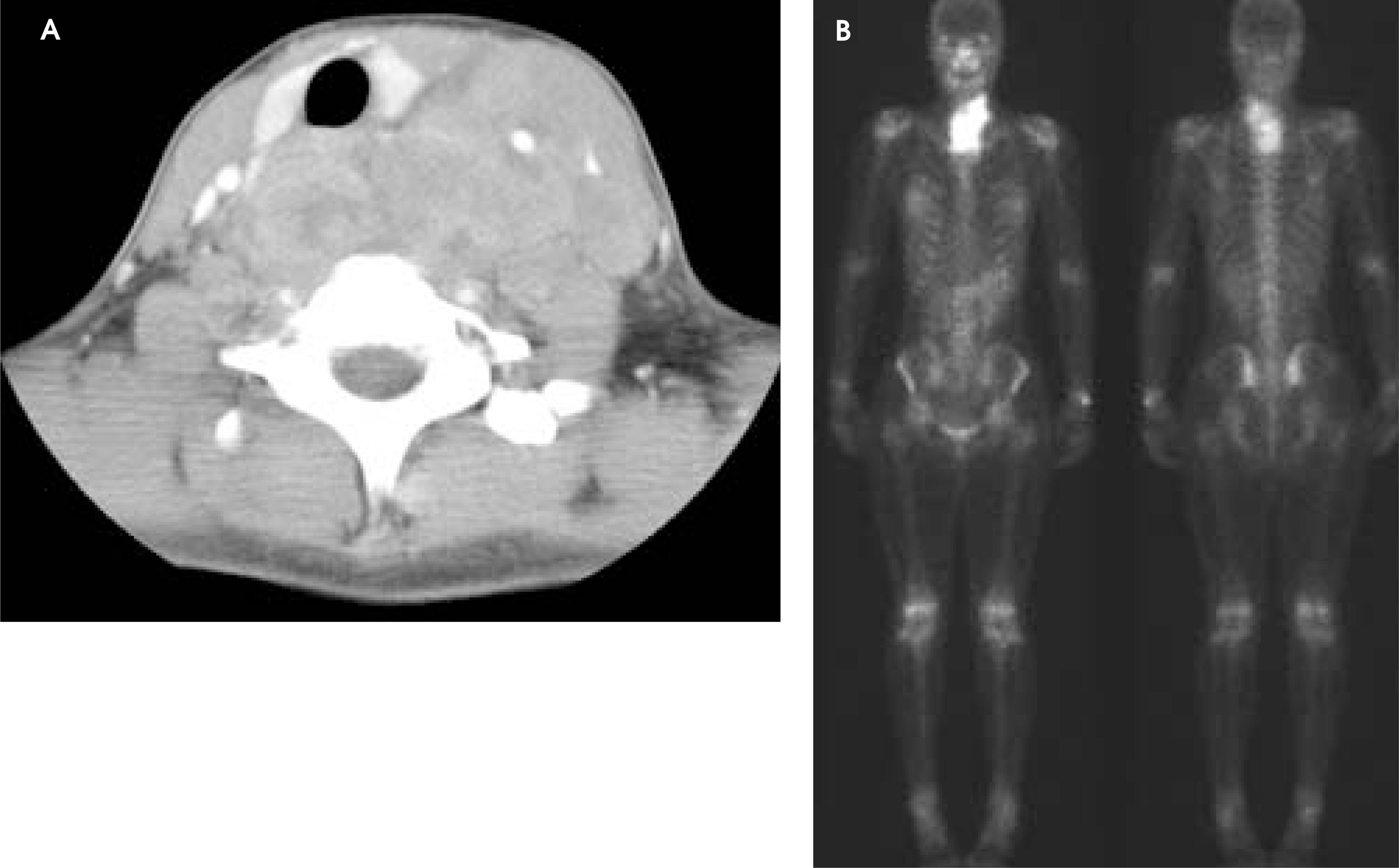
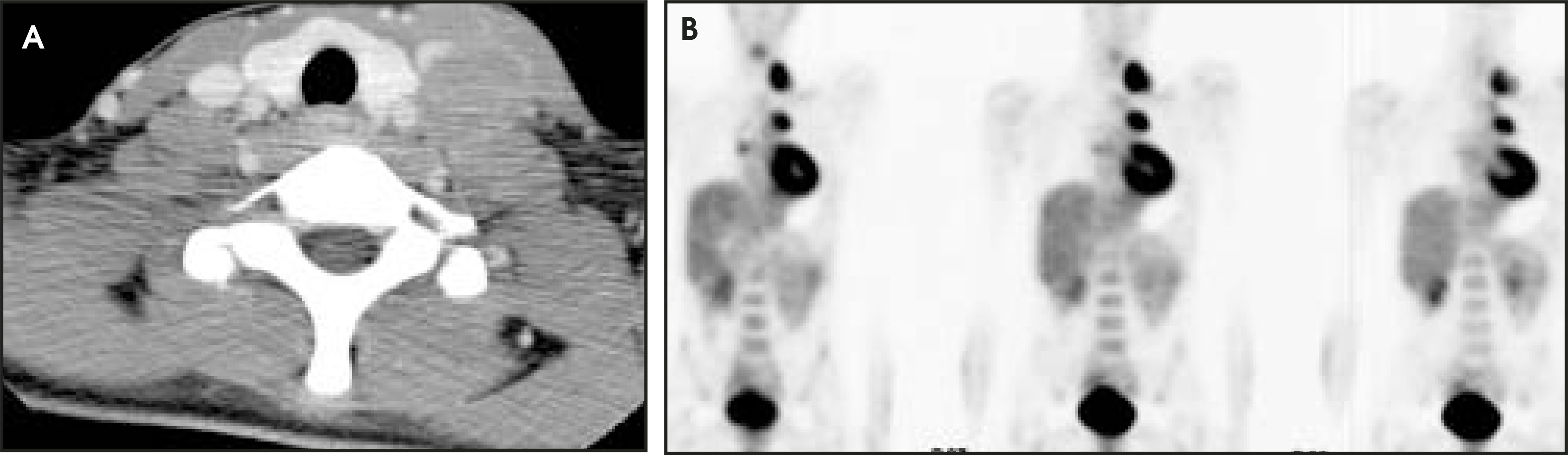
Malignant lymphoma accounts for approximately 50% of head and neck malignancies in children. Approximately 50% of cervical involvement with lymphoma is due to Hodgkin’s disease and 50% due to non-Hodgkin’s lymphoma. Imaging characteristics cannot distinguish between the two. Patients may present with unilateral or bilateral disease in both Hodgkin’s and non-Hodgkin’s lymphoma. Lymphomatous nodes have a similar appearance to inflammatory and metastatic nodes from other primary malignancies. However, lymphomatous nodes are frequently larger and more extensive than inflammatory adenopathy. The differential diagnosis in children with bulky cervical adenopathy includes mononucleosis (particularly if there is associated enlargement of the adenoids and palatine tonsil), metastatic disease (rarely, from primary malignancy such as nasopharyngeal carcinoma, rhabdomyosarcoma, or neuroblastoma), and lymphoproliferative disease (particularly in the posttransplant patients). Imaging of patients with head and neck lymphoma should include CT of the involved area and adjacent lymph nodes as well as the chest, abdomen, and pelvis. In addition, gallium-67 citrate scintigraphy is frequently used for initial staging and follow-up (Figure 22). Increasingly, FDG-PET is being used in evaluation of these patients (Figure 23).
Rhabdomyosarcoma
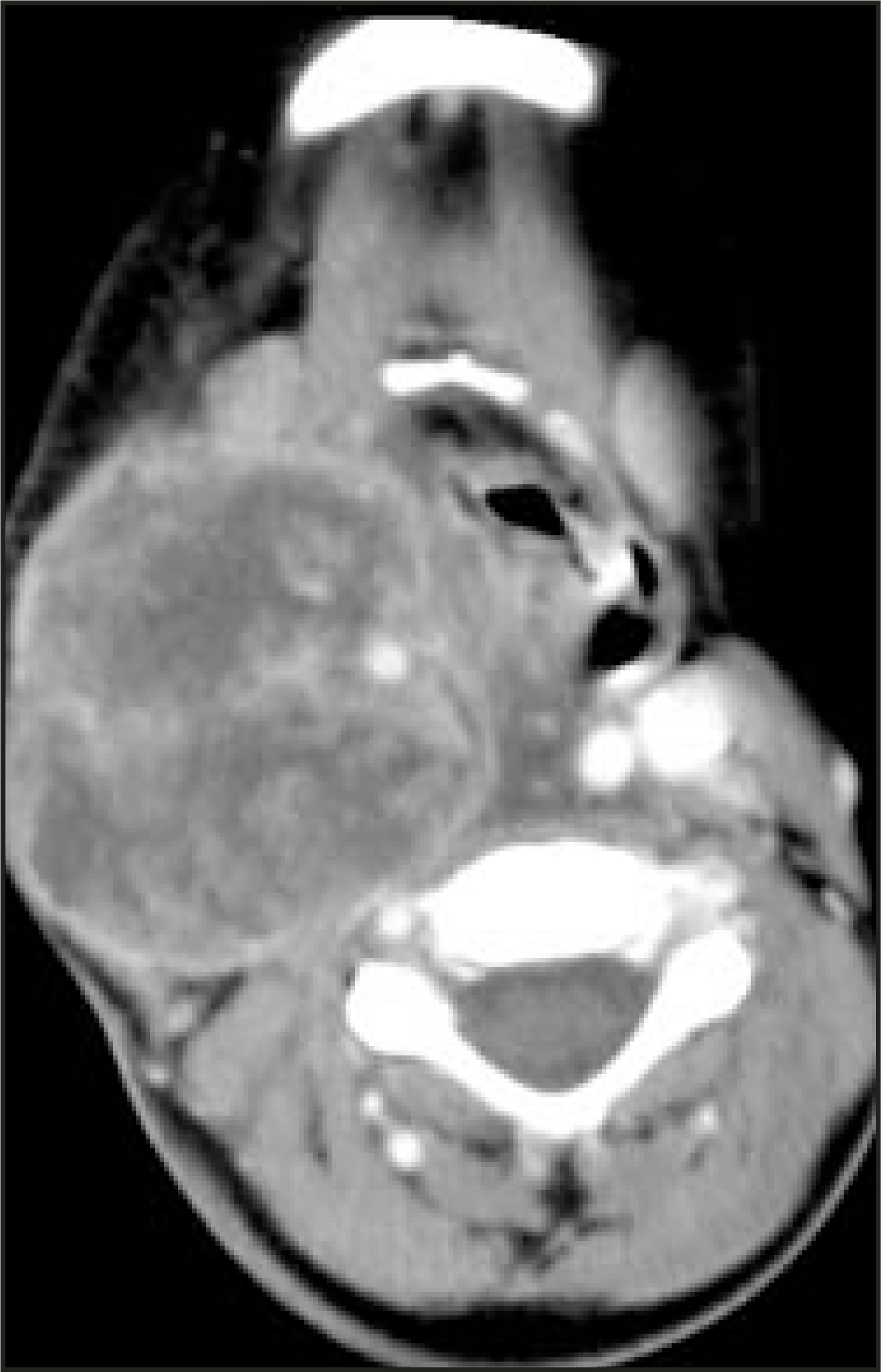
Rhabdomyosarcoma is the most common childhood soft-tissue sarcoma and involves the head and neck in up to 40% of patients.34,35 Rhabdomyosarcoma is divided by sites of origin into orbit, parameningeal (middle ear, paranasal sinus, nasopharynx), and all other sites. Up to 55% of parameningeal rhabdomyosarcomas have intra-cranial extension. In these patients, CT is helpful to assess bony destruction and MRI is complimentary to evaluate for intracranial extension. Rhabdomyosarcoma is typically heterogeneous on all imaging modalities (Figure 24), with or without osseous erosion.34
Neuroblastoma
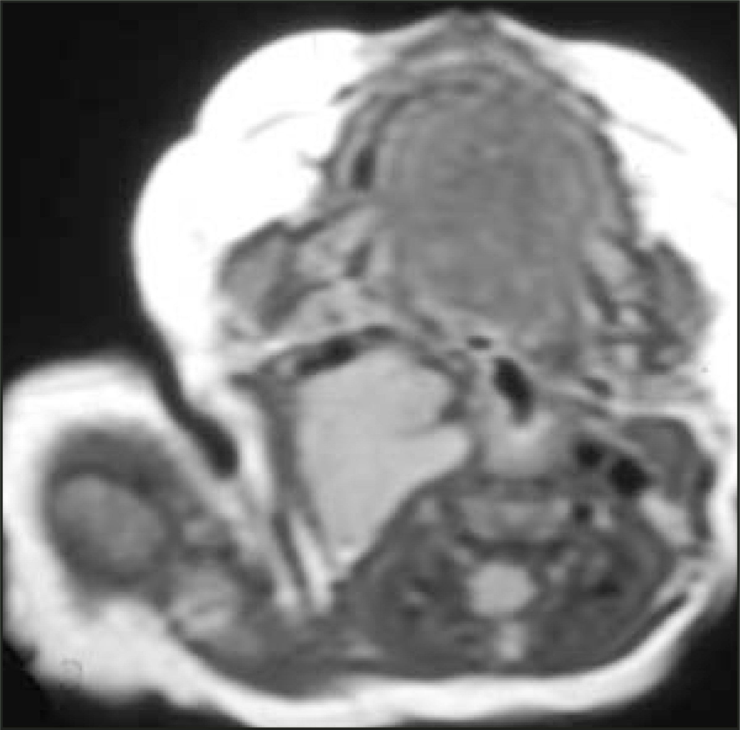
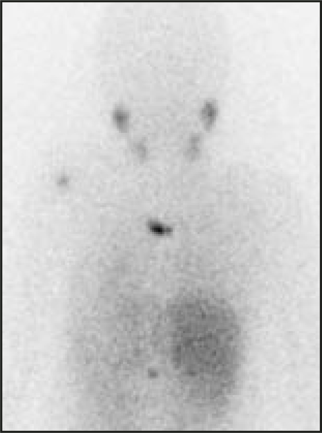
Neuroblastoma is the most common malignant tumor in children <1 year of age; primary lesions are usually located in the adrenal gland and retroperitoneum. Cervical lymphadenopathy from neuroblastoma is most often metastatic disease. Less than 5% of primary lesions are located in the neck. In addition to presenting with a palpable neck mass, patients may present with feeding difficulties, airway symptoms, or opsomyoclonus (opsoclonus, myoclonus, and cerebellar ataxia), which is thought to be a paraneoplastic syndrome. Most patients with cervical primary lesions present with a well- defined mass posterior to the carotid sheath vessels with or without intraspinal extension and with or without calcification.36-38 CT and MRI nicely show the primary mass (Figure 25), occasionally with intraspinal extension. In addition to CT or MRI, I-123-metaiodobenzylguanidine (MIBG) is indicated in the workup of these children to assess for metastatic disease (Figure 26).
Metastatic adenopathy
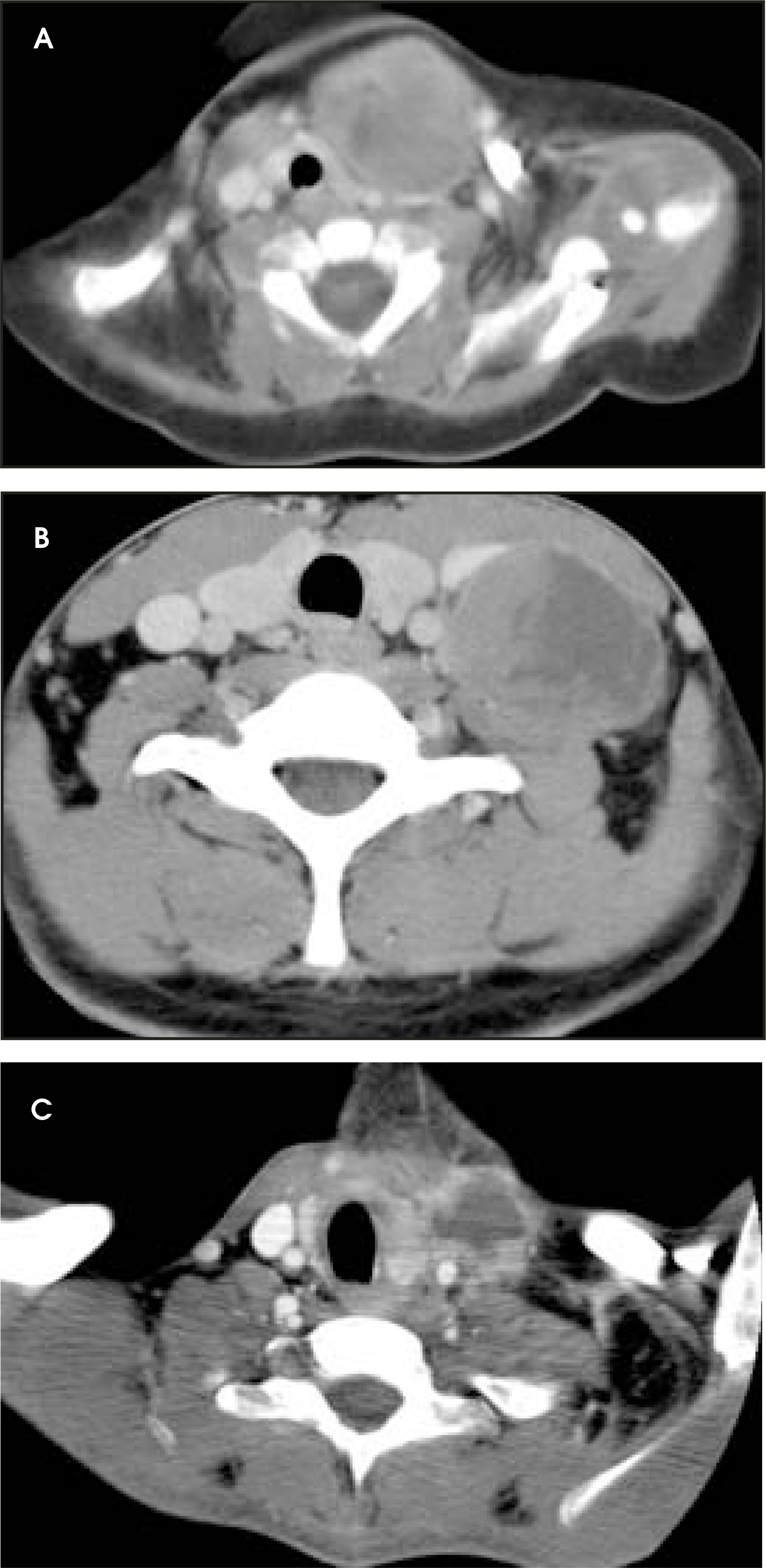
Metastatic cervical adenopathy unrelated to lymphoma is very uncommon in children. Potential etiologies include neuroblastoma, rhabdomyosarcoma, nasopharyngeal carcinoma, and thyroid carcinoma (Figure 27).
Conclusion
The majority of cystic neck masses in children are congenital. When midline or paramidline in position, thyroglossal duct cyst and dermoid cyst are the primary differential considerations. When off midline, branchial apparatus cysts and lymphatic malformations should be considered. Based on location and appearance, the majority of branchial apparatus cysts can be differentiated from lymphatic malformations.
Most solid neck masses in children are benign, including fibromatosis colli, in-flammatory adenopathy, hemangioma of infancy, neurofibroma, and benign teratoma. Using the clinical history, physical examination and imaging characteristics, most of these can be diagnosed with some certainty. Malignant lesions include lymphoma, rhabdomyosarcoma, neuroblastoma, and, rarely, metastatic adenopathy. Although it may be more difficult to make a definitive diagnosis in these children, with the above-mentioned imaging characteristics and clinical history, a reasonable differential diagnosis should be achievable.
References
- Lim-Dunham J, Feinstein K, Yousefzadeh D. Sonographic demonstration of a normal thyroid gland excludes ectopic thyroid in patients with thyroglossal duct cyst. AJR Am J Roentgenol. 1995;164:1489-1491.
- Youkilis R, Koch B, Myer C. Ultrasonographic imaging of sternocleidomastoid tumor of infancy. Ann Otol Rhinol Laryngol. 1995;104:323-325.
- Chan Y, Cheng J, Metreweli C. Ultrasonography of congenital muscular torticollis. Pediatr Radiology. 1992;22:356-360.
- Screaton N, Ravenel J, Lehner P. Lemierre syndrome: Forgotten but not extinct––report of four cases. Radiology. 1999;213:369-374.
- Ramirez S, Hild T, Rudolph C. Increased diagnosis of Lemierre syndrome and other Fusobacterium necrophorum infections at a Children’s Hospital. Pediatrics. 2003;112.
- Thomas J. Thyroglossal-duct cysts. Ear Nose Throat J. 1979;58:510-514.
- Pounds L. Neck masses of congenital origin. Pediatr Clin North Am. 1981;28:841-844.
- Solomon J, Rangecroft L. Thyroglossal-duct lesions in childhood. J Pediatr Surg. 1984;19:555-561.
- Wadsworth D, Siegel M. Thyroglossal duct cysts: Variability of sonographic findings. AJR Am J Roentgenol. 1994;163:1475-1477.
- Sistrunk W. The surgical treatment of cysts of the thyroglossal tract. Ann Surg. 1920;71:121-123.
- Olsen K, Maragos N, Weiland L. First branchial cleft anomalies. Laryngoscope. 1980;90:423-436.
- Benson M, Dalen K, Mancuso A. Congenital anomalies of the branchial apparatus: Embryology and pathologic anatomy. RadioGraphics. 1992;12:943-960.
- Mukherji S, Fatterpekar G, Castillo M. Imaging of congenital anomalies of the branchial apparatus. Neuroimaging Clin N Am. 2000;10:75-93.
- Bailey H. Branchial Cysts and Other Essays on Surgical Subjects in the Faciocervical Region. 1929.
- Zarbo R, McClatchey K, Areen R, Baker S. Thymopharyngeal duct cyst: A form of cervical thymus. Ann Otol Rhinol Laryngol. 1983;92(3 Pt 1):284-289.
- Guba A, Adam A, Jaques D, Chambers R. Cervical presentation of thymic cysts. Am J Surg. 1978;136:430-436.
- Burrows P, Fellows K. Techniques for management of pediatric vascular anomalies. In Cope E, ed. Current Techniques in Interventional Radiology. 1995:12.
- Baker L, Dillon W, Hieshima G. Hemangiomas and vascular malformations of the head and neck: MR characterization. AJNR Am J Neuroradiol. 1993;14:307-314.
- Fordham L, Chung C, Donnelly L. Imaging of congenital vascular and lymphatic anomalies of the head and neck. Neuroimaging Clin N Am. 2000;10:117-136.
- Meyer J, Hoffer F, Barnes P, Mulliken J. Biological classification of soft-tissue vascular anomalies: MR correlation. AJR Am J Roentgenol. 1991;157:559-564.
- Dubois J, Garel L, Abela A. Lymphangiomas in children: Percutaneous sclerotherapy with an alcoholic solution of zein. Radiology. 1997;204:651-654.
- Turner C, Gross S. Treatment of recurrent suprahyoid cervicofacial lymphangioma with intravenous cyclophosphamide. Am J Pediatr Hematol Oncol. 1994;16:325-328.
- Molitch H, Unger E, Witte C, vanSonnenberg E. Percutaneous sclerotherapy of lymphangiomas. Radiology. 1995;194:343-347.
- Yamaguchi M, Takeuchi S, Matsuo S. Ultrasonic evaluation of pediatric superficial masses. J Clin Ultrasound. 1987;15:107-113.
- Hudgins P, Dorey J, Jacobs I. Internal carotid artery narrowing in children with retropharyngeal lymphadenitis and abscess. AJNR Am J Neuroradiol. 1998;19:1841-1843.
- Nadel D, Bilaniuk L, Handler S. Imaging of granulomatous neck masses in children. Int J Pediatr Otorhinolaryngol. 1996;37:151-62.
- Hazra R, Robson C, Perez-Atayde A, Husson R. Lymphadenitis due to nontuberculous mycobacteria in children: Presentation and response to therapy. Clin Infect Dis. 1999;28:123-129.
- Robson C, Hazra R, Barnes P. Nontuberculous mycobacterial infection of the head and neck in immunocompetent children: CT and MR findings. AJNR Am J Neuroradiol. 1999;20:1829-1835.
- Stone M, Walner D, Koch B. Correlation between computed tomography and surgical findings in retropharyngeal inflammatory processes in children. Int J Pediatr Otorhinolaryngol. 1999;49:121-125.
- Mulliken J, Glowacki J. Hemangiomas and vascular malformations in infants and children: A classification based on endothelial characteristics. Plast Reconstr Surg. 1982;69:412-422.
- Burrows P, Mulliken J, Fellows K. Childhood hemangiomas and vascular malformation: Angiographic differentiation. AJR Am J Roentgenol. 1983;141:483-488.
- Suh J, Abenoza P, Galloway H. Peripheral (extracranial) nerve tumors: Correlation of MR imaging and histologic findings. Radiology. 1992;183:341-346.
- Batsakis J. Tumors of the Head and Neck: Clinical and Pathological Considerations. . 1979.
- Castillo M, Pillsbury H. Rhabdomyosarcoma of the middle ear: Imaging features in two children. AJNR Am J Neuroradiol. 1993;14:730-733.
- Latack J, Hutchinson R, Heyn R. Imaging of rhabdomyosarcomas of the head and neck. AJNR Am J Neuroradiol. 1987;8:353-359.
- Abrahmson S, Berdon W, Ruzal-Shapiro C. Cervical neuroblastoma in eleven infants: A tumor with favorable prognosis. Clinical and radiologic findings. Pediatr Radiol. 1993;23:253-257.
- Casselman J, Smet M, Van Damme B, Lemahieu S. Primary cervical neuroblastoma: CT and MR findings. JComput Assist Tomogr. 1988;12:684-686.
- Smith M, Smith R, Bailey C. Primary cervical neuroblastoma in infants. J Laryngol Otol. 1985;99:209-214.
Citation
. The child with a neck mass. Applied Radiology. 2005;34(8):8-22. doi:10.37549/AR1359.