The role of MRI and “second-look” ultrasound for evaluation of breast cancer
Applied Radiology — Vol. 35 , Issue 10 , pp. 10 -20
DOI: 10.37549/AR1454
Published: October 1, 2006
Categories
Magnetic resonance imaging (MRI) of the breast has been shown to be helpful, specifically in women with dense breasts, for preoperative assessment of the local extent of recently diagnosed breast cancer or for the exclusion of breast cancer in high-risk patients. Our practice at The Elizabeth Wende Breast Clinic (EWBC) has been evaluating patients with benign and malignant breast disease for more than 26 years. In our experience, breast tumors can frequently be occult, or tumor size can be underestimated in patients with dense breasts. In general, this subset of patients may not be evaluated as thoroughly as possible, because of the density of their breast parenchyma, and thus they require special consideration.
In the literature, sensitivity of mammography is reported to vary from 60% to 98% with some experts reporting it as low as 30% to 50% in women with dense breast tissue. Specificity is reported to range from 30% to 70% with variation related in part to imaging techniques and methods of patient selection.1-3
Today, as more patients choose breast-conserving therapy instead of mastectomy, complete tumor removal is mandatory to avoid recurrence. After a lumpectomy for breast cancer treatment, residual tumor remains in 30% to 60% of patients.4 Accurate preoperative assessment of tumor size, location, and identification of multifocal disease is imperative. The evaluation of the contralateral breast to detect possible synchronous bilateral breast cancers is just as important. In a study by Fisher et al,5 4% of patients (15 of 336) had synchronous bilateral tumors found by MRI alone. Typically, 2% to 3% of the patients present with bilateral breast cancer based on mammography and physical examination; this percentage is higher in younger patients and in those with invasive lobular cancer.5 Breast cancers can be multifocal (more than one focus in the same quadrant) or multicentric cancers (involving more than one quadrant). Holland et al6 found significant additional foci of cancer, many outside a radius of 2 cm, relative to the index lesion.
Use of ancillary breast imaging studies
Breast ultrasound, in addition to mammography, has been shown to be helpful for the preoperative assessment of the extent of disease. Across large series, a reported change in patient management occurs in roughly 15% of cases when additional imaging, including ultrasound and MRI, are performed after standard mammography.5,7,8 Approximately 30% of these patients harbor undiagnosed additional foci of malignancy that are found when breast MRI is performed. Using MRI, Liberman et al7 discovered 30% to 48% additional foci of malignancy that were not detected by mammography. MRI uses both morphologic and dynamic features to diagnose otherwise occult tumors that are not visualized using standard diagnostic techniques, such as mammography, ultrasound, and physical ex- amination. The reported specificity of MRI varies widely, ranging from 27% to 90%.7 Tumor is detected principally based upon development of neovascularity, so that well-differentiated tumors may be falsely negative on MRI. Ductal carcinoma in situ (DCIS) enhancement and detection are variable, with sensitivity ranging from 40% to 100%.9
When MRI is incorporated into the evaluation of breast disease, our group believes that it is imperative to provide biopsy capability. Once a lesion is found by the radiologist who initiates breast imaging and performs breast MR, the diagnostic loop must be closed with biopsy of any suspicious lesion. Lack of biopsy capability creates anxiety for the patient and inconvenience for referring physicians who must manage the work-up to its conclusion.
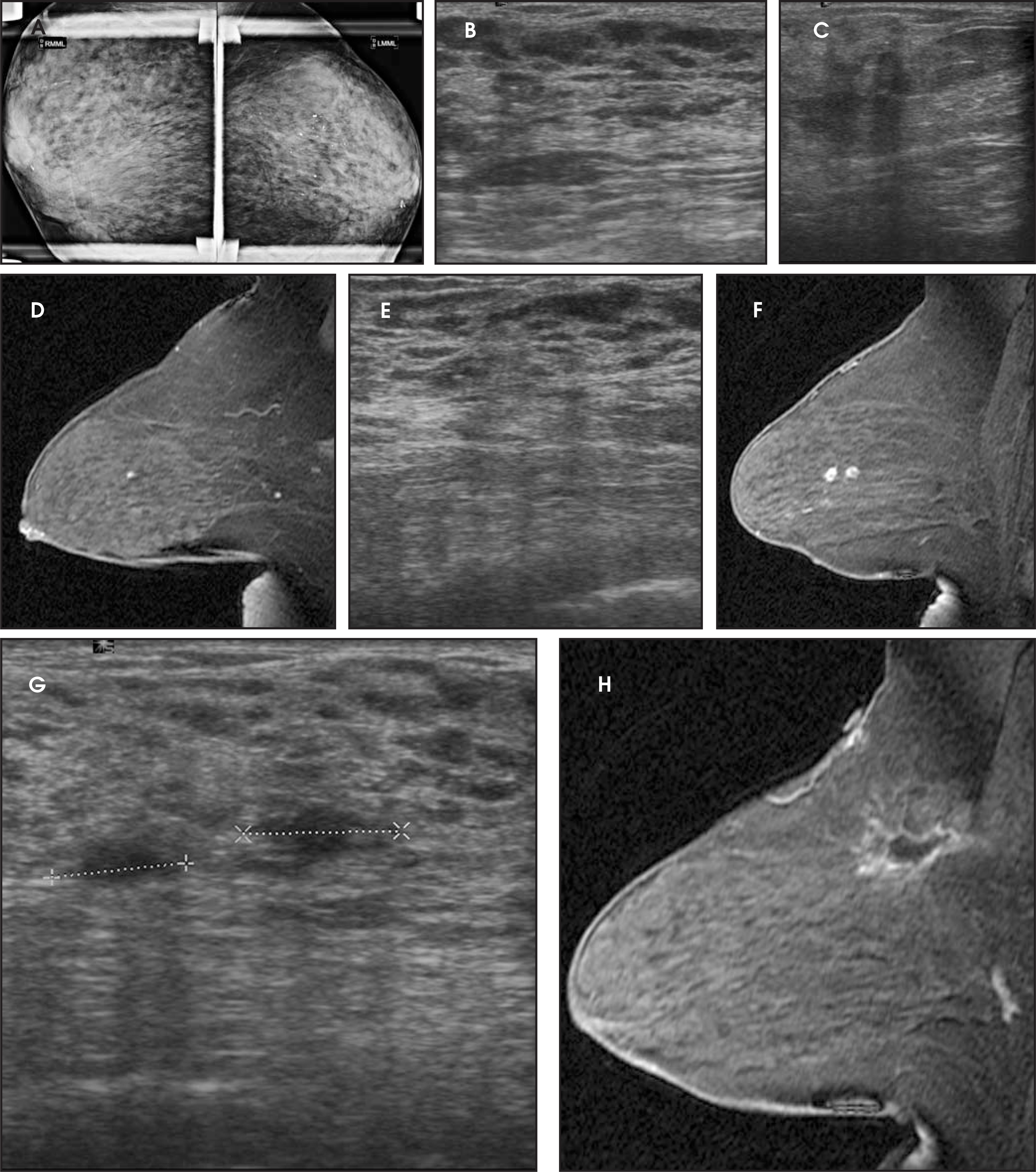
When patients are referred to our clinic from another facility where breast imaging was performed, all prior breast imaging studies and pathology results should be reviewed prior to the patient’s undergoing the MRI. Unilateral MRI is obtained at our site prior to attempting biopsy to exclude false-positive findings. False-positive results may occur particularly in premenopausal women, depending on menstrual cycle fluctuations and hormonal changes, in the presence of fibrocystic disease, and in benign proliferative changes. Fibroadenomas may show contrast enhancement and be a source of false-positive results (Figure 1).
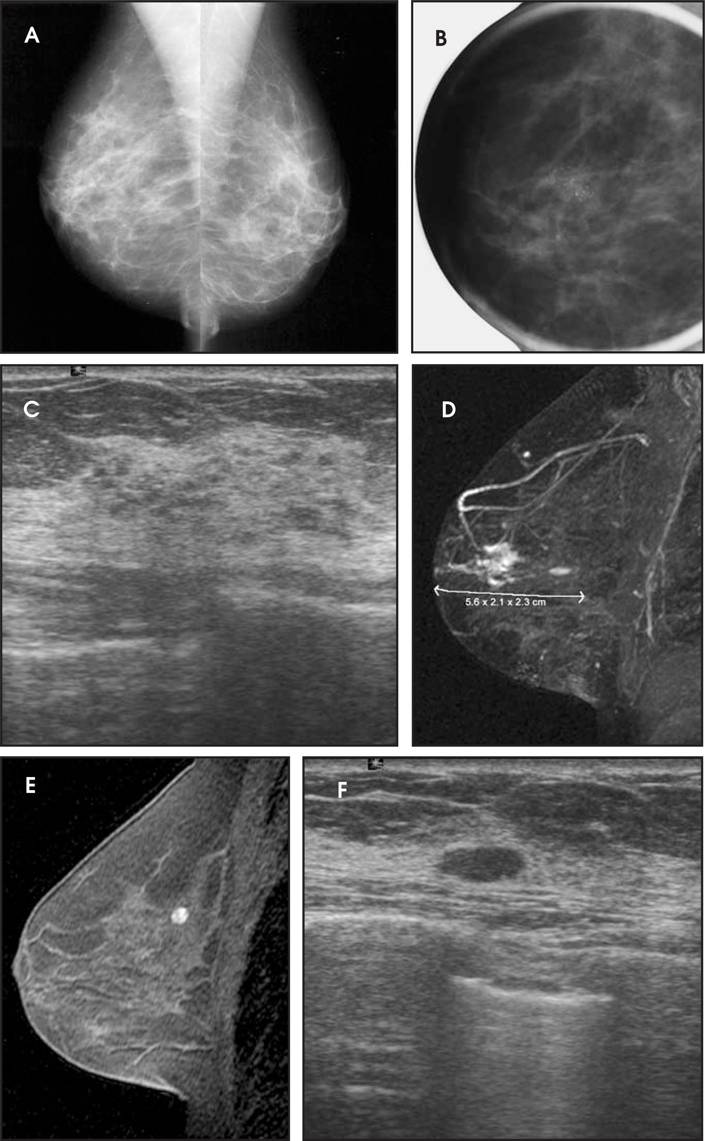
At our facility, all breast MR studies are double read. The first radiologist interprets the MRI in detail, while the second typically performs a quick- review second read. The group’s radiologists confer over difficult cases. A patient with an abnormality on MRI returns within the next few days for a “second-look” ultrasound and possible further mammographic imaging. If the patient was already seen by our practice and was diagnosed with cancer at our facility, we typically have already performed mammography and an initial ultrasound. Even in that scenario, a repeat ultrasound can be very helpful, as subtle lesions are discovered once MRI has noted a lesion in a specific area. In approximately one third of these cases, the second-look ultrasound locates the lesion seen on MRI, and an ultrasound-guided biopsy can then be performed for diagnosis, as needed. In the other two thirds of cases, in which ultrasound does not detect the abnormal area, the patient may be required to return for a scheduled MRI-guided biopsy to further evaluate the abnormality seen on MRI.
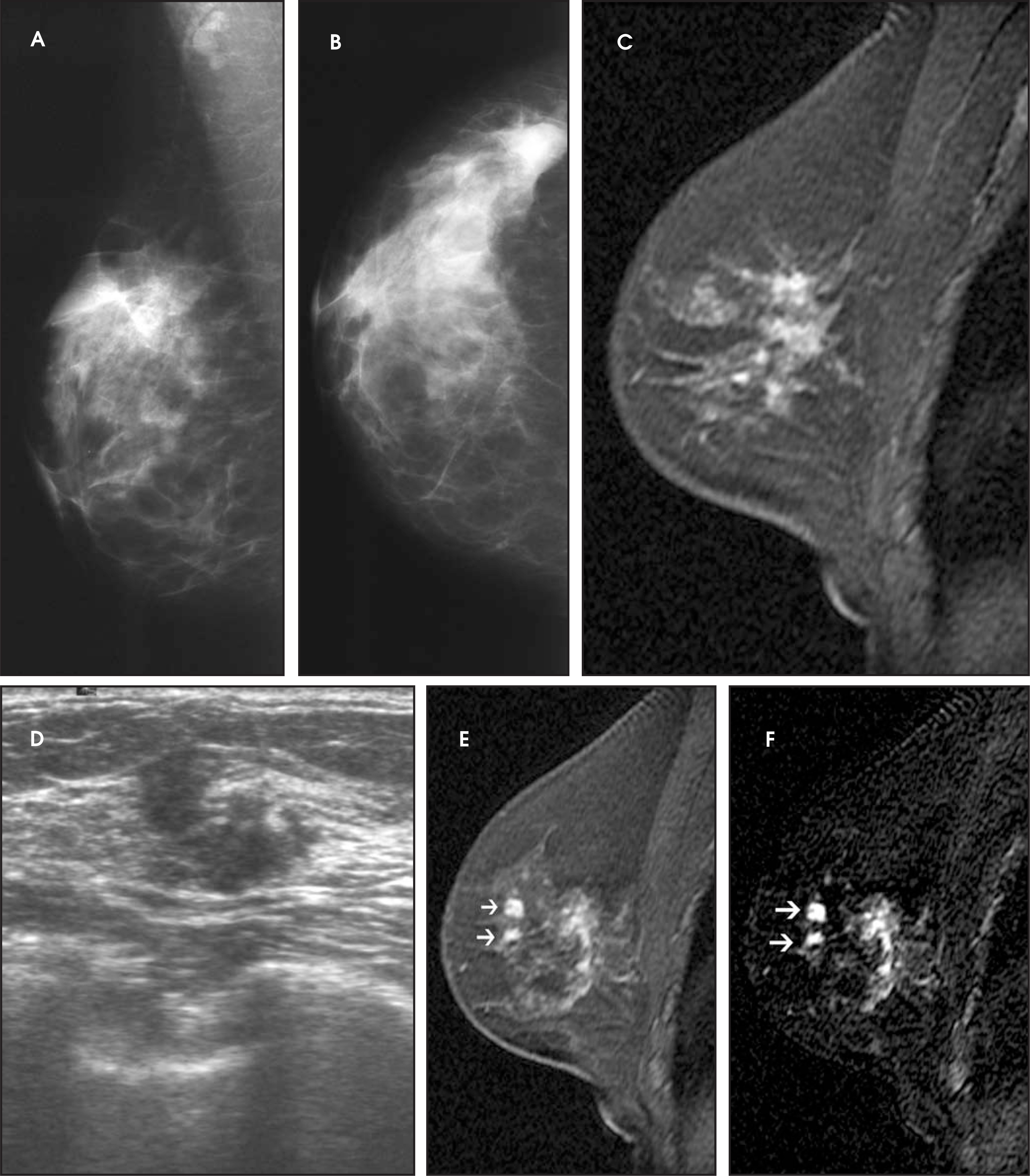
MRI often delineates an area of subtle breast parenchymal abnormality that may have been overlooked on the initial ultrasound study (Figure 2). From June 2003 to December 2005, we imaged 723 patients with MRI at our facility. Among those studies, 241 (33%) patients had suspicious lesions identified that warranted evaluation with second-look ultrasound. Among those 241 lesions, a total of 203 biopsies were performed (by fine-needle aspiration or core biopsy). The resultant pathology revealed 53 cancers (26% of the total biopsies). A retrospective internal review of MRI patients from 2003 and 2004, performed by Dr. Patricia Somerville, revealed that MRI examination changed management, on average, in 12% (27 of 226) of cases. In this review, 72 patients were recalled for a second-look ultrasound that revealed findings in 36% (26 of 72). Among this group, ultrasound-guided core biopsies were positive in 9, accounting for 35% of the total ultrasound biopsies performed and 12% (9 of 72) of all patients recalled for second-look ultrasound (Figure 3). (P. Somerville, internal review, EWBC MRI outcome data 2003-2004.)
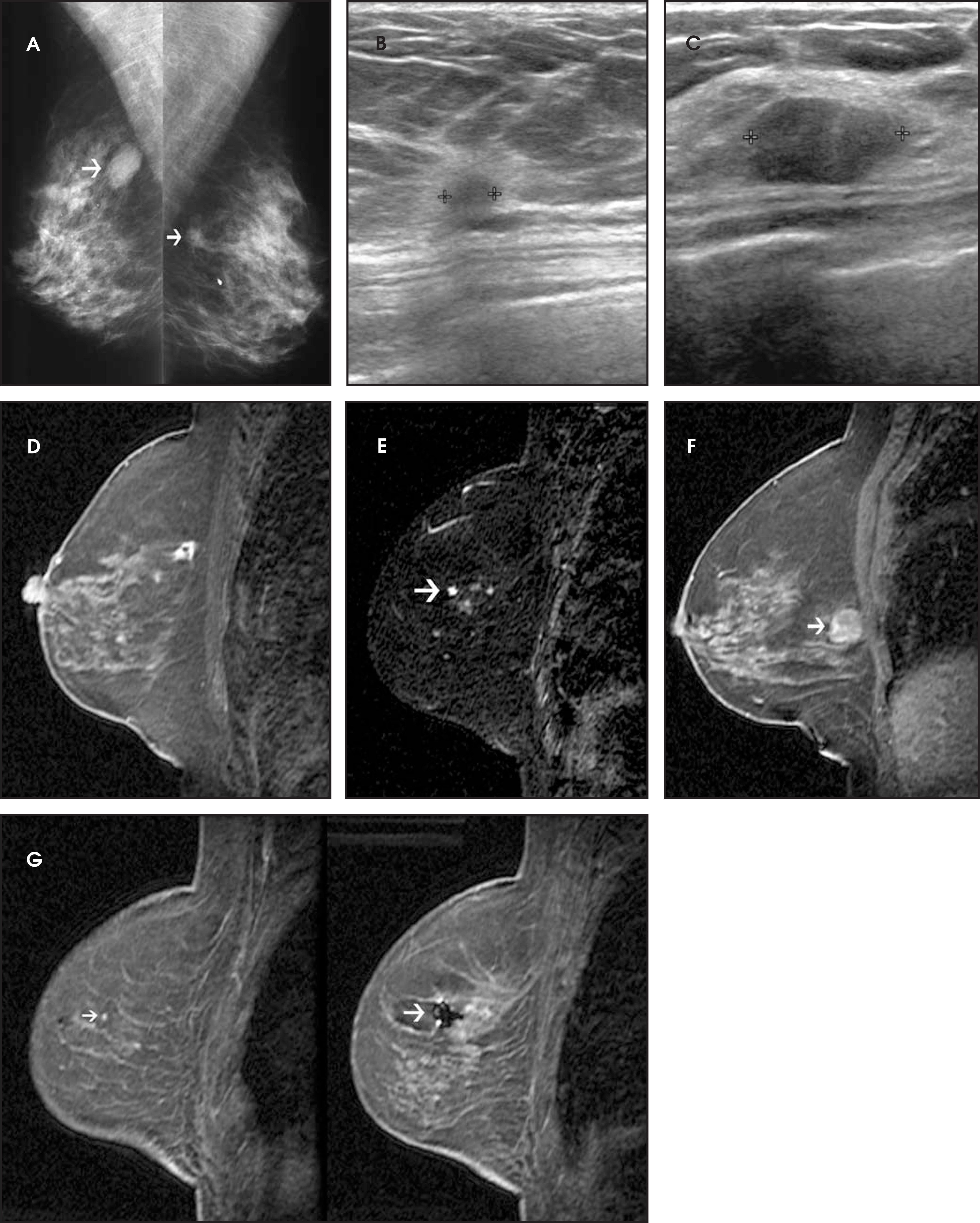
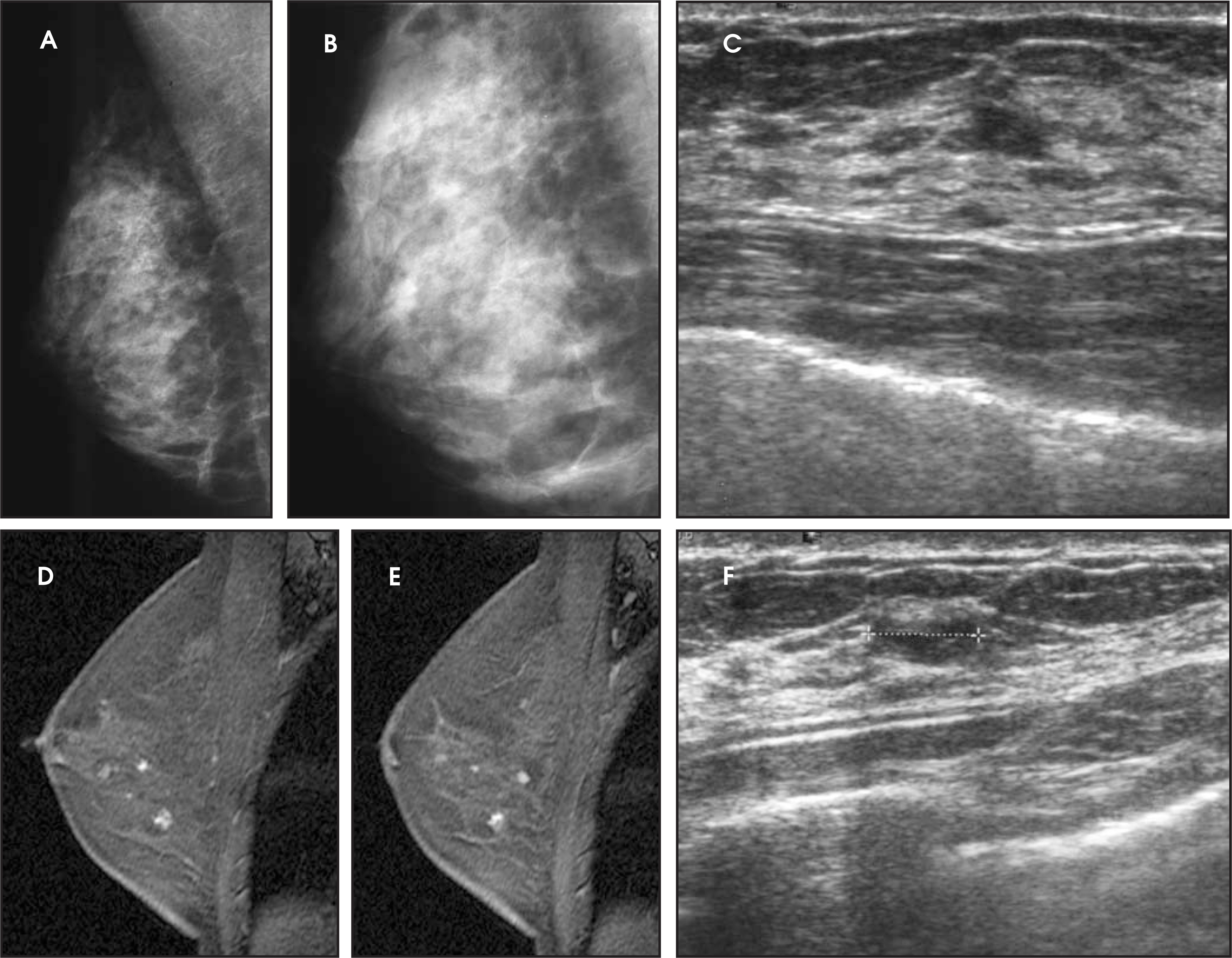
The practice performs MRI in premenopausal patients in the first half of their menstrual cycle to diminish hormonal effects and then studies post-menopausal patients as soon as the schedule allows. Most patients are studied for extent of disease after a recent breast cancer diagnosis. Lesion size and the presence of additional ipsilateral or contralateral disease are determined (Figure 4). The literature has shown a considerable number of additional mammographically occult cancers found using MRI, particularly with invasive lobular cancers.10 A disadvantage of MRI is a reported false-positive rate that varies from 8% to 57% according to some authors.1112 These findings lead to unnecessary biopsies that are expensive to perform and anxiety-provoking for patients.
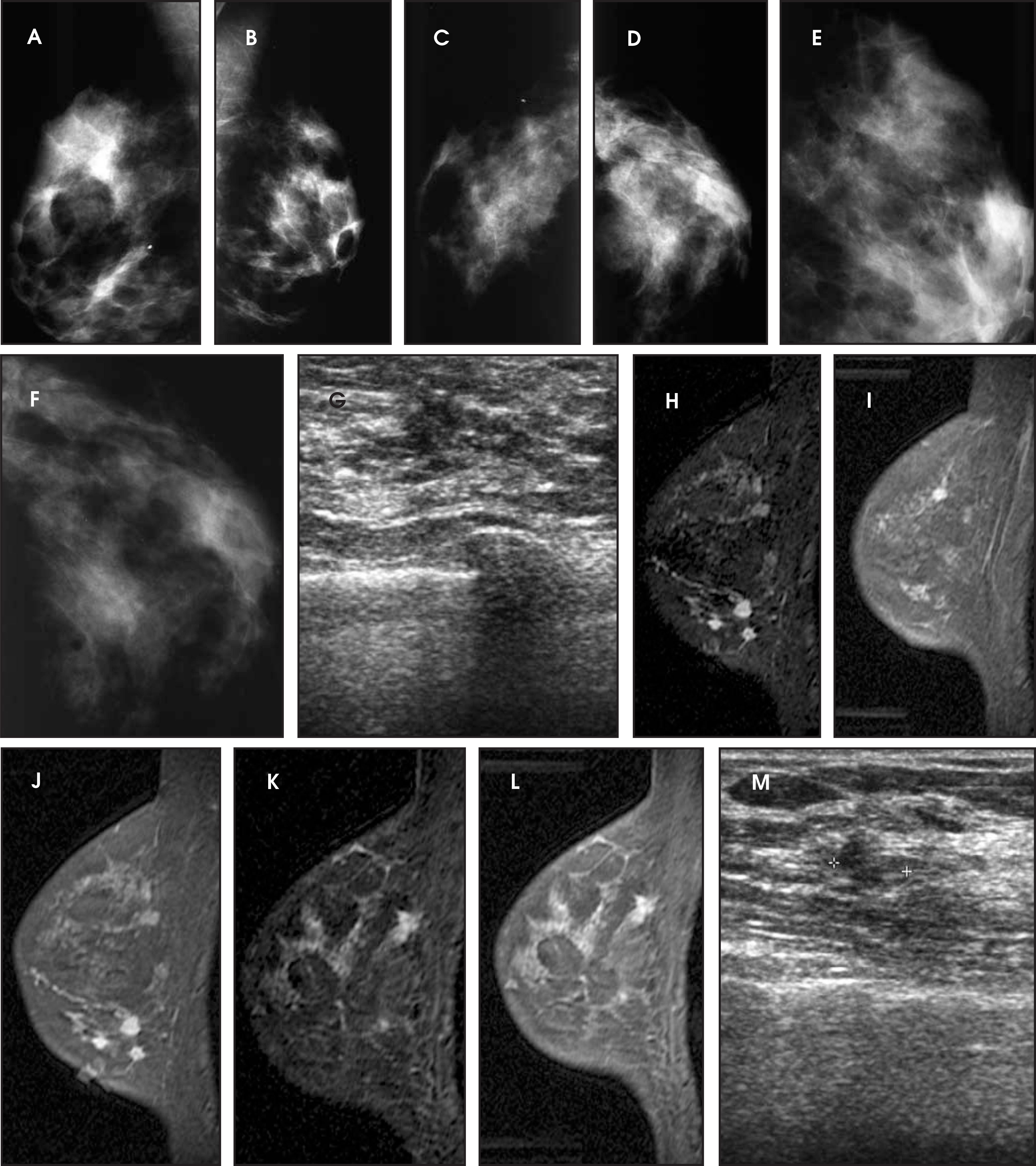
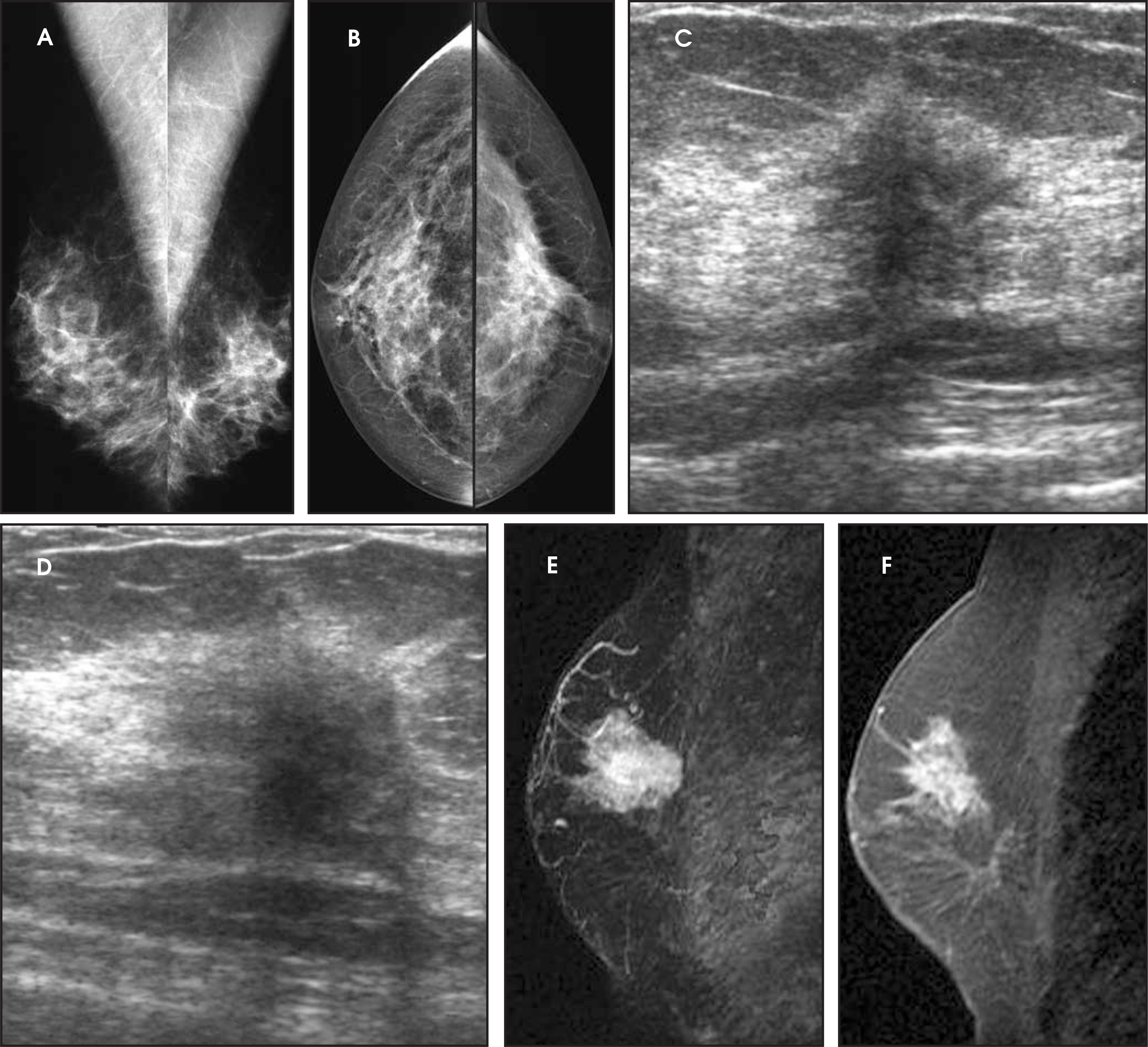
Our practice recommends short-term follow-up for benign-appearing lesions such as fibroadenomas, fibrocystic disease, sclerosing adenosis, and proliferative changes. Abnormalities (such as lobular carcinoma in situ, atypical ductal hyperplasia, and other precancerous lesions) may demonstrate MRI enhancement. To further evaluate indeterminate MRI findings, second-look ultrasound is performed. Since biopsies on MRI are cumbersome to perform, we attempt to identify and biopsy lesions using sonography (Figure 5).
Breast MRI
The mammogram is the first-line screening tool for breast imaging. MRI and ultrasound are additional studies that supplement mammography for selected cases. MRI is an expensive and time-consuming endeavor that adds considerable cost to a breast imaging practice. However, as described above, MRI can often identify lesions missed by other methods in some patients and can improve accuracy in defining extent of disease in others. Breast imagers have a short learning curve for a new technology, as they already interpret the other breast imaging studies and will easily be able to perform MRI-guided interventional procedures. Based on our practice’s experience with breast MRI, and our experiences with insurance carriers and their approved indications, the following are recommended guidelines for performing breast MRI:
For evaluation of extent of disease (Figure 6) in patients who have recently been diagnosed with breast cancer.
For preoperative surgical planning or postoperative study if margins are close or involved after lumpectomy.
For assessment of tumor response after chemotherapy treatment and prior to definitive surgery.
To evaluate patients with prior cancer if there is concern of recurrence.
In patients with bilateral silicone implants if there is concern of rupture (contour deformity, pain, lumps, etc., where the silicone implant makes mammographic diagnosis difficult).
To search for an occult primary in patients with metastatic carcinoma to axillary lymph nodes or elsewhere.
Following an indeterminate work-up after standard imaging techniques, including mammogram, ultrasound, physical examination, or core biopsy.
As a screening for high-risk patients (personal history of breast cancer, strong family history, or BRAC1/ BRAC2-gene-positive) (Figure 7).
Conclusion
Approximately 250,000 women are diagnosed each year with breast cancer, and more than half are candidates for breast conservation. To accurately choose the patients for breast conserving surgery, preoperative assessment of extent of disease in the breast is crucial. MRI can identify known tumor extent as well as additional sites of ipsilateral cancer in up to one third of women diagnosed with breast cancer through needle-core biopsy or another method.7 The presence of additional cancer sites is important to plan surgical treatment and prevent recurrence. MRI is a very sensitive tool for breast cancer diagnosis, but it is not the most specific. False positives occur, and patients do undergo unnecessary biopsies and further evaluation for benign lesions. At this time, MRI cannot stand alone as a screening tool for breast cancer evaluation but is an important additional tool for diagnosis and accurate subsequent treatment.
Acknowledgments
Special thanks to Dr. Wende Logan-Young for her support and Dr. Patricia Somerville for her in-depth review of our MRI outcomes of 2003 and 2004. Also, thanks to our former partner, Dr. Ermelinda Bonaccio, for initiating MRI in our clinical practice; our partner, Dr. Posy Seifert, for the continued support of MRI in our practice; and Dr. Philip Murphy and Dr. Margarita Zuley for case contributions.
References
- Burhenne H, Burhenne L, Goldberg F. Interval breast cancers in the Screening Mammography Program of British Columbia: Analysis and classification. AR Am J Roentgenol. 1994;162.
- Robertson C. A private breast imaging practice: Medical audit of 25,788 screening and 1,077 diagnostic examinations. Radiology. 1993;197:75-79.
- Kerlikowske K, Grady D, Barclay J. Effect of age, breast density and family history on the sensitivity of first screening mammography. JAMA. 1996;276:33-38.
- Gwin J, Eisenberg B, Hoffman J. Incidence of gross and microscopic carcinoma in specimens from patients with breast cancer after re-excision lumpectomy. Ann Surg. 1993;218:729-734.
- Eight-year results of a randomized clinical trial comparing total mastectomy and lumpectomy with or without irradiation in the treatment of breast cancer. N Engl J Med N Engl J Med.
- Holland R, Veling S, Mravunac M, Hendricks J. Histologic multifocality of Tis, T1-2 breast carcinomas. Implications for clinical trials of breast-conserving surgery. Cancer. 1985;56:979-990.
- Liberman L, Morris E, Dershaw D. MR imaging of the ipsilateral breast in women with percutaneously proven breast cancer. AJR Am J Roentgenol. 2003;180:901-910.
- Hlawatsch A, Teifke A, Schmidt M. Preoperative assessment of breast cancer: Sonography vs. MR Imaging. AJR Am J Roentgenology. 2002;179:1493-1501.
- Orel S, Mendonca M, Reynolds C. MR imaging of ductal carcinoma in situ. Radiology. 1997;202:413-420.
- Weinstein S, Orel S, Heller R. MR Imaging of the breast in patients with invasive lobular carcinoma. AJR Am J Roentgenology. 2001;176:399-406.
- Berg W, Gutierrez L, NessAiver M. Diagnostic accuracy of mammography, clinical examination, US, and MR imaging in preoperative assessment of breast cancer. Radiology. 2004;233:830-849.
Citation
. The role of MRI and “second-look” ultrasound for evaluation of breast cancer. Applied Radiology. 2006;35(10):10-20. doi:10.37549/AR1454.