Tracking the Silent Pathway: Imaging the Great Auricular Nerve - Insights for Radiologists
Applied Radiology — Vol. 55 , Issue 4
Published: July 1, 2026
1 Department of Radiology, Division of Neuroradiology, University of California Davis Medical Center, Sacramento, California
2 California Maternal Fetal Medicine, Sacramento, California
3 Department of Radiology, Division of Neuroradiology, University of Texas Health, San Antonio, Texas
4 Department of Pathology and Laboratory Medicine, University of California Davis Health System, Sacramento, California
5 Department of Otolaryngology – Head & Neck Surgery, University of California Davis, Sacramento, California
6 Department of Radiology, Stanford University School of Medicine, Stanford, California
* Corresponding author: Osama A. Raslan (oraslan@health.ucdavis.edu)
Abstract
We present 2 cases of uncommon perineural tumor spread involving the great auricular nerve (GAN), provide a detailed description of the radiologic anatomy of the nerve in relation to surrounding organs and major nerve connections with a focus on key imaging findings of perineural spread, and describe major conditions that can involve the GAN through a comprehensive review of the literature. The clinical relevance of this work spans from enhanced detection of perineural spread in cutaneous cancers—whose incidence is rising—to improved familiarity with emerging treatment strategies involving the GAN.
Keywords
great auricular nerve, perineural tumor spread, head and neck cancer, auriculotemporal nerve, ultrasound imaging
Categories
Introduction and Radiologic Anatomy
The great(er) auricular nerve (GAN) is the largest of the pure sensory superficial branches of the cervical plexus, receiving contributions mainly from the C2 and C3 ventral rami.1-3 The GAN is most reliably identified—both clinically and radiologically—as it emerges superficially with a characteristic looping appearance at the posterior border of the sternocleidomastoid muscle (SCM) (Figure 1). It ascends superficially, deep to the cervical fascia overlying the SCM, where it is particularly vulnerable to surgical injury.
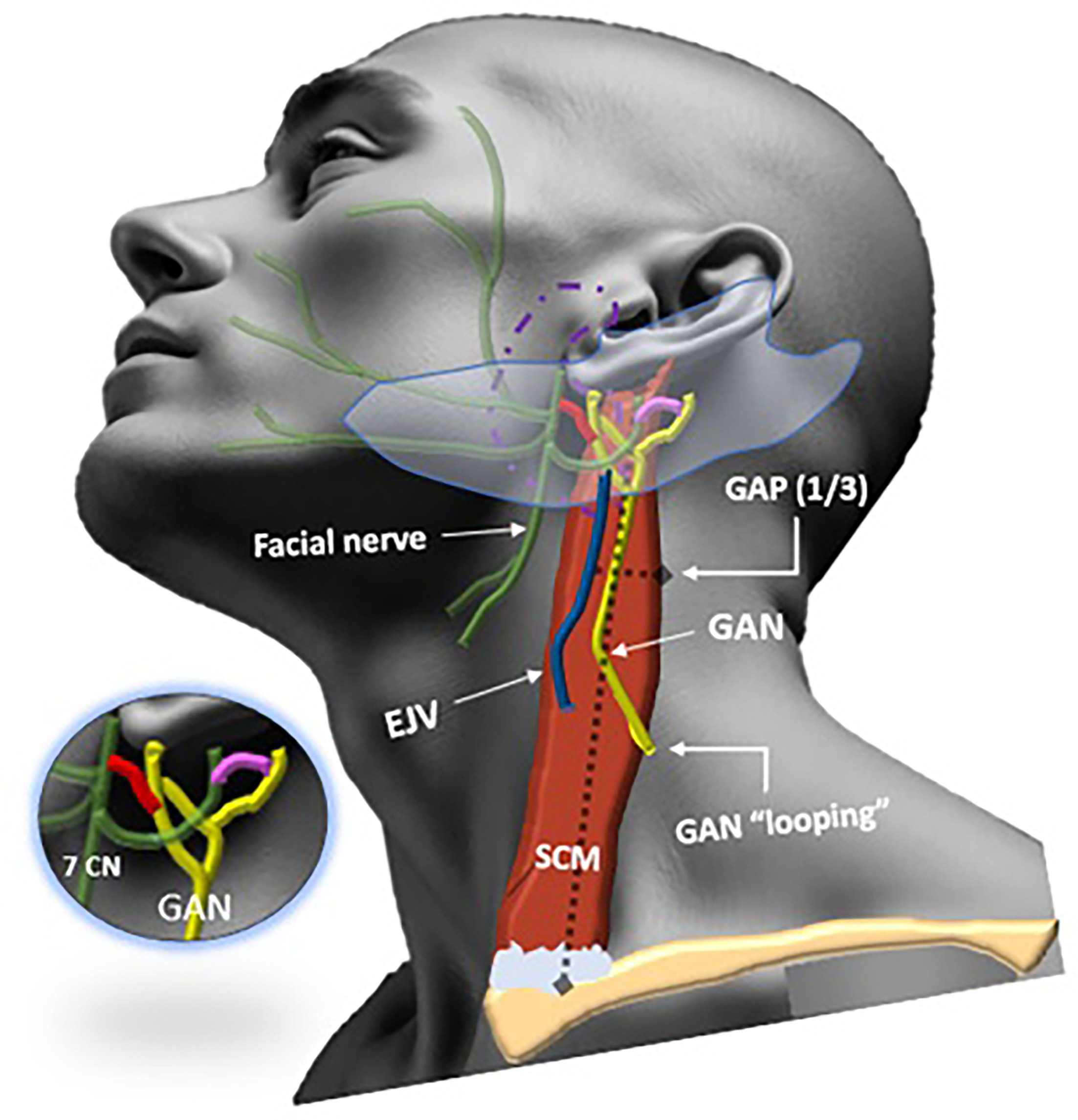
To facilitate identification, several anatomical landmarks have been described, including the nerve point of the neck (punctum nervosum), Erb’s point, and McKinney’s point. However, for greater anatomical precision, we prefer to use the “great auricular point” (GAP) to describe the optimal location for locating the GAN.
The GAP was consistently identified at approximately one-third the distance from either the mastoid process or the external auditory canal to the clavicular origin of the SCM, and about 1 cm posterior to the external jugular vein (Figure 1).
The GAN then divides into an anterior (facial) branch supplying the skin over the parotid gland and anteroinferior aspect of the auricle, and a posterior (mastoid) branch supplying the skin over the mastoid process and the posteroinferior aspect of the auricle.2,4,5
In the parotid gland, the anterior branch connects with the facial nerve (Figure 1, red). Posterior to the ear, the posterior branch connects with both the auricular branch of the vagus nerve and the posterior auricular branch of the facial nerve (Figure 1, pink). These connections serve as conduits for perineural tumor spread (PNS) between the GAN, the facial nerve, upper cervical nerve roots (C2 and C3), and the vagus nerve.2,3,5-17
US Imaging of the GAN
The GAN can be readily visualized using high-resolution US (HRUS) and is frequently identified in US-guided GAN nerve blocks.18-22 Lieba-Samal et al were able to consistently visualize the GAN with an 18-MHz HRUS probe throughout its course, both in anatomical specimens and in vivo.21 Reddy et al also used a 15‐7-MHz hockey-stick transducer for GAN identification.20
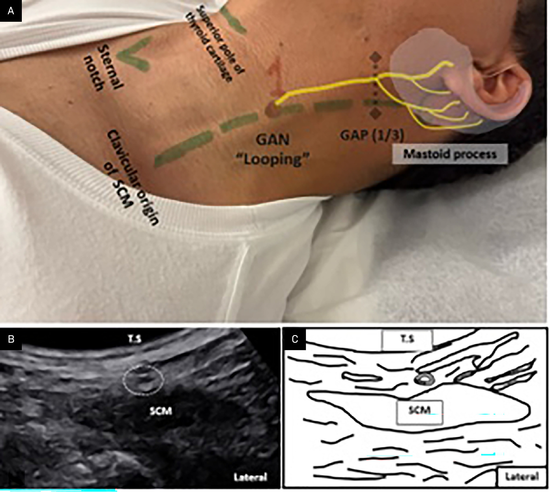
The GAN is identified using the anatomical landmarks illustrated in Figure 2. A linear HRUS transducer is positioned at the GAN looping point, located at the level of the thyroid cartilage along the posterolateral border of the SCM. The normal GAN appears just superficial to the SCM as a small, rounded hypoechoic structure with internal linear hyperechoic fascicles, best visualized on longitudinal scans. When followed inferiorly, the GAN courses deep to the SCM to join the superficial cervical plexus. Tracing the nerve superiorly along the SCM reveals its bifurcation point.18
Pathological Conditions of the GAN
Schwannoma
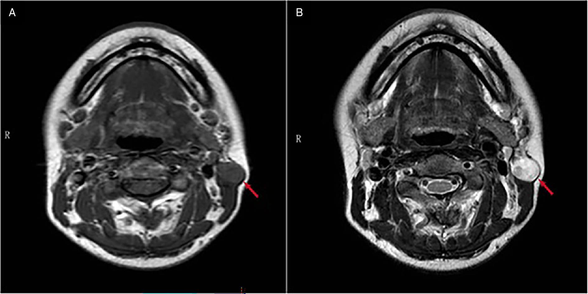
Schwannoma of the GAN is extremely rare, with <10 cases reported in the literature.1,24-26 The lesions are often diagnostically challenging to the clinicians as they usually present as a slowly growing asymptomatic unilateral neck lump with broad differential considerations, including lymphadenopathy, and sebaceous cyst, among others, with schwannoma often finally diagnosed intraoperatively during an excisional biopsy, putting the surgeons at litigation risk due to the ensuing permanent sensory disturbance.24 In their literature review, Oh et al highlighted the striking similarities in the presentation and clinical course of the 4 reported GAN schwannomas, with a median age of 49 years at presentation, a male:female ratio of 3:1, presentation as an asymptomatic neck lump, and an imaging of a well-defined benign-looking inhomogeneous lesion on the surface of the SCM or along the course of GAN (Figure 3).1 To our knowledge, no cases of primary malignant GAN nerve sheath tumors have been reported in the literature as of July 14, 2025.
Perineural Tumor Spread
PNS is a well-described phenomenon of head and neck cancer by which there is retrograde or antegrade extension of the tumor from the primary site along the nerve tissues.27 It is an important finding indicating a worse prognosis, increased recurrence rate, and decreased 5-year survival by up to 30%.27 PNS along the cranial nerves is a relatively well-known and well-described phenomenon in the literature28; however, only a few articles described PNS along the spinal nerves, and to our knowledge, <15 articles have described PNS along the GAN.2,13-17,29-31 As PNS can be asymptomatic, radiologists are often the ones who alert clinicians to the presence of GAN PNS, underscoring the importance of radiologists being familiar with this rare but significant tumor extension. Such awareness can help avoid delayed or misdiagnosis, as seen in our first case.
Case 1
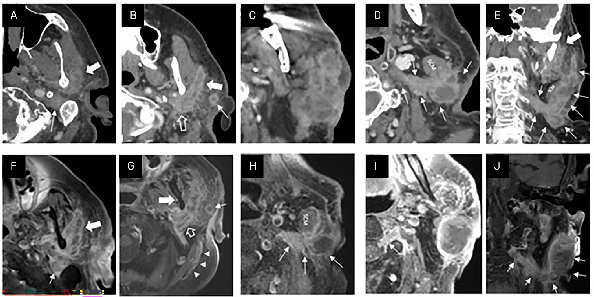
An 83-year-old man with a history of left jawline squamous cell carcinoma, treated by Mohs resection 3 months prior, presented with persistent pain at the surgical site and a 10-day history of slurred speech. Neurological workup and imaging were negative for stroke. Clinical examination revealed tenderness over the scar. PET/CT showed no recurrence but noted mildly increased FDG uptake in a presumed dilated left posterior auricular vein, likely inflammatory (phlebitis/thrombophlebitis) (Figure 4). MRA was inconclusive; venous Doppler supported superficial thrombophlebitis.
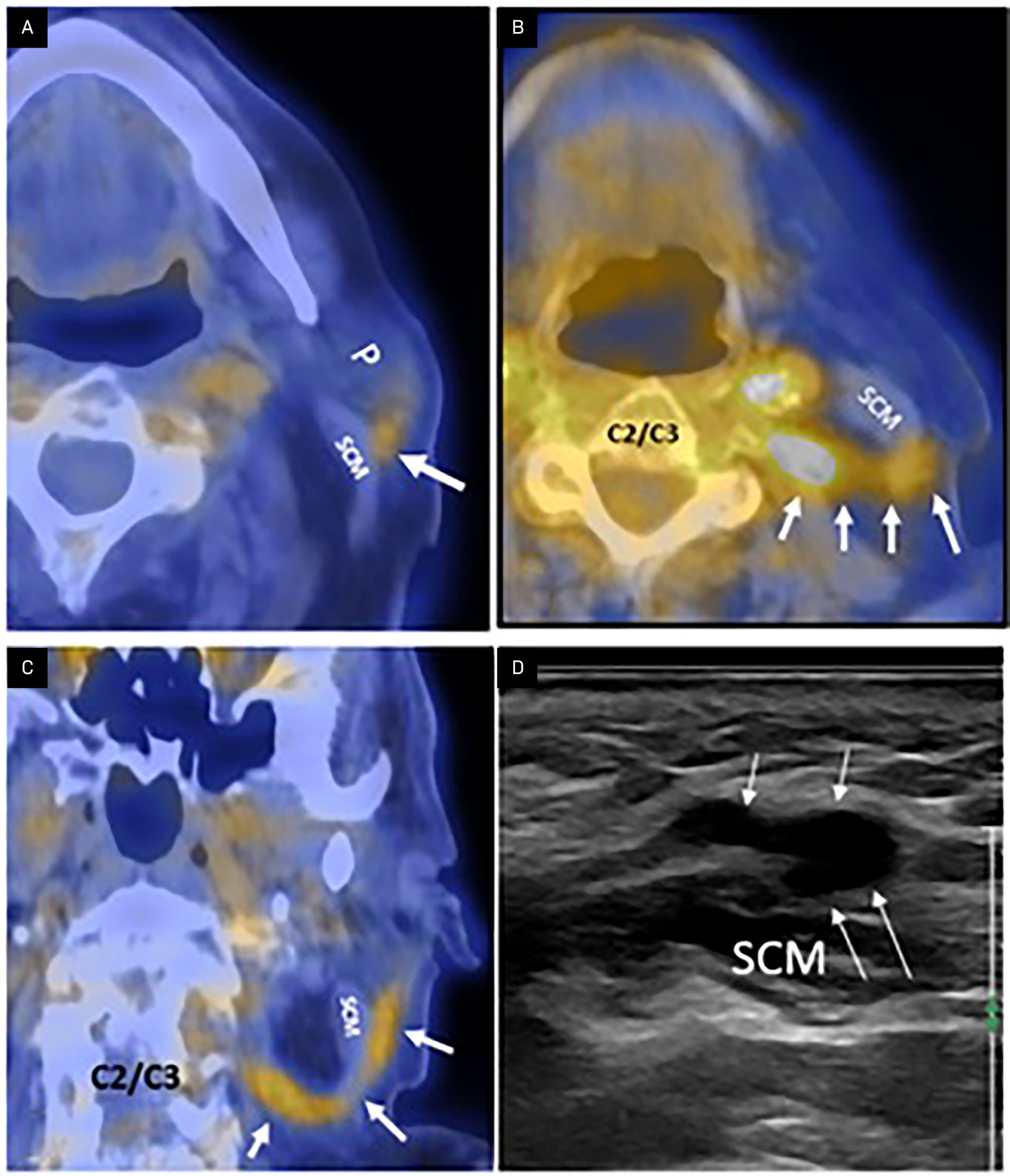
Despite treatment, symptoms progressed to left ear numbness, chin pain, facial droop, and incomplete eye closure, consistent with Bell’s palsy and trigeminal neuralgia; managed with prednisone, valacyclovir, carbamazepine, and eyelid weight. The patient later developed periauricular/parotid swelling; CT and MRI revealed that the presumed thrombophlebitis was, in fact, PNS along the greater auricular nerve, with invasion of the left SCM and intraspinal extension through the left C2-C3 neural foramen. Imaging also identified a metastatic left parotid mass with additional perineural spread along the facial nerve (CN VII), mandibular division of the trigeminal nerve (V3), and the auriculotemporal nerve (Figure 5).
Case 2
This case involves an 81-year-old male patient with a pathologically confirmed ulcerative cutaneous squamous cell carcinoma of the right cheek, demonstrating the same distinct pattern of perineural extension (Figure 6).
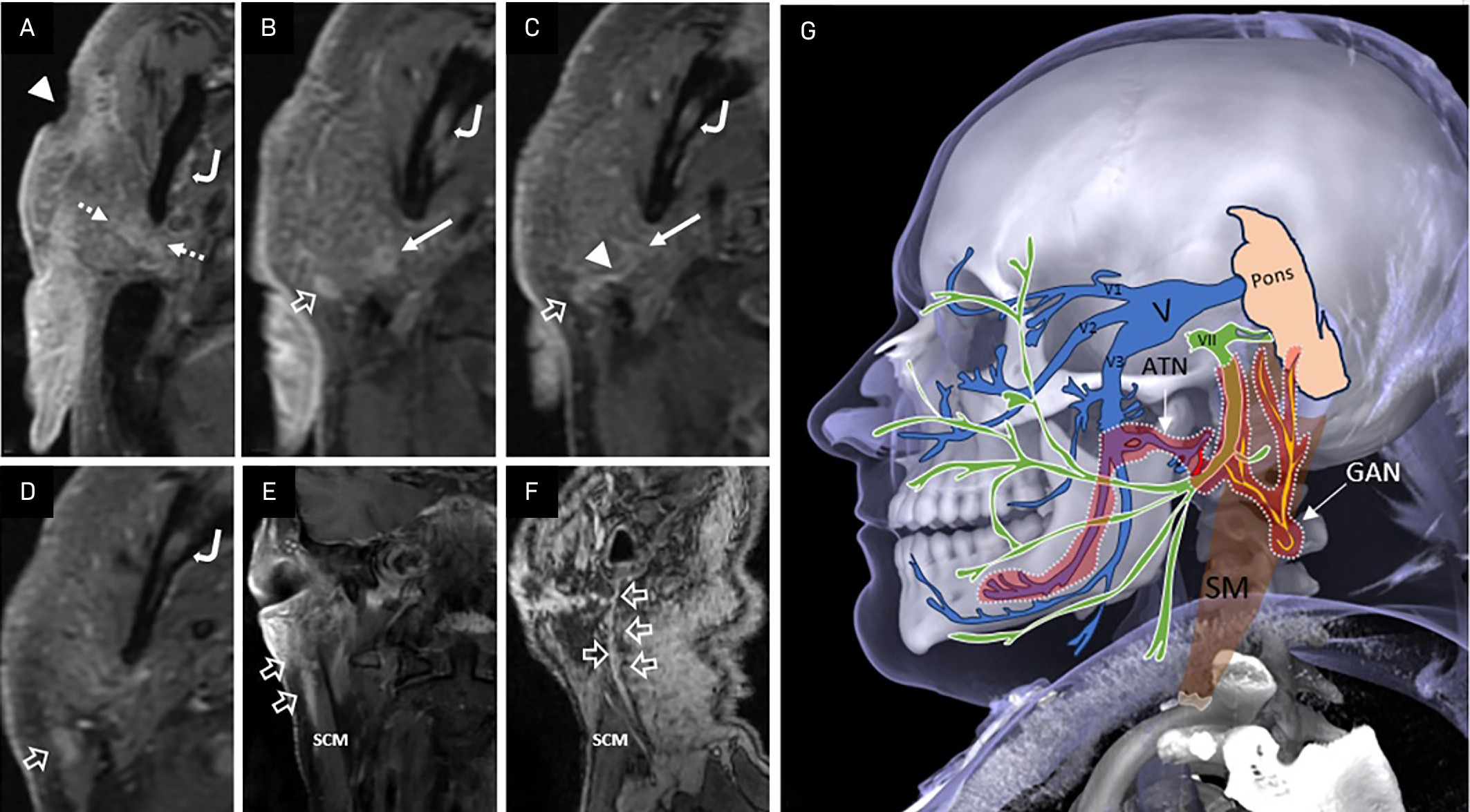
These 2 cases highlight a distinct pattern of perineural spread in which cutaneous malignancies near the parotid gland invade the GAN, extend along its connection to the facial nerve (CN VII), and subsequently spread via the auriculotemporal nerve to the mandibular division (V3) of the trigeminal nerve. Radiologists should be aware of this uncommon pathway and actively evaluate its presence in appropriate clinical contexts.
Lymphoma
On our extensive literature search, only one case of low-grade GAN lymphoma was reported by Duvall et al in a 56-year-old woman with a history of lung sarcoidosis, presenting with intermittent pain involving the GAN distribution. MRI showed an enhancing soft tissue lesion in the superficial right parotid gland directly inferior to the ear pinna, suspecting sarcoidosis; however, the surgeon noted a visible enlargement of the GAN intraoperatively, with surgical pathology revealing low-grade lymphoma.32
Leprosy
Mycobacterium leprae has a propensity to involve Schwann cells and grows best in cool temperatures; hence, its great predication for the superficially located peripheral nerves, most commonly the ulnar and peroneal nerves, with approximately 21 reports describing lepromatous GAN involvement.33-35
The disease is common in the tropics and subtropics, mainly in India and Brazil; however, in the United States, it is more commonly associated with contact with armadillos.35,36 The patients usually present with cord-like swelling along the course of the GPN with corresponding sensory loss.
GAN enlargement can be clinically misdiagnosed as a thrombosed vessel, lymphadenopathy, IgG4-related disease, peripheral nerve sheath tumor, or PNS. US imaging typically shows a hypoechoic, fusiform lesion, and CT/MRI will show an enhancing cord-like lesion following the course of the GPN. Imaging may also reveal the development of nerve abscesses requiring prompt surgical decompression to avoid irreversible damage.35
Tuberculosis (TB)
Warpe et al described an unusual case of cervical tuberculous granulomatous lymphadenopathy with tuberculous granulomas involving the adjacent GAN in a 69-year-old woman presenting with painful tubular neck swelling. Neck ultrasonography and CT again misdiagnosed the GAN involvement as a thrombosed retro-mandibular/external jugular vein precluding FNA cytology, and the lesion was finally accurately diagnosed by surgical pathology. Neuropathy in patients with tuberculosis is more commonly iatrogenic from the antituberculous medications; however, this case highlights the possibility of a primary tuberculous.37
Krabbe’s Disease
Krabbe’s disease is an autosomal-recessive lysosomal storage disorder that impairs myelin turnover, affecting both the central and peripheral nervous systems, including all cranial nerves—most notably the optic nerve. In addition to the well-documented intracranial findings of Krabbe’s disease of nonenhancing symmetric areas of T2 prolongation within the periventricular white matter, and spinal findings of abnormal enhancement of the lumbosacral nerve roots,38,39 Markes et al described a 13-year-old female patient with Krabbe’s disease who presented with bilateral GAN hypertrophy in addition to the distal lower extremity weakness and sensory loss.40
IgG4-Related Disease
IgG4-related disease is a systemic idiopathic fibroinflammatory condition rich in IgG4+ plasma cells affecting many organs, with predilection to the orbits, salivary and thyroid glands in the head neck region.41 In our extensive literature search, 2 cases of IgG4-related GAN perineural disease have been reported, with one presenting as a palpable neck mass and resected as an enlarged lymph node only to discover a pathologically proven GAN IgG4-related disease with preferential epineurium involvement, and the second discovered intraoperatively during open cervical lymph node dissection.42 Both cases also had orbital Ig-G4 disease and postoperatively were responsive to steroid treatment and subsequently suffered sensory loss.42
Great Auricular Neuralgia
GAN neuralgia is described as unilateral brief stabbing pain of abrupt onset and termination, in the distribution of the GAN, which is commonly provoked by neck rotation and may remit and relapse like other craniocervical neuralgias. It is often confused with GAN neuropathy, which is usually a continuous or near-continuous pain, and commonly described as a burning, paresthesia, or dysesthesia and often accompanied by reduced or absent sensation somewhere within the GAN distribution.22,32,43-46
GAN neuralgia can be either idiopathic or secondary to an underlying etiology; hence, imaging using US, CT, or MRI is warranted to evaluate for secondary causes such as inflammation, neoplasm, trauma, or iatrogenic injury, especially with a history of primary malignancyor worsening symptoms.32
Traumatic/Iatrogenic Injury and GAN Sacrifice
Anatomically, the GAN is most vulnerable for traumatic and iatrogenic injury as it emerges from the posterior border of the SCM and runs in the superficial fascia of the SCM.5,32,47,48 GAN is the most injured nerve with face lift with a complication rate of 6-7%.5,48 Injury of the GAN has also been described following mandibular condylar fractures,49,50 carotid endarterectomy,49,51,52 pacemaker placement,43 parotidectomy or submandibulectomy,47 cervical lymph node dissection, and shoulder arthroscopy.53 GAN is also frequently sacrificed during parotidectomy.54 GAN injury can lead to pure anesthesia, paresthesias, and even painful neuromas, and can lead to vexing functional impairments during shaving, combing hair, wearing earrings, and using the telephone. However, some authors also found that the quality of life in such patients was progressively less affected after 1 year.5,55-57
Targeted GAN Interventions
GAN Nerve Grafting58
Since its description by Alberti in 196259 and owing to its proximity to the surgical field, easier harvesting using constant surgical landmarks, and its similar diameter to the facial nerve, the GAN has become the nerve graft donor of choice for reconstructing injured facial nerves and has also been used for inferior alveolar nerve grafting.58
In their review of 128 patients of published GAN-Facial nerve grafting, Werner et al found that facial nerve tumors were the leading cause for grafting (72%) and that the outcome was mostly successful, with most (65%) patients regaining facial nerve function to House-Brackmann grade III (defined as moderate deficits).58
Nerve Blocks and Neuromodulation
GAN block was classically accomplished via superficial cervical plexus block using large volumes of local anesthetics; however, Jeon et al described one of the earliest cases of highly selective US-guided GAN block using smaller volume of local anesthetics and steroids in a 25-year-old man presenting with GAN neuralgia resistant to medications.
In addition, Elahi et al were able to demonstrate excellent pain relief not only on the ipsilateral but also on the contralateral side following GAN neuromodulation for a 32-year-old woman complaining of chronic, intractable headache, adding GAN neuromodulation as a viable treatment option for patients with medically refractory primary chronic headache.60
Conclusion
GAN is an often-overlooked site of pathology, reflecting a significant gap in radiologic awareness. This article reviews the radiologic anatomy, imaging techniques, and spectrum of GAN-related lesions, with a focus on a distinct pattern of perineural spread. Specifically, cutaneous malignancies adjacent to the parotid gland that invade the GAN can result in locoregional extension through both the facial nerve (CN VII) and the auriculotemporal nerve to the mandibular division (V3) of the trigeminal nerve. Increased awareness of this uncommon route will result in improved diagnostic accuracy, more appropriate and earlier patient-specific management, and better outcomes.
References
- Oh S, Abou-Foul A, Patel S, Wilson P. Great auricular nerve schwannoma: a rare presentation and literature review. BMJ Case Rep. 2021;14(7). doi:10.1136/bcr-2021-242972.
- Ginsberg L, Eicher S. Great auricular nerve: anatomy and imaging in a case of perineural tumor spread. AJNR Am J Neuroradiol. 2000;21(3):568-571.
- Raikos A, English T, Yousif O, Sandhu M, Stirling A. Topographic anatomy of the great auricular point: landmarks for its localization and classification. Surg Radiol Anat. 2017;39(5):535-540. doi:10.1007/s00276-016-1758-y.
- Berry M, Bannister L, Standring S. In: Gray’s Anatomy: The Anatomical Basis of Clinical Practice. 1995:901-1398.
- Lefkowitz T, Hazani R, Chowdhry S. Anatomical landmarks to avoid injury to the great auricular nerve during rhytidectomy. Aesthet Surg J. 2013;33(1):19-23. doi:10.1177/1090820X12469625.
- Stambuk H. Perineural tumor spread involving the central skull base region. Semin Ultrasound CT MR. 2013;34(5):445-458. doi:10.1053/j.sult.2013.09.002.
- Sand T, Becser N. Neurophysiological and anatomical variability of the greater auricular nerve. Acta Neurol Scand. 1998;98(5):333-339. doi:10.1111/j.1600-0404.1998.tb01744.x.
- Brennan P, Webb R, Kemidi F, Spratt J, Standring S. Great auricular communication with the marginal mandibular nerve - a previously unreported anatomical variant. Br J Oral Maxillofac Surg. 2008;46(6):492-493. doi:10.1016/j.bjoms.2007.12.005.
- Brennan P, Gholmy M, Ounnas H. Communication of the anterior branch of the great auricular nerve with the marginal mandibular nerve: a prospective study of 25 neck dissections. Br J Oral Maxillofac Surg. 2010;48(6):431-433. doi:10.1016/j.bjoms.2009.07.015.
- Schmitd L, Scanlon C, D’Silva N. Perineural invasion in head and neck cancer. J Dent Res. 2018;97(7):742-750. doi:10.1177/0022034518756297.
- Diamond M, Wartmann C, Tubbs R. Peripheral facial nerve communications and their clinical implications. Clin Anat. 2011;24(1):10-18. doi:10.1002/ca.21072.
- Lynch P, Johnston J. The greater auricular nerve in presumably non-leprous british soldiers. Trans R Soc Trop Med Hyg. 1980;74(1):136. doi:10.1016/0035-9203(80)90034-6.
- Nguyen J, Barber T, Cameron R, Haydon A, Mar V. Findings and resolution of melanoma perineural spread along the greater auricular nerve on FDG PET/CT and MRI. Clin Nucl Med. 2021;46(6). doi:10.1097/RLU.0000000000003534.
- Dhillon M, Mackie G, Singh D. 18F-FDG PET, contrast CT and MRI to comprehensively diagnose and assess rare perineural spread of squamous cell carcinoma to the greater auricular nerve. Radiol Case Rep. 2022;17(6):2106-2110. doi:10.1016/j.radcr.2022.03.080.
- Repanos C, Mitchell D, Gandhi M, Wood M, Panizza B. Great auricular nerve perineural spread of squamous cell carcinoma. ANZ J Surg. 2012;82(3):179-180. doi:10.1111/j.1445-2197.2011.05988.x.
- Beaty S, Colome-grimmer M, Wagner R. Bilateral auricular squamous cell carcinomas with perineural invasion. Dermatol Surg. 2001;27(2):203-206. doi:10.1097/00042728-200102000-00023.
- Mehanna H, John S, Morton R, Chaplin J, McIvor N. Facial palsy as the presenting complaint of perineural spread from cutaneous squamous cell carcinoma of the head and neck. ANZ J Surg. 2007;77(3):191-193. doi:10.1111/j.1445-2197.2006.04007.x.
- Thallaj A, Marhofer P, Moriggl B. Great auricular nerve blockade using high resolution ultrasound: a volunteer study. Anaesthesia. 2010;65(8):836-840. doi:10.1111/j.1365-2044.2010.06443.x.
- Curcean A, Rusu G, Dudea S. Ultrasound appearance of peripheral nerves in the neck: vagus, hypoglossal and greater auricular. Med Pharm Rep. 2020;93(1):39-46. doi:10.15386/mpr-1273.
- Reddy Y, Pidaparthi L, Tourani V, Penneru A, Murthy J. High-resolution ultrasound features of greater auricular nerve in leprosy. Postgrad Med J. 2020;96(1137):443. doi:10.1136/postgradmedj-2020-137703.
- Lieba-Samal D, Pivec C, Platzgummer H. High-resolution ultrasound for diagnostic assessment of the great auricular nerve--normal and first pathologic findings. Ultraschall Med. 2015;36(4):342-347. doi:10.1055/s-0034-1366354.
- Jeon Y, Kim S. Treatment of great auricular neuralgia with real-time ultrasound-guided great auricular nerve block: a case report and review of the literature. Medicine. 2017;96(12). doi:10.1097/MD.0000000000006325.
- Benton L, Trescot A, Trescot A. Peripheral Nerve Entrapments: Clinical Diagnosis and Management. 2016:117-126. doi:10.1007/978-3-319-27482-9_16.
- Xu C, Sun Q. Great auricular nerve schwannoma in neck region: a case report with the risk of medical disputes. BMC Neurol. 2019;19(1):308. doi:10.1186/s12883-019-1532-y.
- Zhang H, Cai C, Wang S. Extracranial head and neck schwannomas: a clinical analysis of 33 patients. Laryngoscope. 2007;117(2):278-281. doi:10.1097/01.mlg.0000249929.60975.a7.
- Kim K, Lee H, Choi J, Hwang J, Lee S. Schwannoma of the posterior branch of the great auricular nerve. Arch Craniofac Surg. 2020;21(6):368-371. doi:10.7181/acfs.2020.00549.
- Moreira M, Dos Santos A, Cintra M. Perineural spread of malignant head and neck tumors: review of the literature and analysis of cases treated at a teaching hospital. Radiol Bras. 2017;50(5):323-327. doi:10.1590/0100-3984.2015.0215.
- Raslan O, Muzaffar R, Shetty V, Osman M. Image findings of cranial nerve pathology on [18F]-2- deoxy-d-glucose (FDG) positron emission tomography with computerized tomography (PET/CT): a pictorial essay. Cancer Imaging. 2015;15:20. doi:10.1186/s40644-015-0054-0.
- Ginat D, Bolotin D, Langerman A. Perineural spread of cutaneous squamous cell carcinoma along the great auricular nerve [corrected]. Ear Nose Throat J. 2015;94(4-5):150-152.
- Panizza B, Warren T, Solares C. Histopathological features of clinical perineural invasion of cutaneous squamous cell carcinoma of the head and neck and the potential implications for treatment. Head Neck. 2014;36(11):1611-1618. doi:10.1002/hed.23509.
- Barrios B, Cederberg C. Squamous cell carcinoma recurring to the great auricular nerve. Oncology. 2014;28(8):687-688.
- Duvall J, Garza I, Kissoon N, Robertson C. Great auricular neuralgia: case series. Headache. 2020;60(1):247-258. doi:10.1111/head.13690.
- Maurya P, Kazmi K, Kulshreshtha D, Singh A, Malhotra K. Isolated thickened greater auricular nerve due to leprosy. Neurol Sci. 2016;37(4):649-650. doi:10.1007/s10072-015-2452-2.
- Gupta N, Vinod K, Singh G, Nischal N. Bilateral greater auricular nerve thickening in leprosy. QJM. 2020;113(8):579. doi:10.1093/qjmed/hcz287.
- Kaya D, Garnes N, Robins A. Leprosy of the great auricular nerve: case report and review of the literature. Clin Imaging. 2022;82:63-66. doi:10.1016/j.clinimag.2021.10.001.
- Rodrigues L, Lockwood D. Leprosy now: epidemiology, progress, challenges, and research gaps. Lancet Infect Dis. 2011;11(6):464-470. doi:10.1016/S1473-3099(11)70006-8.
- Warpe B, Poflee S, Pande N, Shrikhande A. Tuberculous neuritis: a rare sequel of a common disease. Indian J Pathol Microbiol. 2014;57(1):69-71. doi:10.4103/0377-4929.130902.
- Given C, Santos C, Durden D. Intracranial and spinal MR imaging findings associated with Krabbe’s disease: case report. AJNR Am J Neuroradiol. 2001;22(9):1782-1785.
- Provenzale J, Peddi S, Kurtzberg J. Correlation of neurodevelopmental features and MRI findings in infantile Krabbe’s disease. AJR Am J Roentgenol. 2009;192(1):59-65. doi:10.2214/ajr.07.3885.
- Marks H, Scavina M, Kolodny E, Palmieri M, Childs J. Krabbe’s disease presenting as a peripheral neuropathy. Muscle Nerve. 1997;20(8):1024-1028. doi:10.1002/(sici)1097-4598(199708)20:8<1024::aid-mus13>3.0.co;2-v.
- Deshpande V. IgG4 related disease of the head and neck. Head Neck Pathol. 2015;9(1):24-31. doi:10.1007/s12105-015-0620-6.
- Wong W, Morton R. Immunoglobulin G4-related disease of the orbital cavity, cervical lymph nodes and greater auricular nerve: case report. Am J Otolaryngol. 2016;37(3):182-185. doi:10.1016/j.amjoto.2016.01.010.
- Blumenthal H. Great auricular neuralgia. Headache. 1992;32(8):413-415. doi:10.1111/j.1526-4610.1992.hed3208413.x.
- Fukushima M, Sakuraba H, Shiraiwa N, Matsushita S. Electroacupuncture therapy for auricular paresthesia. Med Acupunct. 2017;29(5):331-334. doi:10.1089/acu.2017.1239.
- Maimone-Baronello M, Piccoli F, La Bella V. Great auricular neuralgia: a case report. Headache. 2003;43(9):1005-1006. doi:10.1046/j.1526-4610.2003.03194.x.
- Hall G. Auricular neuralgia. Arch NeurPsych. 1933;29(3):615. doi:10.1001/archneurpsyc.1933.02240090185011.
- Nusair Y, Dickenson A. Great auricular causalgia: an unusual complication of excision of the submandibular gland. Br J Oral Maxillofac Surg. 2003;41(5):334-335. doi:10.1016/s0266-4356(03)00116-5.
- Lewin M, Tsur H. Injuries of the great auricular nerve in rhytidectomy. Aesth Plast Surg. 1976;1(1):409-417. doi:10.1007/BF01570275.
- Verta M, Applebaum E, Mcclusky D, Yao J, Bergan J. Cranial nerve injury during carotid endarterectomy. Ann Surg. 1977;185(2):192-195. doi:10.1097/00000658-197702000-00010.
- Vesnaver A, Ahčan U, Rozman J. Evaluation of surgical treatment in mandibular condyle fractures. J Craniomaxillofac Surg. 2012;40(8):647-653. doi:10.1016/j.jcms.2011.10.029.
- Ballotta E, Da Giau G, Renon L. Cranial and cervical nerve injuries after carotid endarterectomy: a prospective study. Surgery. 1999;125(1):85-91. doi:10.1016/S0039-6060(99)70292-8.
- Schauber M, Fontenelle L, Solomon J, Hanson T. Cranial/cervical nerve dysfunction after carotid endarterectomy. J Vasc Surg. 1997;25(3):481-487. doi:10.1016/s0741-5214(97)70258-1.
- Park T, Kim Y. Neuropraxia of the cutaneous nerve of the cervical plexus after shoulder arthroscopy. Arthroscopy. 2005;21(5):631. doi:10.1016/j.arthro.2005.02.003.
- Yan F, Desiato V, Nguyen S, Lentsch E. Impact of greater auricular nerve sacrifice during parotidectomy on quality of life. Head Neck. 2021;43(1):70-78. doi:10.1002/hed.26452.
- Patel N, Har-El G, Rosenfeld R. Quality of life after great auricular nerve sacrifice during parotidectomy. Arch Otolaryngol Head Neck Surg. 2001;127(7):884-888.
- Ryan W, Fee W. Great auricular nerve morbidity after nerve sacrifice during parotidectomy. Arch Otolaryngol Head Neck Surg. 2006;132(6):642-649. doi:10.1001/archotol.132.6.642.
- Grammatica A, Perotti P, Mancini F. Great auricular nerve preservation in parotid gland surgery: long-term outcomes. Laryngoscope. 2015;125(5):1107-1112. doi:10.1002/lary.25025.
- Werner C, D’Antoni A, Iwanaga J. A comprehensive review of the great auricular nerve graft. Neurosurg Rev. 2021;44(4):1987-1995. doi:10.1007/s10143-020-01426-9.
- Alberti P. The greater auricular nerve. donor for facial nerve grafts: a note on its topographical anatomy. Arch Otolaryngol. 1962;76:422-424. doi:10.1001/archotol.1962.00740050434007.
- Elahi F, Reddy C, Bellinger A, Manolitsis N. Neuromodulation of the great auricular nerve: a case report. Neuromodulation. 2014;17(8):784-787. doi:10.1111/ner.12114.
Disclosures
The authors have no conflicts of interest to disclose. None of the authors received outside funding for the production of this original manuscript, and no part of this article has been previously published elsewhere.
Acknowledgments
The authors of this manuscript declare that the patient image in Figure 1 is artificial intelligence (AI)-generated (magichour.ai); the remainder of the illustration was created by Osama Raslan, MD. No generative AI or AI-assisted technologies were used to generate content, ideas, or theories. We utilized AI solely to ensure anonymity in Figure 1 without compromising image quality or detail. This use was under strict human oversight and control. After the application of AI technologies, the authors carefully reviewed and edited the manuscript to ensure its accuracy and coherence.
Citation
. Tracking the Silent Pathway: Imaging the Great Auricular Nerve - Insights for Radiologists. Applied Radiology. 2026;55(4). doi:10.37549/AR-D-26-0011.