Trilateral Retinoblastoma
Journal of Pediatric Case Reports — Vol. 1 , Issue 7
Published: October 1, 2026
1 Department of Medicine, Kettering Health, Dayton, Ohio
2 Department of Radiology, Phoenix Children’s Hospital, Phoenix, Arizona
3 Department of Radiology, Cincinnati Children’s Hospital, University of Cincinnati College of Medicine, Cincinnati, Ohio
* Corresponding author: Richard B. Towbin (rtowbin@gmail.com)
Abstract
Trilateral retinoblastoma is a rare condition occurring in the setting of retinoblastoma with an associated pineal or suprasellar mass. Baseline brain MRI at the time of retinoblastoma diagnosis is recommended to identify synchronous intracranial tumors. Ongoing surveillance may be warranted for intracranial cystic lesions with suspicious features, such as wall irregularity, nodularity, or atypical size. While isolated retinoblastoma carries a favorable prognosis with curative potential, the development of trilateral retinoblastoma is associated with significantly poorer outcomes, even with aggressive treatment.
Keywords
orbit, brain, neoplasm, genetic
Categories
Case Summary
A male infant presented with signs of cellulitis of the right eye and parental concerns of worsening vision. On ophthalmoscopic examination, leukocoria was present in the right eye.
Imaging Findings
MRI (Figure 1) obtained at the time of diagnosis showed a large heterogeneous mass occupying the right globe. The mass covered the optic disk, and there was mild enhancement of the optic nerve near its insertion at the disk. Small nodules were visible within the right anterior chamber. 2 small nodules were visible within the left globe. In addition to the masses of each globe, there was a 1.9 cm mass of the pineal gland. The mass was slightly hyperintense to the adjacent deep white matter structures on T2-weighted images and hypointense on T1-weighted images. It had several small internal cystic spaces and enhanced avidly. The lateral and 3rd ventricles were dilated.
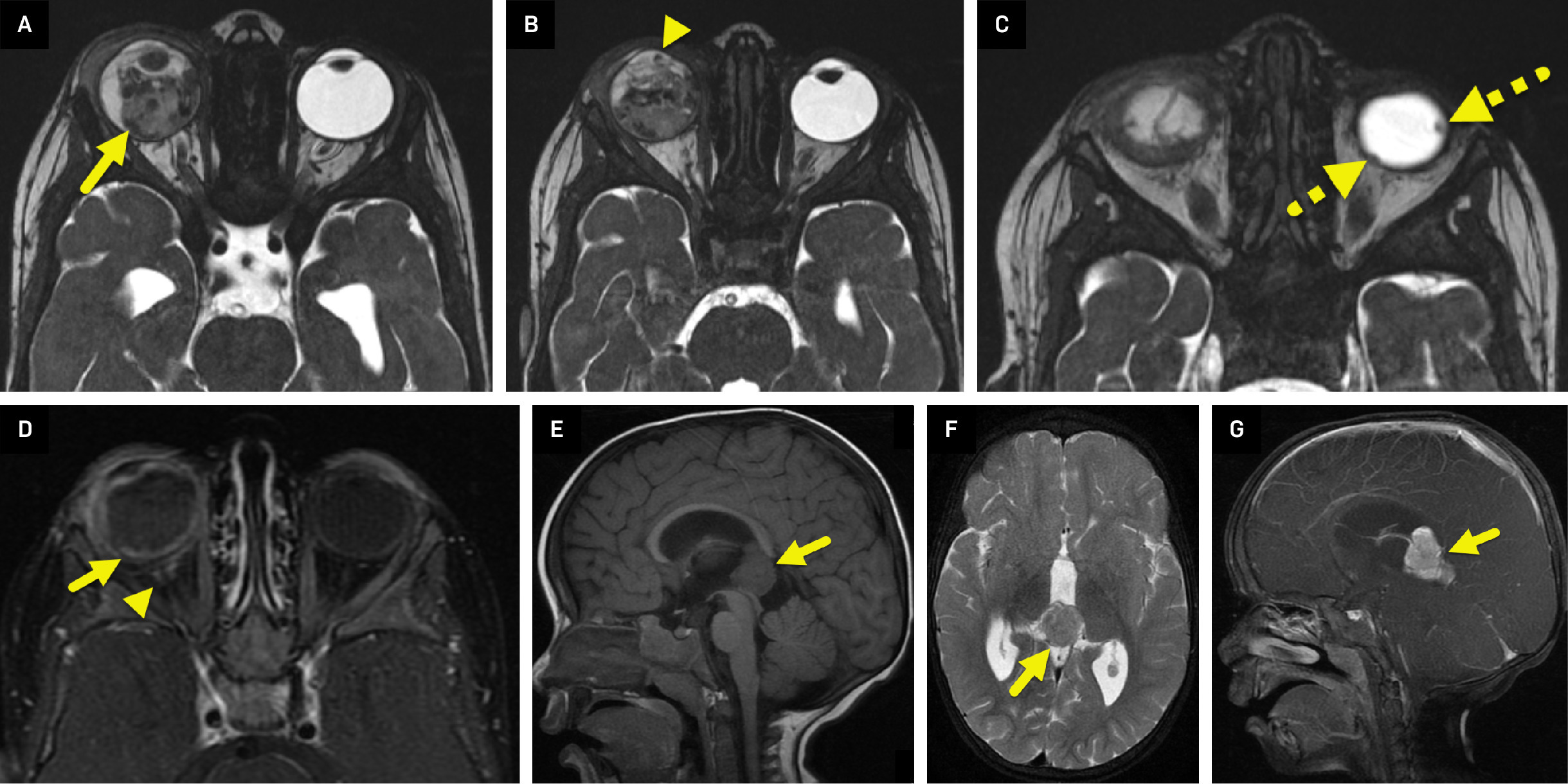
CT (Figure 2) performed after right eye enucleation showed the pineal mass to be partially calcified.
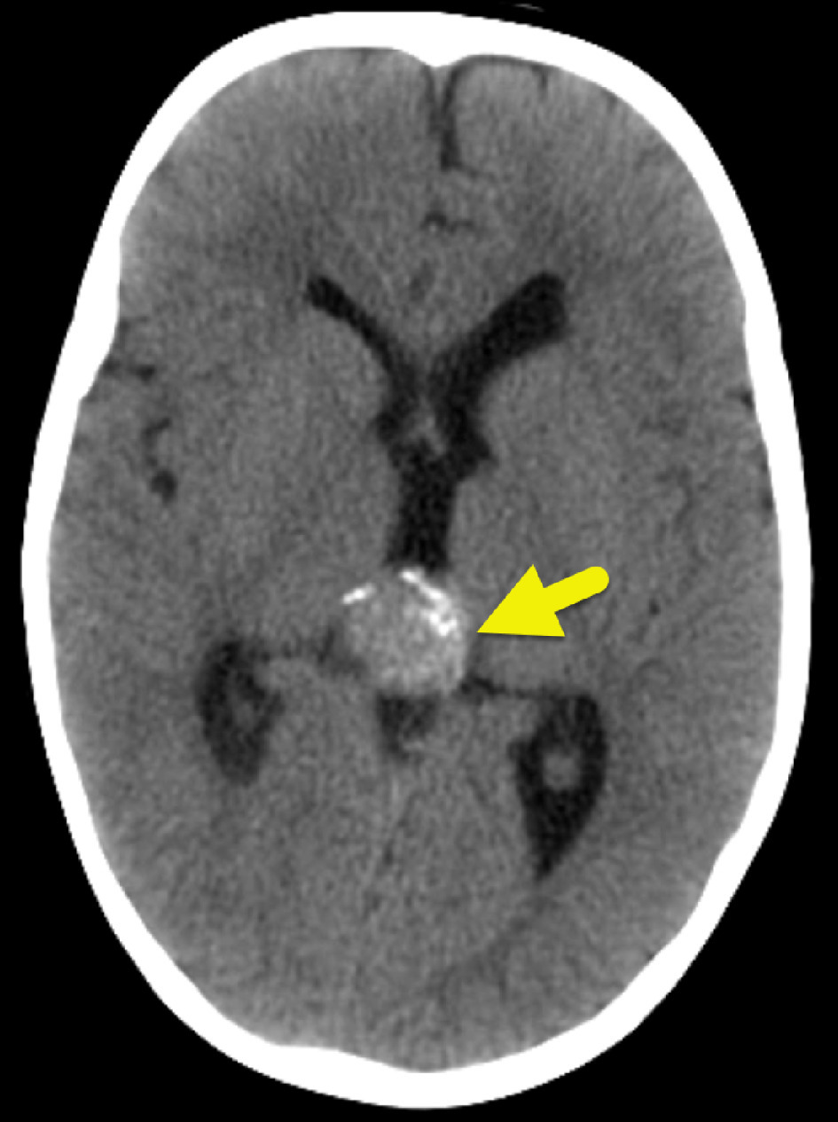
Diagnosis
Retinoblastoma in both eyes with a pineoblastoma—so-called trilateral retinoblastoma.
The differential diagnosis for leukocoria in an infant includes congenital cataracts and Coats disease.
Discussion
Trilateral retinoblastoma is a rare malignancy defined by the presence of a midline intracranial primitive neuroectodermal tumor in patients with unilateral or bilateral retinoblastoma. A germline mutation in the RB1 gene plays a central role in the pathogenesis of retinoblastoma, including trilateral retinoblastoma. In one study, RB1 mutations were identified in all patients with trilateral retinoblastoma. In 70-75% of patients with trilateral retinoblastoma, the intracranial tumor arises from the pineal gland. In the remaining patients, tumors develop in the suprasellar or parasellar region.1 When both pineal and suprasellar tumors are present alongside ocular retinoblastoma, the condition is referred to as quadrilateral retinoblastoma, although this presentation is exceedingly rare.2,3
The reported incidence of trilateral retinoblastoma varies across studies of patients with retinoblastoma.4-7 In an analysis of large cohorts of patients with bilateral retinoblastoma, de Jong et al identified an overall incidence of 3.8%. Of these, 82% had pineal trilateral retinoblastoma and 18% had nonpineal tumors. Among patients with hereditary retinoblastoma—defined as all bilateral cases and unilateral cases with either a family history or a germline RB1 mutation—the incidence of trilateral retinoblastoma was slightly lower at 3.5%.4
Historically, the pineal tumor component of trilateral retinoblastoma was detected metachronously, often months or years after the initial diagnosis of retinoblastoma. However, with the widespread adoption of routine baseline brain imaging, particularly MRI, synchronous detection of intracranial tumors has become more common. These synchronous tumors are often asymptomatic and smaller in size at the time of diagnosis, enabling earlier intervention. Notably, survival has been reported in cases of asymptomatic pineoblastoma less than 15 mm in diameter, as well as in nonpineal trilateral retinoblastoma regardless of tumor size or the presence of symptoms.1,4
The historical incidence of synchronous trilateral retinoblastoma may have been underestimated due to inconsistent use of baseline neuroimaging. In a study by Rodjan et al, patients who underwent baseline brain MRI were diagnosed with trilateral retinoblastoma at a median age of 12 months, typically before the onset of intracranial symptoms. In contrast, patients who did not receive baseline imaging were diagnosed an average of 27 months after retinoblastoma diagnosis and often presented with signs of intracranial hypertension. Tumors in the baseline imaging group were significantly smaller (mean diameter 18 mm, range 9-34 mm) compared with those without baseline imaging (mean diameter 35 mm, range 11-59 mm). Hydrocephalus and positive cerebrospinal fluid cytology were also more common in the latter group.
On MRI, intracranial tumors in trilateral retinoblastoma, whether pineal or suprasellar, typically demonstrate well-defined borders and heterogeneous enhancement due to cystic components or necrosis. These lesions are generally isointense to gray matter on both T1- and T2-weighted sequences. Cystic components are seen in up to 57% of pineal tumors.5-11 The increased detection of smaller cysts in more recent studies may reflect advances in MRI resolution and technique.1
Because trilateral retinoblastoma is a rare malignancy, there is no uniform consensus regarding follow-up frequency for cystic pineal lesions. Recommendations often depend on lesion characteristics. Rodjan et al advised repeat MRI 6 months after detection of “probably benign pineal cysts,” which they defined as cystic lesions with thin, smooth walls and discrete rim enhancement. In contrast, de Bloeme et al recommended a more selective approach, reserving follow-up imaging for lesions with suspicious features such as irregular wall thickening greater than 2 mm, nodularity, or when the size of the solid or cystic component exceeds the 99th percentile for age-based norms. Even when using baseline screening and selective surveillance for suspicious lesions, a high number of follow-up MRIs may still be required to detect malignant transformation. This strategy increases the burden of imaging, contributes to patient and caregiver anxiety, and introduces risks associated with repeated anesthesia exposure.1
Once diagnosed, trilateral retinoblastoma typically requires aggressive treatment. Intensive chemotherapy followed by autologous stem cell rescue has been associated with longer survival compared with conventional chemotherapy alone.6 Despite these efforts, overall prognosis remains poor. In one study that included patients with metastatic retinoblastoma, the 1-year event-free survival rate following treatment was 28.3%.12 Another study reported a 5-year mortality rate of 56% for patients with pineal trilateral retinoblastoma.6
Our patient was successfully treated with chemotherapy, right eye enucleation, left eye thermotherapy, and pineoblastoma resection.
Conclusion
Trilateral retinoblastoma is a rare condition occurring in the setting of retinoblastoma with an associated pineal or suprasellar mass. Baseline brain MRI at the time of retinoblastoma diagnosis is recommended to identify synchronous intracranial tumors. Ongoing surveillance may be warranted for intracranial cystic lesions with suspicious features, such as wall irregularity, nodularity, or atypical size. While isolated retinoblastoma carries a favorable prognosis with curative potential, the development of trilateral retinoblastoma is associated with significantly poorer outcomes, even with aggressive treatment.
References
- De Bloeme C, Jansen R, De Haan J. Follow-up of cystic pineal glands in retinoblastoma patients does not increase detection of pineal trilateral retinoblastoma. Am J Ophthalmol. 2024;268:199-211. doi:10.1016/j.ajo.2024.07.005.
- Silvera V, Guerin J, Brinjikji W, Dalvin L. Retinoblastoma: what the neuroradiologist needs to know. AJNR Am J Neuroradiol. 2021;42(4):618-626. doi:10.3174/ajnr.A6949.
- Kebudi R, Akdeniz D, Bayramova J. RB1 gene mutations in turkish retinoblastoma patients. JCO. 2022;40(16_suppl). doi:10.1200/JCO.2022.40.16_suppl.e22007.
- De Jong M, Kors W, de Graaf P. The incidence of trilateral retinoblastoma: a systematic review and meta-analysis. Am J Ophthalmol. 2015;160(6):1116-1126. doi:10.1016/j.ajo.2015.09.009.
- Kivelä T. Trilateral retinoblastoma: a meta-analysis of hereditary retinoblastoma associated with primary ectopic intracranial retinoblastoma. J Clin Oncol. 1999;17(6):1829-1837. doi:10.1200/JCO.1999.17.6.1829.
- De Jong M, Kors W, De Graaf P. Trilateral retinoblastoma: a systematic review and meta-analysis. Lancet Oncol. 2014;15(10):1157-1167. doi:10.1016/S1470-2045(14)70336-5.
- Yamanaka R, Hayano A, Takashima Y. Trilateral retinoblastoma: a systematic review of 211 cases. Neurosurg Rev. 2019;42(1):39-48. doi:10.1007/s10143-017-0890-4.
- Rodjan F, de Graaf P, Brisse H. Trilateral retinoblastoma: neuroimaging characteristics and value of routine brain screening on admission. J Neurooncol. 2012;109(3):535-544. doi:10.1007/s11060-012-0922-4.
- Whitehead M, Oh C, Choudhri A. Incidental pineal cysts in children who undergo 3-t MRI. Pediatr Radiol. 2013;43(12):1577-1583. doi:10.1007/s00247-013-2742-x.
- Lacroix-Boudhrioua V, Linglart A, Ancel P. Pineal cysts in children. Insights Imaging. 2011;2(6):671-678. doi:10.1007/s13244-011-0117-0.
- Ramasubramanian A, Kytasty C, Meadows A. Incidence of pineal gland cyst and pineoblastoma in children with retinoblastoma during the chemoreduction era. Am J Ophthalmol. 2013;156(4):825-829. doi:10.1016/j.ajo.2013.05.023.
- Dunkel I, Piao J, Chantada G. Intensive multimodality therapy for extraocular retinoblastoma: a children’s oncology group trial (ARET0321). J Clin Oncol. 2022;40(33):3839-3847. doi:10.1200/JCO.21.02337.
Disclosures
The authors have no conflicts of interest to disclose. None of the authors received outside funding for the production of this original manuscript and no part of this article has been previously published elsewhere.
Citation
. Trilateral Retinoblastoma. Journal of Pediatric Case Reports. 2026;1(7). doi:10.37549/JPCR-26-0117.