Tubal Infertility: A Review for Interventional Radiologists
Applied Radiology — Vol. 55 , Issue 2
Published: March 9, 2026
1 Division of Interventional Radiology, Department of Radiology, University Hospital, Rutgers New Jersey Medical School, Newark, New Jersey
2 Division of Interventional Radiology, Department of Radiological Sciences, David Geffen School of Medicine at UCLA, Los Angeles, California
Abstract
Proximal fallopian tube obstruction (pFTO) is a common and potentially reversible cause of female subfertility. Image-guided techniques have established interventional radiology as a central discipline in the diagnosis and treatment of this condition. This review summarizes the pathophysiology, imaging evaluation, and management of pFTO, with a primary focus on fluoroscopy-guided transcervical fallopian tube recanalization (FTR). Normal tubal anatomy, mechanisms of proximal obstruction, and the diagnostic and therapeutic roles of hysterosalpingography are reviewed. Technical aspects of FTR, including patient selection, procedural technique, periprocedural care, and follow-up, are described. Published outcomes demonstrate technical success rates exceeding 90% with favorable pregnancy outcomes in appropriately selected patients and low complication rates. Given its safety profile, cost-effectiveness, and ability to immediately restore tubal patency, fluoroscopic FTR represents an effective first-line, minimally invasive treatment for women with isolated pFTO and an important application of image-guided therapy in infertility care.
Keywords
tubal infertility, fallopian tube obstruction, fallopian tube recanalization
Categories
Introduction
Subfertility at a Glance
Subfertility is defined as the inability to conceive within 12 months of frequent, unprotected intercourse in women younger than 35 years, or within 6 months in women 35 years and older, and can be either primary (never achieved pregnancy) or secondary (unable to achieve subsequent pregnancy). 1,2 More than 15% of child-seeking couples are affected by infertility globally, 2 and the incidence is thought to be increasing, in part due to a trend in couples rearing offspring at a later age, when female fertility and male sperm counts are both in decline, and cumulative exposure to sexually transmitted infections is increased. 3 Most subfertility is attributable to a male factor (~25%), ovarian factor (~25%), or fallopian tube factor (~25%). Another 10% arise from a uterine or peritoneal cause, and in 40% of cases, both male and female factors are thought to contribute. Despite a wealth of knowledge on the causes of infertility, still up to one-third of workups yield no identifiable male or female factor, known as unexplained infertility. 4-6
The American College of Obstetricians and Gynecologists, American Society for Reproductive Medicine (ASRM), and National Institute for Health and Care Excellence (NICE) recommend that any child-seeking woman who meets criteria for infertility, or who has a condition known to cause infertility, should be offered an evaluation. 2,4,7 The workup consists of 4 essential components: (1) a comprehensive medical history of both partners (if one exists), (2) targeted physical examination of the female partner, (3) semen analysis, (4) and testing to exclude common ovarian, tubal, and uterine causes of infertility. The latter generally includes, but is not limited to, fluoroscopic hysterosalpingography (HSG) to assess the uterine and tubal structures.
Fallopian Tube Obstruction
Tubal factor infertility accounts for 25-35% of all female subfertility worldwide, among which 10-25% is due to proximal fallopian tube obstruction (pFTO). 3,5,8,9 There are many causes of pFTO, including inspissated debris (eg mucus, blood), tubal spasm (pseudo-obstruction), salpingitis isthmica nodosa (SIN), pelvic inflammatory disease (PID), endometriosis, polyps, and congenital Müllerian anomalies. However, it is estimated that tubal spasm and mucus plugs alone account for up to 40% of cases. 6,10 It is also estimated that half of patients with bilateral obstructions have isolated short segment disease and no pelvic adhesions. 11 The remaining causes relate to distal fallopian tube obstruction (dFTO), which is virtually always caused by chronic adhesions from PID or other inflammatory processes. 9 It is important to distinguish this entity from pFTO, as dFTO leads to hydrosalpinges and is managed surgically. 9
Before discussing treatment options for pFTO, it is important to first understand normal tubal anatomy, physiology, and underlying mechanisms that lead to subfertility. Fallopian tubes are 7‐14 cm in length, trumpet-shaped, musculomembranous conduits between the ovaries and the uterus. Each tube is comprised of an inner mucosal membrane made up of 2 simple columnar epithelial cell types, a middle muscularis comprised of 2 smooth muscle layers, and an outer serosa that is contiguous with the mesosalpinx of the broad ligament. 10 Their course is straight or slightly curved in 60% of women and tortuous in 40%. This nonlinear anatomy is thought to assist in preventing access of vaginal flora into the peritoneum. However, this also makes the oviduct prone to accumulation of secretions and inflammation. 12
The proximal fallopian tube comprises roughly the first 4 cm of duct with a diameter of about 1 mm and consists of a proximal 1.5‐2.5 cm intramural segment (also known as the uterine or interstitial segment) and distal 2‐3 cm isthmic (or extrauterine) segment. The intramural segment has a thick muscular wall relatively devoid of ciliated epithelium and empties into the uterus through an ostium at the apex of the uterine cornu or horn. This combination of features renders the intramural segment prone to spasm and transient obstruction by upstream secretions generated at the junction of the intramural and isthmic segments, or uterotubal junction, during the follicular phase of the menstrual cycle. 6
The distal fallopian tube is comprised of an ampulla, infundibulum, and fimbriae that gradually increase in caliber toward the ovary. The distal fimbriated end is open to the peritoneal cavity, where it performs a sweeping motion over the ovary allowing an ovulated egg to be drawn into the oviduct. Once in the duct, the ovum becomes available for fertilization, where a coordinated effort between ciliary action, secretory flow, and smooth muscle contractions optimizes the environment for sperm ascent, ovum descent, and transportation of a developing embryo to the endometrium. These multiple essential functions of the fallopian tube are modulated by an intricate network of sympathetic and parasympathetic nerve fibers, estrogen, progesterone, and prostaglandins, all of which play a vital role in achieving spontaneous pregnancy. 10,13 The focus of this article is to discuss the role of the interventional radiologist (IR) in the workup and management of pFTO.
Treatment of Proximal FTO: Then and Now
Since its inception in the early 20th century, diagnostic transcervical HSG was recognized for having therapeutic value in the management of pFTO. 14,15 Its therapeutic effect on infertility was first described in 1923 when a report noted increased pregnancy rates following successful intrauterine insufflation of carbon dioxide to overcome tubal occlusions. 16 In 1931, a landmark paper detailed a similar phenomenon using oil-based contrast media. 17 The therapeutic advantage of lipid-soluble contrast over aqueous-based agents was subsequently determined. 5,18 Today, the safety and efficacy of HSG using lipid-soluble contrast is well established. Current evidence from randomized clinical trials comparing HSG to no treatment suggests at least 3-fold increased odds of achieving both clinical pregnancy and live birth in the setting of pFTO. 5
In the 1980s, with the advent of improved low-profile coaxial catheter and wire technologies, selective salpingography and tubal recanalization emerged as a superior treatment option for tubal infertility. 19-21 The contemporary fluoroscopy-guided transcervical fallopian tube recanalization (FTR) technique used today combines standard HSG and coaxial angiography techniques from the IR’s armamentarium. 21 The FTR procedure augments the therapeutic benefit of conventional HSG via selective tubal flushing of contrast and catheter- and wire-directed mechanical clearance of tubal debris and adhesions. Moreover, selective salpingography has a greater positive predictive value for diagnosing tubal obstruction compared to HSG. 5,22 While there are many underlying etiologies and nuances of tubal obstruction, histopathologic findings in the proximally occluded fallopian tube include only 3 entities: fibrotic adhesions, amorphous intraluminal debris, or normal histology (in which case transient spasm or dislodged debris is presumed). 23 Several mechanisms have been proposed to explain the improved fertility outcomes from FTR, which include not only direct mechanical effects, but also indirect effects on the endometrium and peritoneum. 5 With regard to the endometrium, there is evidence to suggest that changes in the local immunological environment may increase endometrial receptivity by favorably modulating leukocyte populations essential to early embryo development. 24 With regard to the peritoneum, oil-based contrast media modulate dendritic and regulatory T cell populations specific to pregnancy. 25 There is also evidence for inhibition of sperm phagocytosis by peritoneal macrophages by altering interleukin and prostaglandin production. 26
Guidelines and Indications for FTR
Current ASRM and NICE infertility guidelines recommend the use of selective salpingography and recanalization as a first-line approach to diagnosis and management of proximal tubal obstruction. 4,7 FTR is indicated for women with primary or secondary infertility who have confirmed unilateral or bilateral proximal tubal obstruction and no other significant infertility factors. 7,27 The procedure can be performed as a standalone therapy, or as a neoadjuvant with microsurgical tubal anastomosis or in vitro fertilization (IVF) and intracytoplasmic sperm injection (ICSI). 6,28 Tubal cannulation can be performed fluoroscopically or hysteroscopically; however, a fluoroscopic approach is preferred as it allows simultaneous diagnosis and treatment. Moreover, fluoroscopy is less costly and mitigates the need for sedation or anesthesia. 7 Contraindications to FTR include active pelvic infection, pelvic malignancy, or pregnancy. 6 Relative contraindications include the presence of concomitant dFTO, cornual fibroids, severely distorted uterine anatomy, Müllerian anomalies, or severe SIN. 7
Patient Evaluation
Patients are preferably seen for preprocedural consultation in a dedicated IR clinic. Key historical factors include duration of infertility, coital frequency and timing, pregnancy history, menstrual history, sexual and gynecologic history, prior contraceptive methods, prior abdominopelvic surgeries, and drug and alcohol use. 2 Current medications and any supplements should be reviewed for potential teratogenic agents. Any prior infertility studies should be reviewed for evidence of other contributing factors to infertility, including results of ovulatory function testing, ovarian reserve testing, and male semen analysis, which will aid in the prognostication of FTR outcomes and appropriate patient counseling. Perhaps the most important role during the clinical encounter is to set expectations and to explain to the patient and her family the risks, benefits, and alternatives to FTR. This ensures an open dialogue that allows for shared decision-making and informed consent. 6
Prior imaging should be reviewed for procedural planning and to identify any findings that could potentially compromise the success of FTR, such as hydrosalpinges, pelvic adhesions, endometriosis, severe SIN, or complex uterine anatomy. 6 The diagnosis of pFTO is established by demonstrating a lack of patency within the first 4 cm of the fallopian tube, evaluated either by HSG, hysterosalpingo-contrast sonography, or diagnostic laparoscopy and chromotubation. Fallopian tube recanalizationis optimally performed during the follicular phase of the ovarian cycle and proliferative phase of the endometrial cycle, that is, days 7-14. This increases the chances of conception and decreases the risk of contrast intravasation. 18 A preprocedural pregnancy test is not routinely required provided that FTR is performed during the follicular phase. 27 Note that ensuring optimal timing requires a detailed menstrual history of the patient’s ovulatory cycle, especially if intervals are irregular.
Technique
Patient Preparation
Five days total of doxycycline antibiotic prophylaxis is recommended for all patients undergoing FTR. Regimens can include 100 mg twice daily beginning 2 days before, or a single 200 mg dose immediately prior to the procedure followed by 100 mg twice daily for 4 additional days. 6,28 Patients should fast for a minimum of 6 hours in the event that sedation is needed. If sedation is administered, standard moderate intravenous sedation with midazolam and fentanyl titrated to comfort is recommended.
In the angiography suite, the patient is placed in the lithotomy position with the lower extremities fastened in table stirrups. The perineum is prepped and draped in the usual sterile fashion. At this stage, patient communication is crucial to minimize discomfort. An appropriately sized, lubricated speculum is then inserted slowly into the vagina and gently opened until the cervix is in clear view. Optional warming of the speculum can be performed prior to insertion by rinsing it under warm water or holding it in a gloved hand. Once in view, the cervix is cleansed using sterile betadine swabs. 6
Intrauterine Access and HSG
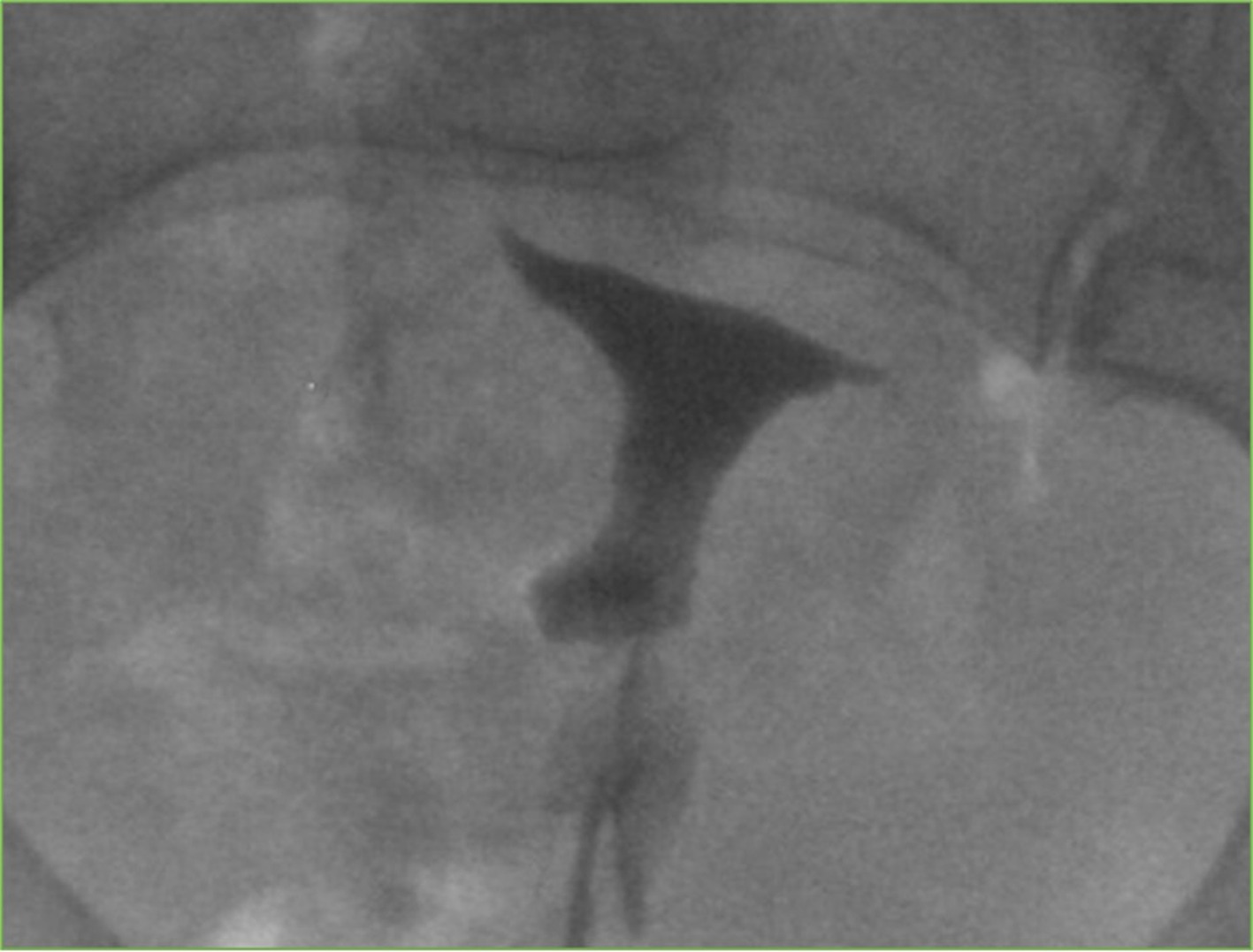
Transcervical uterine access is obtained using a balloon occluding sheath or catheter, such as a 9-French intrauterine access balloon catheter (Cook Medical, Bloomington, Indiana) or similar device that can accommodate the passage of a 5- or 6-French diagnostic catheter while occluding the internal os. A tenaculum should be on standby in the event that excessive mobility or difficult anatomy prohibits successful cervical cannulation. If this step is required, a topical anesthetic applied to the cervix is encouraged prior to engaging the tenaculum at the 12 o’clock position to fix the cervix in place. The tenaculum can also help stabilize the uterus for tubal cannulation. Next, a conventional hysterosalpingogram is performed to confirm the presence of pFTO; gentle back tension can be applied to the balloon catheter to prevent leakage of contrast through the os. 6,12 Water- or lipid-soluble contrast is injected through the catheter into the uterine cavity until all its contours are well-delineated, and fluoroscopic runs are reviewed for evidence of tubal patency (Figure 1). Contrast should be prepared as a 50% volume dilution in normal saline to ensure adequate visualization. A slow and steady injection is recommended to minimize tubal spasm and prevent a false positive result. 6 If the tubes are not visualized and spasm is suspected, turning the patient prone will occasionally result in their opacification. 12
Selective Salpingography and Recanalization
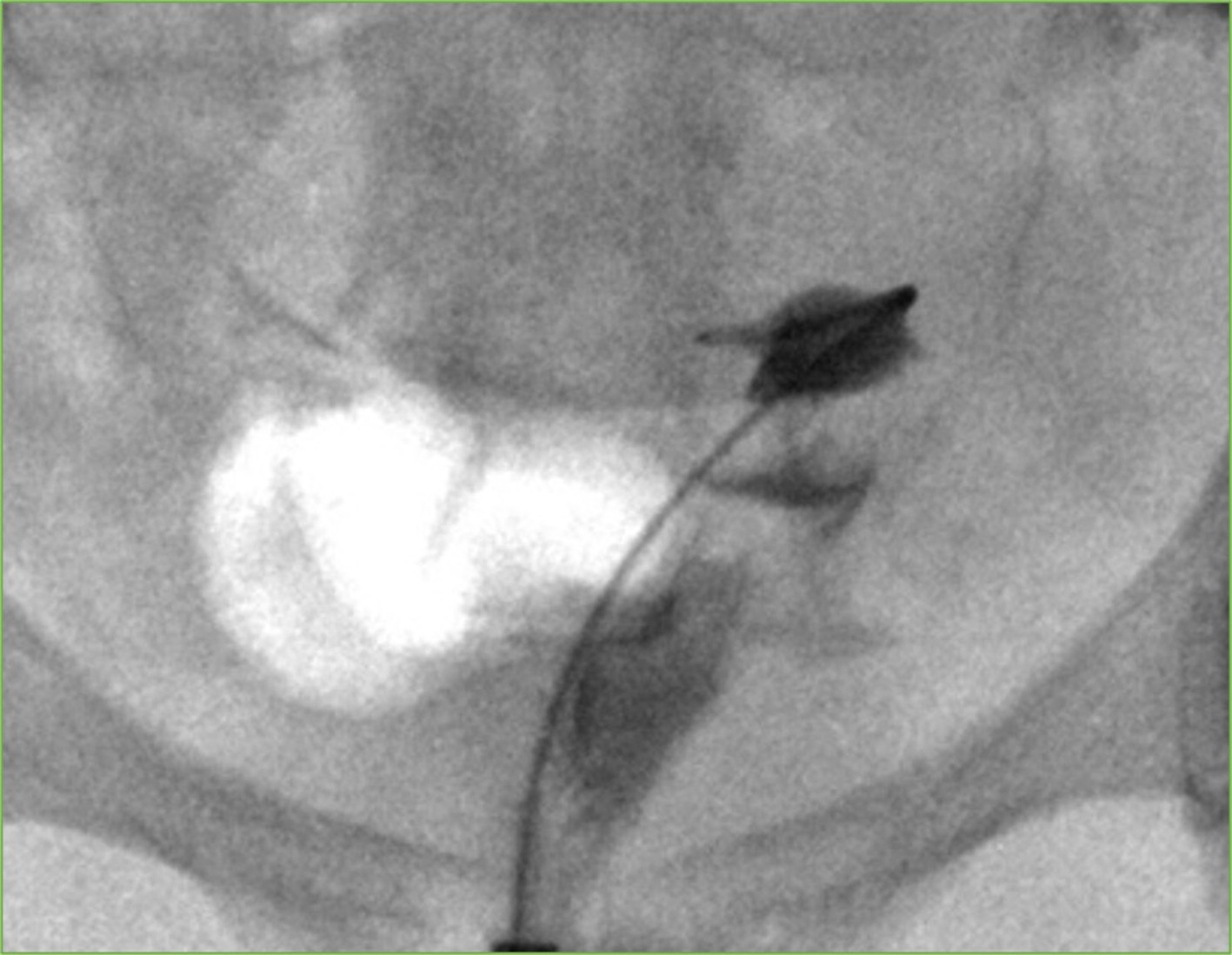
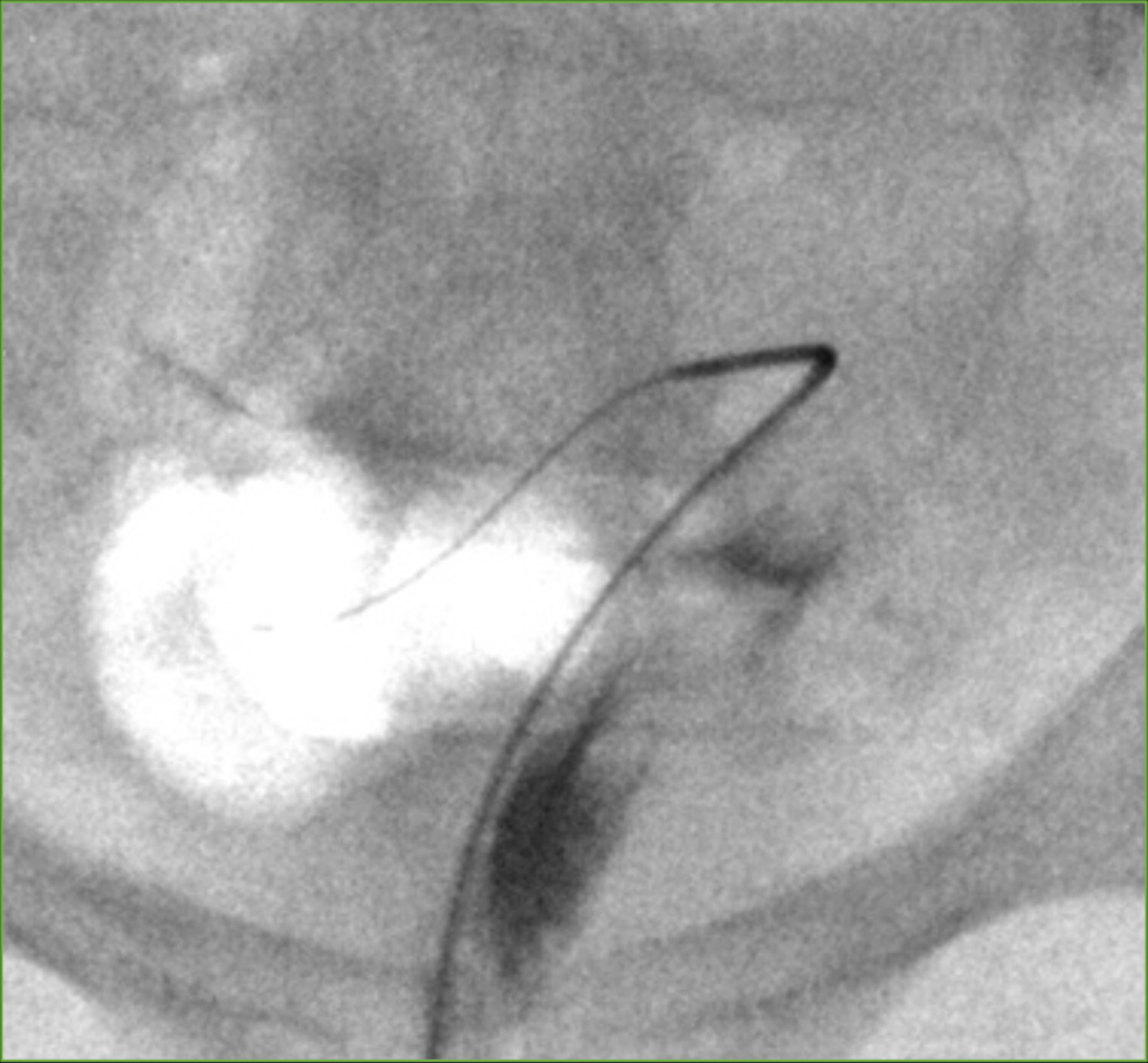
Selective catheterization of the tubal ostium is then preferably performed with a triaxial system that includes an angled 5-French catheter, 0.035-inch hydrophilic guidewire, 3-French microcatheter, and 0.018 microwire in order to engage the laterally oriented cornu and cross the obstruction. The Rösch-Thurmond fallopian tube catheterization set (Cook Medical, Bloomington, Indiana) is ideal for this purpose. First, the 5-French catheter is advanced over the 0.035-inch guidewire into the uterine cornu (Figure 2), and a salpingogram is performed. In a minority of cases, a patent tube will be revealed, and no further intervention is needed. 28 Assuming persistent tubal obstruction, the microcatheter and microwire are then coaxially advanced across the proximal occlusion, with gentle probing movements of the microwire as necessary to clear the obstruction (Figure 3). If the proximal tube is acutely angulated or the site of obstruction is within the isthmic segment, exchanging the system for a softer, tapered guidewire and catheter may be preferred. 6,12
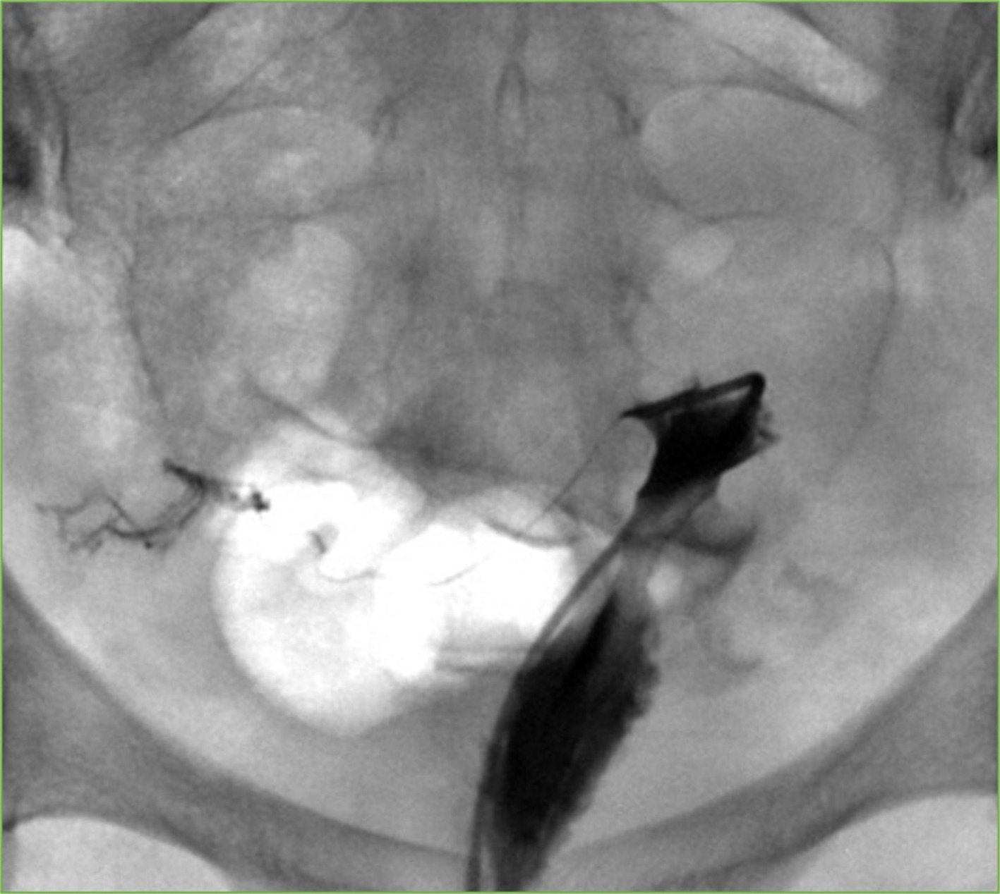
Following recanalization, the crossing wire is removed and selective salpingography through the 3-French catheter is performed to confirm luminal patency (Figure 4). Lastly, with the 5-French catheter still wedged in the tubal ostium, the 3-French catheter is removed, and a final salpingogram is performed (Figure 5). Full-strength oil-based contrast is preferred over water-soluble contrast due to its association with higher clinical pregnancy rates. 5 Rarely, tubal perforation may give the false impression of a patent tube, in which case enough of the agent should be injected to ensure spillage of contrast through the distal fimbriated end of the tube.
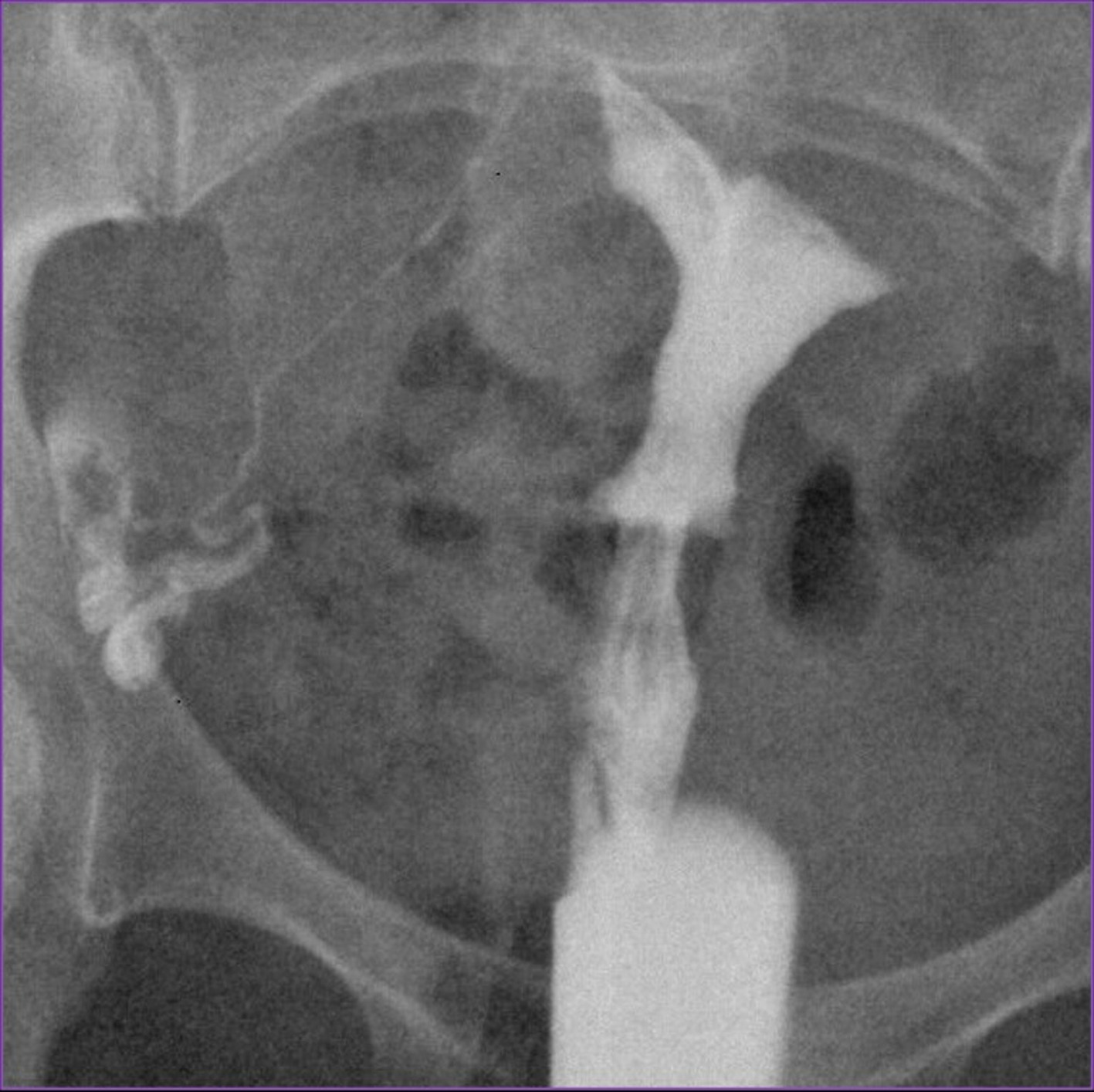
If bilateral obstruction is present, attention is then turned to the contralateral side, and the procedure is repeated in the same manner. All devices are then removed from the uterus and vagina, the patient is cleaned, and the procedure is concluded. Total procedure time is approximately 30 minutes, with fluoroscopy time usually under 10 minutes. 12
Postprocedural Care and Follow-Up
Patients are discharged the same day following a brief observation period to monitor pain levels and ensure no complications. Postprocedural expectations and return precautions should be reviewed with patients prior to discharge. Mild uterine cramping and vaginal bleeding or discharge is expected within the first 24 hours and may continue for up to 3 days. Oral nonsteroidal anti-inflammatory agents are prescribed to be taken as needed for pain control. Patients are encouraged to abstain from sexual intercourse for 24 hours but can otherwise resume their usual physical activity after the procedure. 6 If not already doing so, patients should be advised to start taking daily prenatal vitamins, as the chance of pregnancy is highest immediately following FTR. Given the diminishing chances of spontaneous conception over time, repeat FTR or an alternative therapy should be considered 6‐12 months after successful recanalization if pregnancy has not yet ensued. 7 In cases of failed FTR, one study revealed that 93% of patients had evidence of SIN, chronic salpingitis, or obliterative fibrosis on excised proximal tube specimens. 29 Lastly, patients should be advised to continue follow-up with their fertility specialist, as couples may have additional fertility treatments planned after tubal patency is restored. 12
Outcomes
Technical success for restoring tubal patency via fluoroscopic FTR ranges from 70% to 100%, 8,23 with most contemporary reports citing success rates of well over 90%. 30,31 However, tubal re-occlusion is common following FTR and may warrant early reintervention in certain cases, as repeat recanalization is feasible and has resulted in successful pregnancies. 11 Among women who do not conceive within one year, rates of tubal re-occlusion are as high as 25%, 50%, and 60% at 3, 6, and 12 months, respectively. 11,32
Due to differences in reporting methods and heterogeneity among study populations, clinical success rates of FTR vary considerably within the literature. One meta-analysis found a clinical pregnancy rate of 27% at 12 months, with a live birth rate of 22% 33 ; however, many women had a combination of infertility factors and therefore did not reflect true pFTO rates. In carefully selected patients who have undergone a full workup with no other identifiable causes of infertility other than proximal tubal occlusion, 12 month clinical pregnancy rates in the range of 41-58% are widely reported. 30-32,34 In general, favorable factors are thought to include younger age (< 35 years), secondary infertility, duration of infertility < 5 years, and pFTO as the sole cause of infertility, 30 although this has yet to be elucidated by large-scale randomized data. At this time, there is no convincing evidence for a difference in outcomes between unilateral and bilateral pFTO. 30,33 While FTR is an effective treatment for unilateral pFTO, it should be noted that similar pregnancy rates have been achieved with ovarian stimulation therapy and intrauterine insemination in this population. 7 Lastly, multiple retrospective studies have demonstrated no significant difference in pregnancy outcomes between fluoroscopic and hysteroscopic recanalization techniques, which is important to consider when counseling patients and communicating with colleagues across disciplines. 7,33
Given the substantial heterogeneity within the literature, it is best to advise patients with a conservative estimate of pregnancy outcomes. Patients should be counseled only after a routine infertility workup has demonstrated that they are a good candidate for FTR. In general, patients should be advised that their chance of conception is at least 30% shortly after recanalization, and that if they do not achieve pregnancy after 6 months, there is a 50% chance that re-occlusion has occurred. 35
Adverse Events
Primary complications include ectopic pregnancy, spontaneous abortion, tubal perforation, and pelvic infection. Ectopic pregnancy has been reported to occur in 3-9% of patients. 4,33 However, the percentage directly attributable to FTR is unclear, as patients with tubal disease have a baseline elevated risk of ectopic pregnancy prior to intervention, and similar ectopic rates are seen following IVF. The rate of spontaneous abortion ranges from 4% to 8% among large cohorts and does not appear to differ from hysteroscopic techniques. 33 Asymptomatic tubal perforation occurs in 2-11% of cases and requires no additional monitoring or intervention. 4,7,11 It is unclear at this time whether perforation has an impact on subsequent pregnancy outcomes, as case numbers are overall low. The rate of pelvic infection, although not reported in the literature, is a theoretical risk of any transcervical procedure and is generally assumed to be < 1% following FTR. 6
Historically, intravasation, or the backflow of contrast into the blood or lymphatic vessels, was considered an important risk factor of HSG, said to occur in approximately 1-7% of cases. 5 While intravasation occasionally occurs, it is nearly always asymptomatic. Nevertheless, there is a potential, although mostly historical, risk of allergic reaction or embolic phenomena. Lastly, there is a potential risk of harm from radiation exposure. However, studies have shown that the dose absorbed by the ovaries during FTR is < 1 Rad (10 Gy), or roughly equivalent to one barium enema, and is within the acceptable limits for women of reproductive age. 11 Importantly, while a fertilized gamete is highly radiosensitive, an unfertilized ovum is relatively radioresistant. 6
Alternatives and Future Applications
It is imperative that IRs performing FTR have fundamental knowledge of both the pathophysiology of and treatments for proximal tubal disease. Microsurgical tubocornual anastomosis is a well-established procedure for pFTO that involves careful resection of the occluded proximal segment and surgical conjoining of the non-diseased tube to the uterine cornu, with pregnancy rates of up to 58% over the course of months to years, and an ectopic rate of 4%. 11 While the procedure has fallen out of favor in recent years due to its invasiveness and lag time in efficacy, it is still used as a first-line treatment for tubal ligation reversal. 7
The number of couples seeking IVF and ICSI treatment for tubal infertility has dramatically increased over the past few decades and continues to increase in popularity and efficacy. Recent global estimates of successful per-cycle pregnancy rates and live birth rates are 24% and 18% for IVF, and 26% and 19% for ICSI, respectively. 36 The main disadvantages of these therapies include high cost, potential need for multiple treatments and several weeks of monitoring, and the risks of twin gestation and ovarian hyperstimulation syndrome. 7,36 Thus, for infertility due to isolated proximal tubal obstruction, FTR is preferred as the first-line option, with microsurgical anastomosis and IVF/ICSI reserved for refractory cases.
A number of recent developments have emerged as potential future treatment options for pFTO. Fertiloscopy and falloposcopy are among such developments. 4 Similar to conventional hysteroscopy, falloposcopy allows direct visualization of the working field, but on a much smaller scale. The new low-profile scopes are designed to navigate into the fallopian tube and can be used to cross tubal obstructions under direct view. However, further study is needed to demonstrate feasibility, safety, and efficacy. Another area of ongoing research is in the development of solutions to prevent re-occlusion after FTR. A number of small studies have used various biochemical agents injected into the fallopian tubes after recanalization. A recent 4-arm randomized controlled trial studied rates of pregnancy and re-occlusion following injection of beta-(1–4)-2-amino-2-deoxy-d-glucosechitosan (chitosan) and Salvia miltiorrhiza (Dan-shen), 2 commonly used Eastern medicinal compounds. 32
At 12 months post-FTR, cumulative pregnancy rates for each group were 31% (control), 47% (chitosan), 43% (Dan-shen), and 64% (chitosan plus Dan-shen), which was statistically significant. For patients who had not conceived by 12 months, tubal patency rates were 39% (control), 78% (chitosan), 77% (Dan-shen), and 94% (chitosan plus Dan-shen). While the mechanisms by which these agents might work to prevent re-occlusion are not well understood, these results show promise for future research into adjuvant agents other than oil-based contrast.
Conclusions
pFTO is a common cause of female infertility in which IRs have a primary role in the diagnosis, treatment, and management of. Due to its favorable safety and efficacy profile, fluoroscopic transcervical FTR has become the first-line treatment of choice for pFTO. In experienced hands, FTR is associated with a technical success rate of up to 100% and 1-year pregnancy rate of up to 60%, with immediate onset of efficacy and extremely low rate of complications. Tubal re-occlusion is common after FTR, occurring in up to 50% of patients after 6 months. Therefore, patients should be closely followed by their IR and infertility specialist in the immediate postprocedural period and offered repeat intervention or prompt referral for appropriate treatment if pregnancy is unlikely to occur.
References
- Farquhar C, Bhattacharya S, Repping S. Female subfertility. Nat Rev Dis Primers. 2019;5(1):7. doi:10.1038/s41572-018-0058-8.
- Infertility workup for the women’s health specialist: ACOG committee opinion, number 781. Obstet Gynecol. 2019;133(6). doi:10.1097/AOG.0000000000003271.
- Evers J. Female subfertility. Lancet. 2002;360(9327):151-159. doi:10.1016/S0140-6736(02)09417-5.
- Fertility Problems [Quality Standard No. QS73]. . 2014.
- Wang R, Watson A, Johnson N. Tubal flushing for subfertility. Cochrane Database Syst Rev. 2020;10(10). doi:10.1002/14651858.CD003718.pub5.
- Kohi M. Interventional radiologist’s approach to fallopian tube recanalization. Tech Vasc Interv Radiol. 2021;24(1):100736. doi:10.1016/j.tvir.2021.100736.
- Role of tubal surgery in the era of assisted reproductive technology: a committee opinion. Fertil Steril. 2021;115(5):1143-1150. doi:10.1016/j.fertnstert.2021.01.051.
- Hou H, Chen Y, Li T. Outcome of laparoscopy-guided hysteroscopic tubal catheterization for infertility due to proximal tubal obstruction. J Minim Invasive Gynecol. 2014;21(2):272-278. doi:10.1016/j.jmig.2013.09.003.
- Role of tubal surgery in the era of assisted reproductive technology: a committee opinion. Fertil Steril. 2015;103(6). doi:10.1016/j.fertnstert.2015.03.032.
- Honoré G, Holden A, Schenken R. Pathophysiology and management of proximal tubal blockage. Fertil Steril. 1999;71(5):785-795. doi:10.1016/s0015-0282(99)00014-x.
- Thurmond A. Fallopian tube recanalization: technique, results, and complications. J Vasc Interv Radiol. 2002;13(2):P252-P254. doi:10.1016/S1051-0443(02)70162-0.
- Thurmond A. Fallopian tube catheterization. Semin Intervent Radiol. 2008;25(4):425-431. doi:10.1055/s-0028-1102995.
- Lyons R, Saridogan E, Djahanbakhch O. The reproductive significance of human fallopian tube cilia. Hum Reprod Update. 2006;12(4):363-372. doi:10.1093/humupd/dml012.
- Rindfleisch W. Darstellung des cavum uteri. Klin Wochenschr. 1910;47:780.
- Smith W. On a new method of treating sterility, by the removal of obstructions of the fallopian tubes. Lancet. 1849;53(1342):529-531. doi:10.1016/S0140-6736(02)65312-7.
- Peterson R. Therapeutic value of transuterine gas inflation. JAMA. 1923;81(12):980. doi:10.1001/jama.1923.02650120012004.
- Robins S, Shapira A. The value of hysterosalpingography. N Engl J Med. 1931;205(8):380-395. doi:10.1056/NEJM193108202050804.
- Gillespie H. The therapeutic aspect of hysterosalpingography. Br J Radiol. 1965;38(448):301-302. doi:10.1259/0007-1285-38-448-301.
- Platia M, Krudy A. Transvaginal fluoroscopic recanalization of a proximally occluded oviduct. Fertil Steril. 1985;44(5):704-706. doi:10.1016/s0015-0282(16)48992-2.
- Jansen R, Anderson J. Catheterisation of the fallopian tubes from the vagina. Lancet. 1987;330(8554):309-310. doi:10.1016/S0140-6736(87)90893-2.
- Thurmond A, Novy M, Uchida B, Rösch J. Fallopian tube obstruction: selective salpingography and recanalization. Radiology. 1987;163(2):511-514. doi:10.1148/radiology.163.2.3562835.
- Woolcott R, Fisher S, Thomas J, Kable W. A randomized, prospective, controlled study of laparoscopic dye studies and selective salpingography as diagnostic tests of fallopian tube patency. Fertil Steril. 1999;72(5):879-884. doi:10.1016/s0015-0282(99)00382-9.
- Thurmond A. Selective salpingography and fallopian tube recanalization. AJR Am J Roentgenol. 1991;156(1):33-38. doi:10.2214/ajr.156.1.1898568.
- Yun A, Lee P. Enhanced fertility after diagnostic hysterosalpingography using oil-based contrast agents may be attributable to immunomodulation. AJR Am J Roentgenol. 2004;183(6):1725-1727. doi:10.2214/ajr.183.6.01831725.
- Izumi G, Koga K, Takamura M. Oil-Soluble Contrast Medium (OSCM) for hysterosalpingography modulates dendritic cell and regulatory T cell profiles in the peritoneal cavity: a possible mechanism by which OSCM enhances fertility. J Immunol. 2017;198(11):4277-4284. doi:10.4049/jimmunol.1600498.
- Johnson J, Montoya I, Olive D. Ethiodol oil contrast medium inhibits macrophage phagocytosis and adherence by altering membrane electronegativity and microviscosity. Fertil Steril. 1992;58(3):511-517. doi:10.1016/s0015-0282(16)55254-6.
- Thurmond A, Machan L, Maubon A. A review of selective salpingography and fallopian tube catheterization. Radiographics. 2000;20(6):1759-1768. doi:10.1148/radiographics.20.6.g00nv211759.
- Thurmond A, Jafri S, Diokno A, Amendola M. Lower Genitourinary Radiology: Imaging and Intervention. 1998:501-507. doi:10.1007/978-1-4612-1648-3_29.
- Letterie G, Sakas E. Histology of proximal tubal obstruction in cases of unsuccessful tubal canalization. Fertil Steril. 1991;56(5):831-835. doi:10.1016/S0015-0282(16)54651-2.
- Al-Omari M, Obeidat N, Elheis M, Khasawneh R, Gharaibeh M. Factors affecting pregnancy rate following fallopian tube recanalization in women with proximal fallopian tube obstruction. J Clin Med. 2018;7(5):110. doi:10.3390/jcm7050110.
- Shen H, Cai M, Chen T. Factors affecting the success of fallopian tube recanalization in treatment of tubal obstructive infertility. J Int Med Res. 2020;48(12). doi:10.1177/0300060520979218.
- Huang C, He X, Luo W, Chen H, Huang Y. Combined chitosan and dan-shen injection for long-term tubal patency in fallopian tube recanalization for infertility. Drug Deliv Transl Res. 2019;9(4):738-747. doi:10.1007/s13346-018-00611-0.
- De Silva P, Chu J, Gallos I. Fallopian tube catheterization in the treatment of proximal tubal obstruction: a systematic review and meta-analysis. Hum Reprod. 2017;32(4):836-852. doi:10.1093/humrep/dex022.
- Hayashi M, Hoshimoto K, Ohkura T. Successful conception following fallopian tube recanalization in infertile patients with a unilateral proximally occluded tube and a contralateral patent tube. Hum Reprod. 2003;18(1):96-99. doi:10.1093/humrep/deg006.
- Thurmond A, Rösch J. Nonsurgical fallopian tube recanalization for treatment of infertility. Radiology. 1990;174(2):371-374. doi:10.1148/radiology.174.2.2296648.
- Adamson G, de Mouzon J, Chambers G. International committee for monitoring assisted reproductive technology: world report on assisted reproductive technology, 2011. Fertil Steril. 2018;110(6):1067-1080. doi:10.1016/j.fertnstert.2018.06.039.
Disclosures
The authors have no conflicts of interest to disclose. None of the authors received outside funding for the production of this original manuscript, and no part of this article has been previously published elsewhere.
Acknowledgments
The authors of this manuscript declare that in the writing process of this work, no generative artificial intelligence (AI) or AI-assisted technologies were used to generate content, ideas, or theories. We utilized AI solely for the purpose of enhancing readability and refining language. This use was under strict human oversight and control. After the application of AI technologies, the authors carefully reviewed and edited the manuscript to ensure its accuracy and coherence.
Citation
. Tubal Infertility: A Review for Interventional Radiologists. Applied Radiology. 2026;55(2). doi:10.37549/AR-D-26-0005.