Tubo-Ovarian Abscess
Applied Radiology — Vol. 55 , Issue 2
Published: January 29, 2026
1 University of Arizona College of Medicine-Phoenix, Phoenix, Arizona
2 Department of Radiology, Phoenix Children’s Hospital, Phoenix, Arizona
Categories
Case Summary
A nonsexually active adolescent with a history of constipation, obesity, recurrent urinary tract infection (UTI), and recent treatment of UTI presented with acute, chronic right-sided abdominal pain, low back pain, and fever. On examination, the abdomen was soft and mildly tender to palpation at the periumbilical and right flank and lower quadrant regions. The patient denied sexual activity/sexual abuse. Gonorrhea and chlamydia testing were negative. Leukocytosis was present at 22.3K, and clean catch urine culture demonstrated > 100K Escherichia coli. The patient was found on CT and US to have an 8 cm right tubo-ovarian abscess (TOA), from which purulent fluid was aspirated and determined to be culture positive for E. coli and Peptoniphilus asaccharolyticus.
Imaging Findings
Pelvic CT coronal and sagittal views (Figure 1) demonstrated 6.0 × 7.3 × 7.0 cm rim-enhancing fluid collections in the right lower pelvis, displacing the uterus slightly to the left of midline, with associated right moderate hydroureteronephrosis and associated reactive adenopathy. US pelvis and power Doppler demonstrated 8.1 cm hypervascular right adnexa structure with cystic component.
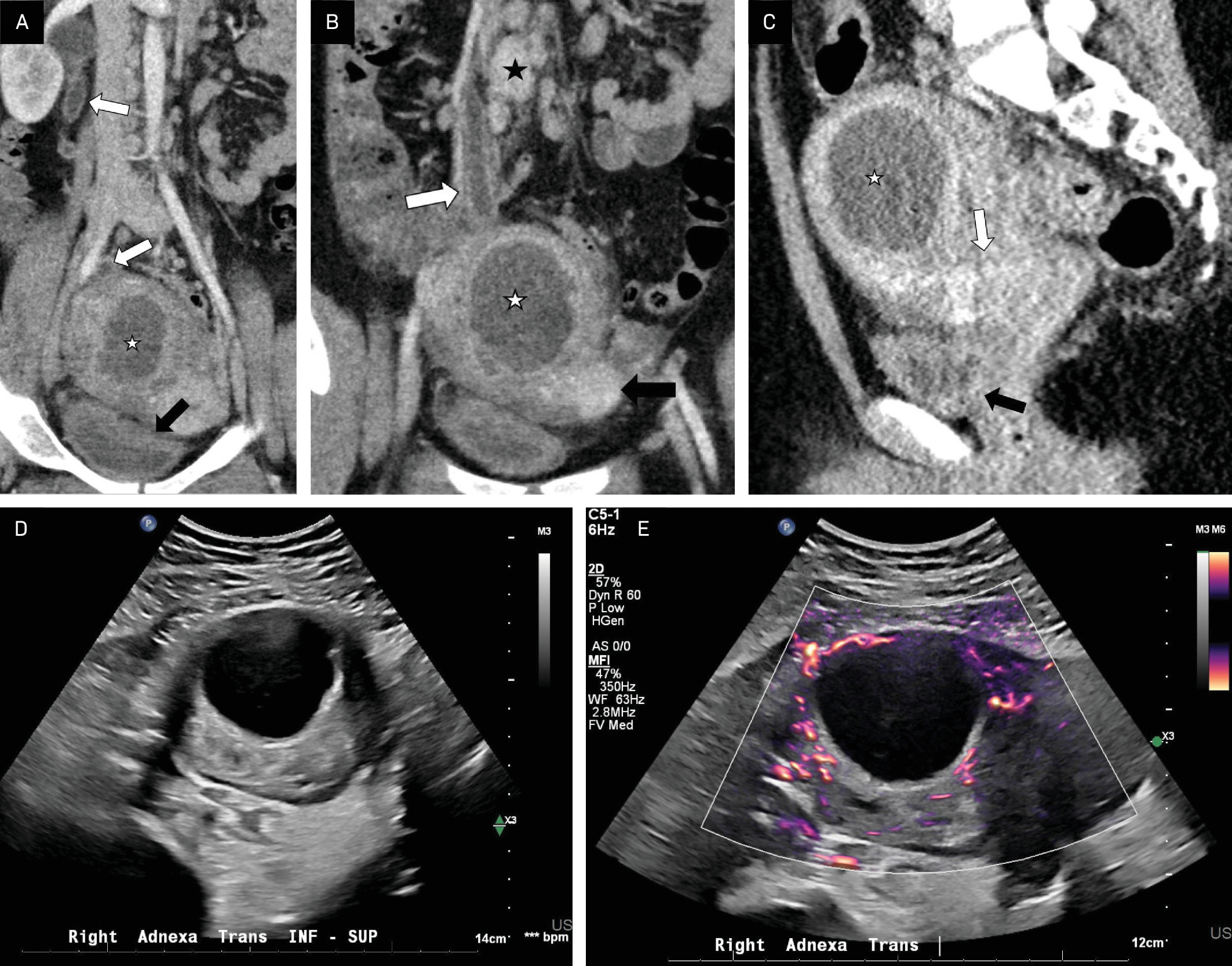
Diagnosis
Tubo-ovarian abscess.
Depending on the clinical scenario, the differential diagnosis may include uterine or ovarian tumor, ovarian or fallopian tube cyst, malignancy, diverticular or appendiceal abscess or mass, endometrioma, ovarian cyst, hematoma, hemorrhagic cysts, hydrosalpinx, ectopic pregnancy, inflammatory bowel disease, pelvic inflammatory disease (PID), ovarian torsion, and pyelonephritis. 1,2
Discussion
TOA is a severe, potentially life-threatening infection of the fallopian tube and ovary, typically affecting sexually active females of reproductive age as a complication of PID. TOA is caused by ascending genital infections, mainly Chlamydia trachomatis and Neisseria, though E. coli, Bacteroides species, and Peptostreptococcus may also be involved. 1,3,4 Symptoms classically include acute lower abdominal pain, vaginal discharge, chills, and fever, though these can vary, complicating diagnosis. 1 Conservative treatment with broad-spectrum antibiotics, and possibly drainage if antibiotics fail, is recommended to preserve fertility. 1,4 Surgery is recommended in cases of large abscesses, hemodynamic instability, antibiotic failure, or signs of rupture (eg, peritonitis and sepsis). 1,3,4 Complications can include rupture, sepsis, and rarely, perihepatitis (Fitz-Hugh-Curtis syndrome). 2,3 Prognosis is generally good with proper treatment, with a mortality rate of 1 in 740 and potential reproductive complications such as infertility and ectopic pregnancy. 3
TOA is rare in nonsexually active females. 1,5 Abdominal pain is the most common symptom, with fever, leukocytosis, nausea, vomiting, diarrhea, and abdominal tenderness also common. 1,5 The mechanism of spread in such cases may include direct translocation through the intestines, ascension through the urogenital tract; for example, from vaginal urine pooling related to obesity, or hematogenous spread with bacteremia. 6 The proposed risk factors include recurrent UTIs, obesity, inflammatory bowel disease, prior pelvic surgery, and poor hygiene, all of which can facilitate ascending infection and bacterial seeding from the gastrointestinal tract. 1,4,5 The organisms found in nonsexually active females include E. coli, Streptococcus milleri, Bacteroides uniformis, coagulase-negative staphylococci, Streptococcus viridans, Peptostreptococcus anaerobius, Streptococcus anginosus, and Streptococcus constellatus. 1,4,5
Endovaginal and transabdominal US are the preferred initial imaging studies for work-up in a patient with symptoms of TOA (notably, at the authors’ institution, vaginal US is not performed in patients who are not sexually active). 2 US is useful to distinguish between TOA and uncomplicated PID, as the clinical features are similar. US typically demonstrates a solid, cystic, or complex adnexal or cul-de-sac mass with an adjacent fluid collection. 7
Additional US findings suggestive of a typical TOA include a multilocular, complex, adnexal mass with debris, septations, and irregular/thick walls/margins, as well as loss of visualized tissue boundaries between pelvic organs, and thick, dilated fallopian tubes. 5,7 These are commonly bilateral with echogenic debris within the pelvis. 2,8 The masses may present as indistinct uterine margins, and there may be loss of midline endometrial echoes. 7 CT or MRI may be required to distinguish a TOA from ovarian malignancies. 2,5,7
CT most commonly demonstrates a pelvic mass with uniformly thick, enhancing abscess walls and internal septations. 7 The pelvic mass typically has high attenuation and may contain fluid-fluid levels or gas. When present, internal gas helps differentiate TOA from other ovarian cystic masses. There may be adjacent or contained pyosalpinx, visualized as a fluid-filled tubular multilocular lesion with enhancing, thick walls. Uterosacral ligament thickening is visible when the inflammation extends posteriorly. Other organ involvement may be seen, most commonly rectosigmoid colon and ureter. 7,9
MRI may be useful when US is inconclusive or if gas content is difficult to distinguish from bowel gas. 10 On MRI, TOA typically appears as a mass with hypointense abscess contents on T1 images and heterogeneous or hyperintense abscess contents on T2 images. 2 Abscess contents may vary in signal intensity depending on viscosity or protein concentration. Occasionally, TOA can contain areas of T1 hyperintensity owing to granulation tissue and hemorrhage products. 7 Dense pelvic adhesions or fibrosis may create mesh-like strands in the pelvic fat planes that appear T2-hypointense but avidly enhance on postcontrast images. 7
While no single finding on CT/MRI is pathognomic for TOA, the condition should be suspected in any febrile female with a complex pelvic fluid collection without an alternative source of infection (such as appendicitis).
In summary, while rare, our case of an 11-year-old girl with a history of obesity, recurrent UTIs, and constipation presenting with abdominal pain, tenderness, and fever has similar factors to other nonsexually active pediatric TOA cases reported in the literature; for example, case 2 in Hartmann et al of a 12-year-old girl with obesity, constipation, and recurrent UTIs. 4 Of note, our case presented with a simpler, cystic appearance than a typical TOA secondary to PID. There does not appear to be a discussion in the literature regarding the differences in appearance on imaging in these cases versus typical TOA secondary to PID.
Conclusion
TOA is most common in reproductive-aged, sexually active females as a complication of PID, but it can also be seen in nonsexually active pediatric patients and should be considered in the differential diagnosis in patients with a concerning clinical presentation and imaging findings, especially in the presence of other risk factors. The proposed risk factors for TOA in nonsexually active females include a history of recurrent UTIs, obesity, inflammatory bowel disease, prior pelvic surgery, and poor hygiene. Modalities such as sonography and CT are useful in arriving at a diagnosis.
Antibiotics are used as the primary treatment, preferably with adequate anaerobic coverage given the large proportion of cases attributable to anaerobic gut flora. Surgical drainage may not always be required but could be used for diagnosis or when conservative measures do not result in improvement.
References
- Bathan L, Mendez Y, Sacks M. Tubo-ovarian abscess in a virgin pediatric patient. J Case Rep Images Obstet Gynecol. 2020;6:100067Z08LB2020. doi:10.5348/100067Z08LB2020CR.
- Weerakkody Y, Sharma R, Nassar I. Tubo-ovarian abscess. doi:10.53347/rID-9818.
- Kairys N, Roepke C. In: StatPearls [Internet]. 2025.
- Hartmann K, Lerand S, Jay M. Tubo-ovarian abscess in virginal adolescents: exposure of the underlying etiology. J Pediatr Adolesc Gynecol. 2009;22(3). doi:10.1016/j.jpag.2008.03.006.
- Fei Y, Lawrence A, McCracken K. Tubo-ovarian abscess in non-sexually active adolescent girls: a case series and literature review. J Pediatr Adolesc Gynecol. 2021;34(3):328-333. doi:10.1016/j.jpag.2020.12.002.
- Fink D, Lim P, Desai A. Recurrent tubo-ovarian abscess in a nonsexually active adolescent. Consultant. 2021;62(1). doi:10.25270/con.2021.04.00010.
- Kim S, Kim S, Yang D, Kim K. Unusual causes of tubo-ovarian abscess: CT and MR imaging findings. Radiographics. 2004;24(6):1575-1589. doi:10.1148/rg.246045016.
- Stanley K, Morato D, Chilstrom M. Ultrasound diagnosis of bilateral tubo-ovarian abscesses in the emergency department. West J Emerg Med. 2013;14(6):641-642. doi:10.5811/westjem.2013.7.18487.
- Hiller N, Sella T, Lev-Sagi A, Fields S, Lieberman S. Computed tomographic features of tuboovarian abscess. J Reprod Med. 2005;50(3):203-208.
- Telischak N, Yeh B, Joe B. MRI of adnexal masses in pregnancy. AJR Am J Roentgenol. 2008;191(2):364-370. doi:10.2214/AJR.07.3509.
Disclosures
The authors have no conflicts of interest to disclose. None of the authors received outside funding for the production of this original manuscript and no part of this article has been previously published elsewhere.
Citation
. Tubo-Ovarian Abscess. Applied Radiology. 2026;55(2). doi:10.37549/AR-D-25-0127.