Ultrasound of the acute pediatric abdomen
Applied Radiology — Vol. 32 , Issue 3 , pp. 13 -19
DOI: 10.37549/AR1171
Published: March 1, 2003
Categories
Acute abdominal pain is a frequent problem in childhood. The list of differential diagnostic possibilities is long and varies significantly with the patient’s age and sex. Because it is often difficult to reach a diagnosis on clinical grounds alone, and several of the possible diagnoses require immediate surgical attention, radiologists are often called upon to assist in the evaluation of these patients.
Historically, standard abdominal roentgenography had been the cornerstone in the evaluation of acute abdominal pain in children. While plain film can be useful for finding free intraperitoneal air, evidence of bowel obstruction, or abnormal abdominal calcifications, results may be negative in up to 50% of patients.1 Cross-sectional imaging techniques, including ultrasonography, have significantly influenced the imaging approach in children with certain abdominal findings. Three common, and important, clinical diagnoses that may present with acute abdominal symptoms in pediatric patients are appendicitis, intussusception, and hypertrophic pyloric stenosis. For all three of these entities, ultrasound (US) has been shown to be a reliable imaging tool for rapid and accurate diagnosis.
Appendicitis
The most frequent surgical diagnosis in children who present with right lower quadrant pain is acute appendicitis. Historically, approximately 10% to 25% of children undergoing appendectomy did not have acute appendicitis at surgery. This false-positive rate has been considered acceptable, because of the relatively low morbidity of laparotomy and the serious consequences of a missed diagnosis of appendicitis. Early diagnosis and surgery are essential because mortality and late complications increase significantly if perforation occurs. Perforation with acute appendicitis is more common in pediatric patients than in adults, occurring in approximately 40% of children <4 years of age and in up to 80% of infants. Also, the progression from onset of symptoms to perforation is more rapid the younger the patient.
The clinical diagnosis of acute appendicitis is usually suggested by the history of abdominal pain followed by anorexia and vomiting. The pain is classically periumbilical initially and then localized to the right lower quadrant. Fever and leukocytosis are often present. The clinical diagnosis can be especially difficult in certain pediatric subgroups, namely infants, adolescent girls, and immunocompromised children.
Plain film of the abdomen can be normal in half of cases of appendicitis. The presence of a calcified appendicolith is considered strongly suggestive of acute appendicitis, although is not specific enough to be the sole basis for diagnosis.2
In 1986, Puylaert3 described the use of ultrasonography in the diagnosis of acute appendicitis. The technique provides direct visualization of the appendix, using high-resolution sonography with graded abdominal compression. A high-frequency linear array transducer is used to evaluate the right lower quadrant, with particular attention to the site of maximal tenderness. As normal overlying small bowel and cecum are compressed and air is displaced, the appendix can be visualized3 (Figure 1).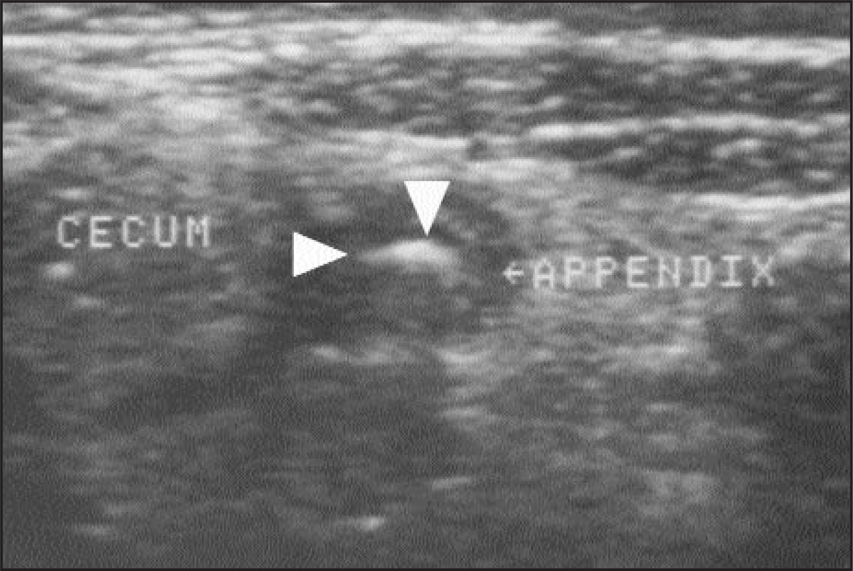
A normal appendix appears as a blind-ending tubular structure with a diameter measuring <6 mm. It can be distinguished from normal small bowel by lack of peristalsis. A normal appendix usually appears as a thin central echogenic region corresponding to the submucosa, surrounded by a hypoechoic outer zone representing the muscularis propria. Usually the lumen is collapsed, although in a small percentage of normal appendices, a small amount of intraluminal fluid may be seen.4
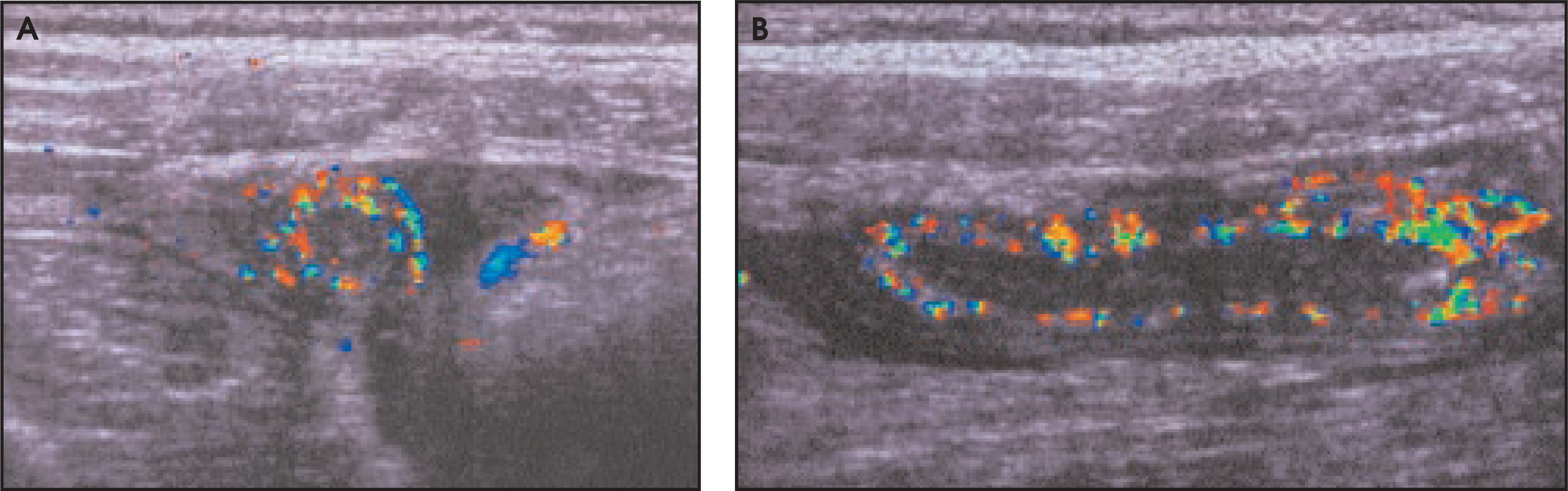
Ultrasound diagnosis of acute appendicitis rests on finding a tender, blind-ending, nonperistaltic tubular structure in the right iliac fossa with a cross-sectional diameter >6 mm (from outer wall to outer wall) with compression (Figure 2). Hyperemia, the presence of an appendicolith, and periappendiceal inflammatory changes are strongly suggestive of the diagnosis, but are nonspecific. Quillin et al5 found that increased blood flow in the appendiceal wall or in a right lower quadrant mass on color Doppler suggests appendicitis. However, absence of flow did not reliably distinguish a normal from an abnormal appendix.5 Other studies confirm the utility of color Doppler sonography in the diagnosis of acute appendicitis,6,7 including a study by Gutierrez et al6 in which 10 of 12 patients with abnormal color Doppler signal in the right lower quadrant had acute appendicitis, while 100 of 105 patients with no abnormal color flow did not (Figure 3).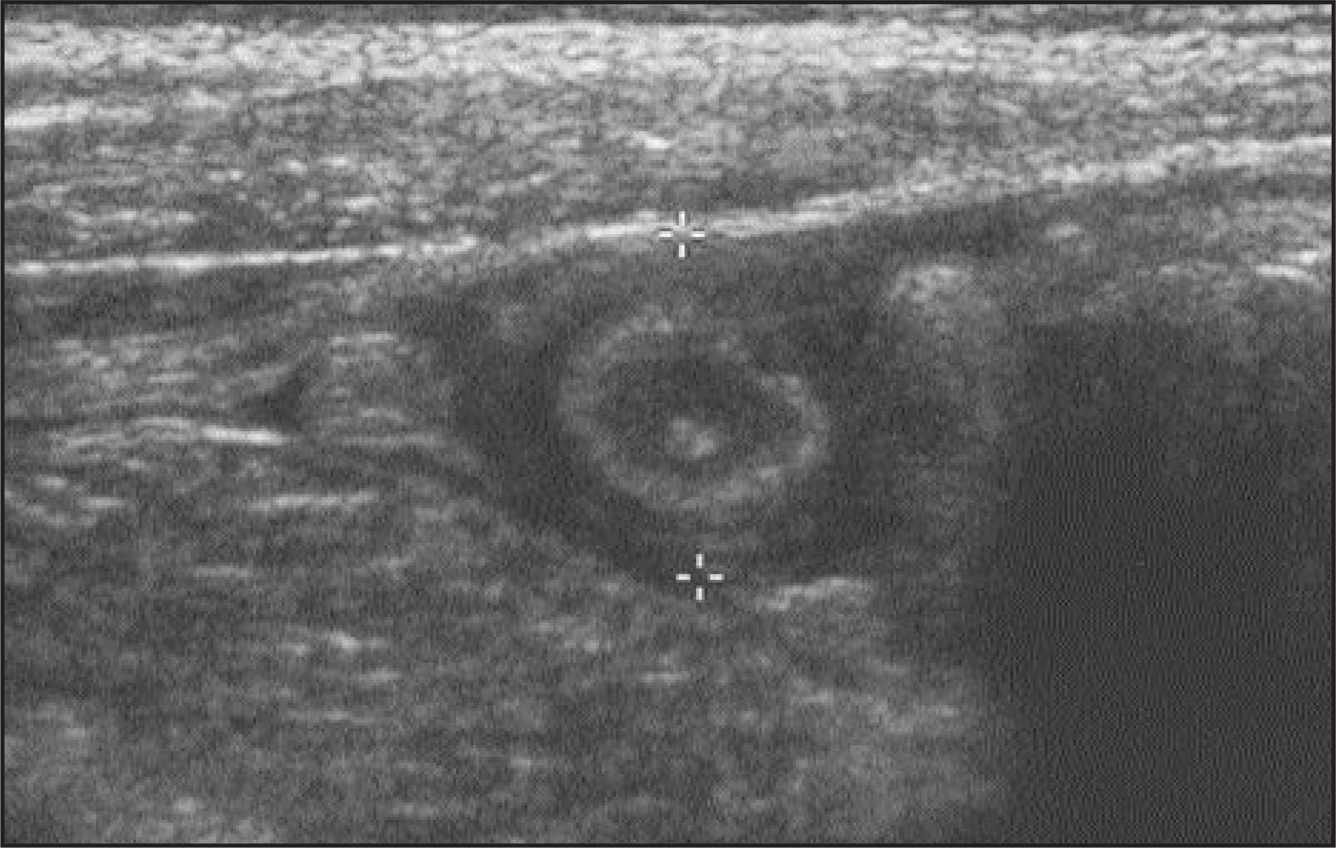
Other findings include loss of the echogenic submucosal layer, fluid-filled appendiceal lumen, increased echogenicity of pericecal fat, or periappendiceal mass.4 In focal appendicitis, inflammation may be localized to the distal tip, highlighting the importance of imaging the entire length of the appendix. In perforated appendicitis, the caliber of the burst appendix may be normal. Thus, in these cases, there may be a higher rate of false-negative studies, and other indicators of appendicitis should be sought, including periappendiceal fluid or fluid confined to the right peritoneal reflection, free pelvic fluid, thickening of adjacent bowel wall, right lower quadrant adenopathy, or a well-defined abscess.8
The reported sensitivity and specificity of the clinical examination in children suspected to have acute appendicitis are 49% and 95%, respectively.9 Numerous studies have shown that the sensitivity of US for acute appendicitis in pediatric populations ranges from 74% to 100% with specificities ranging from 74% to 95%.10-15 One group found the sensitivity of US to be remarkably lower, at 44%.16 Negative laparotomy rates ranging from 0.7% to 8.9% have been reported when US was used.9,12,17
Investigators have compared computed tomography (CT) scan and US in the diagnosis of acute appendicitis in children. These studies find higher sensitivities with CT (sensitivities 84% to 97%, specificities 89% to 100%) versus US (sensitivities 44% to 95%, specificities 88% to 100%), although in some studies the difference is not statistically significant. Some authors have argued that US is suitable as a first step in diagnosis, since US has relatively high sensitivity and specificity compared with clinical examination, and can triage most patients effectively. In those cases that are equivocal, the use of limited CT scan with rectal contrast (CTRC) may be useful for further evaluation. This combination of US and CTRC in equivocal cases was found in one study to increase overall sensitivity from 44% (US alone) to 94% (US followed by limited CTRC).16
The advantages of US over CT scanning include lack of ionizing radiation, lack of need for contrast material, wide availability, and lower cost. Garcia Pena et al16 found that using US as the first-line imaging in the diagnosis of acute appendicitis, followed by limited CTRC in equivocal cases, there was an overall reduction in morbidity and acute care costs. Some studies have found that US can aid in reducing unnecessary admissions to the hospital for observation and decreasing the number of unnecessary appendectomy procedures, without increasing the risk of perforation.13,18
There are some limitations of US in the evaluation of acute appendicitis. In early appendicitis, the sonogram may have a false-negative result. When clinical suspicion remains high despite a negative US, serial US examinations can be used to follow the patient in conjunction with serial clinical evaluation. In patients with perforation, diagnosis can be made more difficult by decompression of the appendix. Retrocecal location of the appendix also makes diagnosis more difficult. Technical factors can also be limiting. These include extreme abdominal tenderness and guarding, obesity, and excessive bowel gas. As in other applications of US, the graded compression technique is operator-dependent; the best results are obtained when the examiner is experienced in this procedure. However, despite these limitations, US has proven to be a rapid and reliable tool for diagnosing acute appendicitis in the pediatric population.
Intussusception
Intussusception is one of the common surgical emergencies in infancy and early childhood. Most cases occur in the first 2 years of life, with a peak occurrence between 3 to 9 months of age. However, intussusception has become common in children older than 2 years of age, suggesting a changing pattern for this disease.19 The classic clinical features include blood in the stool and a palpable abdominal mass. Approximately 15% to 20% of children may experience no pain. In up to 50% of children, the presenting sign is listlessness.
In up to 95% of cases, there is no leading point for the intussusception. The older the child is, the more likely there is a pathologic leading point, the differential diagnosis of which includes lymphoma, Meckel’s diverticulum, lymphadenopathy, and Henoch-Schonlein purpura. In those cases without an identifiable leading point, it is thought that viral infection leading to hypertrophy of Peyer’s patches may be the cause.
The most common location for intussusception is at the ileocecal junction, with up to 85% of cases occurring here. Most of the remaining cases occur in the colon, with rare cases in the small bowel.
Plain films of the abdomen are often obtained in children suspected of having intussusception. These may be normal in up to 25% of patients20; however, their main utility is in demonstrating free intraperitoneal air indicative of perforation, for which the patient would be immediately taken to surgery. Up to 50% of patients may have an abnormal bowel-gas pattern and suggestion of a soft-tissue mass; 25% may have a pattern suggesting a small bowel obstruction. The overall accuracy of plain radiography in diagnosing or excluding intussusception ranges from 40% to 90%.21
The gold standard for the diagnosis of intussusception is the contrast enema, which is reported to have an accuracy of 100%.22 In addition, performance of this study may be therapeutic. However, for screening of patients suspected of having intussusception, many of who will not have the condition, US has been proposed as an alternate method. The main advantages of US include its speed and accuracy, its noninvasiveness, and the absence of ionizing radiation. Also, US is well suited for investigation of other causes of abdominal complaints in children, many of which would not be seen on contrast enema examination.
In addition to diagnosing intussusception, US can be used to guide reduction. In one series, US-guided hydrostatic reduction was performed successfully in 91% (42 of 46) of intussusceptions.23
The typical sonographic finding is a mass, approximately 3 to 5 cm in diameter, with a hypoechoic intestinal wall surrounding an echogenic central area, representing submucosa and intussuscepted fat24 (Figure 4). On transverse section, this has a “target” or “doughnut” appearance. Multiple concentric rings or the bull’s-eye sign of alternating rings of hypo- and hyperechogenicity are even more characteristic (Figure 5). On longitudinal section, the hypo-echoic layers of swollen intestinal wall are seen on either side of the centrally located intussusceptum (Figure 6). This has been called the “pseudokidney” or “sandwich” sign.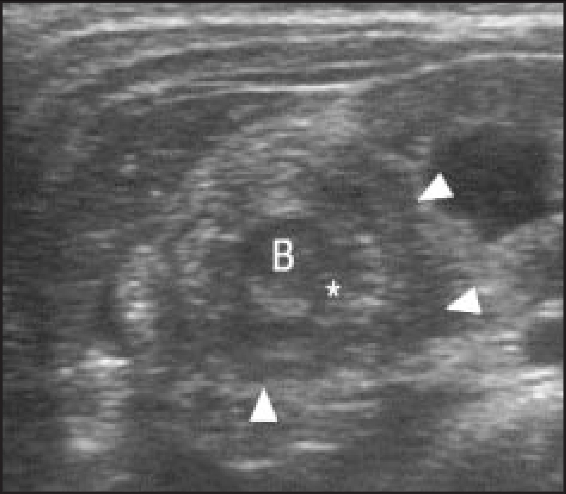
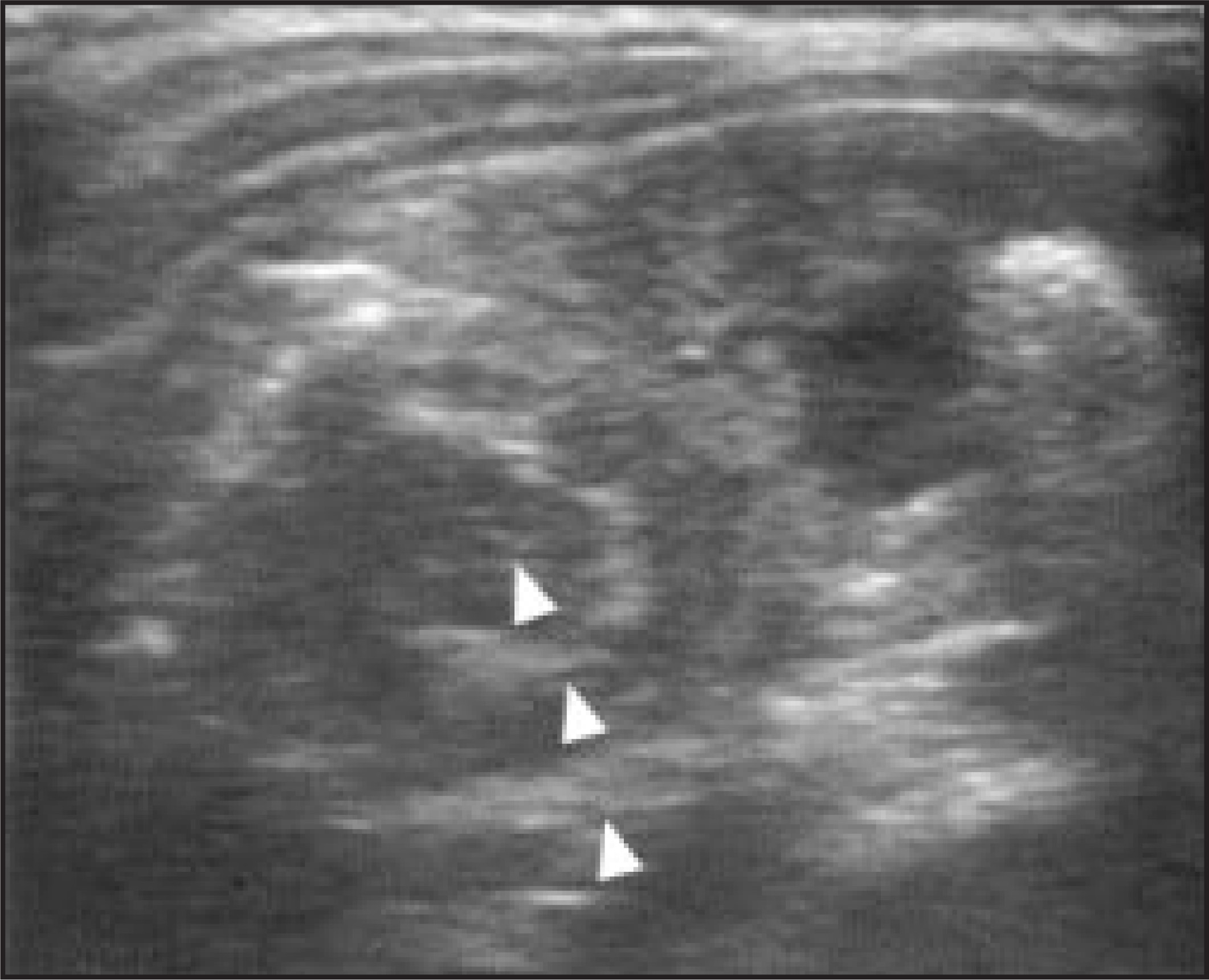
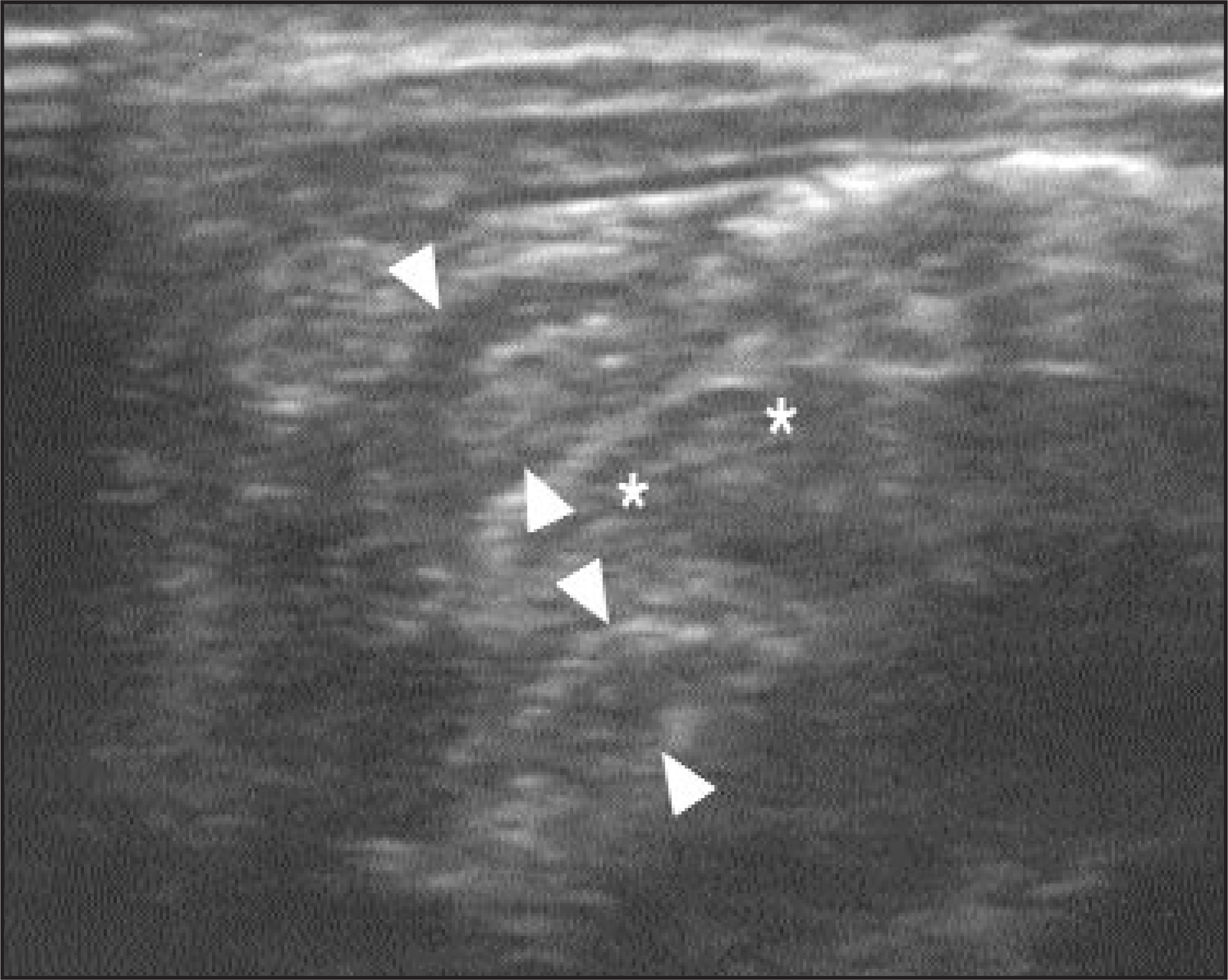
Ultrasound has been shown to be highly accurate in the diagnosis of intussusception, with sensitivities in the range of 98% to 100% and specificities from 88% to 100%.22,25-28 Some studies report the ability to offer alternate diagnoses based on the US examinations, ranging from 6% to 27% of cases negative for intussusception.27,29
The use of color Doppler ultrasonography (CDU) has been described as a useful tool to assess for bowel necrosis and to predict reducibility of an intussusception by enema reduction. In a series of 125 patients with a total of 134 intussusceptions who underwent both CDU and air enema, the rate of successful reduction was 91% (109 of 121) in those intussusceptions with blood flow, and only 31% (4 of 13) in intussusceptions with no flow seen on CDU.30
Similar correlation between the presence of color flow and reducibility have been found by other authors.31,32 In these smaller studies, bowel necrosis was seen only in those patients with no color flow by CDU, suggesting a strong correlation between lack of color flow and bowel gangrene.
Hypertrophic pyloric stenosis
The classic presentation of hypertrophic pyloric stenosis (HPS) is non-bilious vomiting that occurs minutes after feeding, which worsens over time and may become forceful and projectile. The infant remains hungry and may feed immediately after vomiting. The onset of symptoms typically occurs between 3 and 6 weeks of age. Boys are affected more than girls by a ratio of 5:1.33 Gastric outlet obstruction is caused by hypertrophy of the antral and pyloric circular muscle, thought to be an acquired rather than a congenital condition.
Clinical diagnosis can be made by palpation of the hypertrophied muscle mass, likened to an “olive.” In skilled hands, palpation is a fairly sensitive and highly specific method of detecting HPS. In one series, physical examination of children with HPS revealed a palpable abdominal mass in 80% to 87% of cases.34 However, the art of clinical diagnosis has given way to imaging diagnosis. There was a decline in the rate of clinical diagnosis between the years 1974 to 1977 and the years 1988 to 1991, with a threefold increase in imaging studies for HPS.34
Plain films are not routinely obtained when the diagnosis of HPS is suspected. Before the use of US for HPS, upper gastrointestinal contrast study (UGI) had been the recommended method to establish the diagnosis. Ultrasound was first introduced as an aid in the diagnosis of HPS by Teele and Smith35 in 1977, and is now widely considered the study of choice.
The patient is examined in the supine and right lateral decubitus positions. A 5.0 or 7.5 MHz transducer is applied to the right upper quadrant, and the pylorus is imaged in its longest dimension and in transverse plane (Figure 7).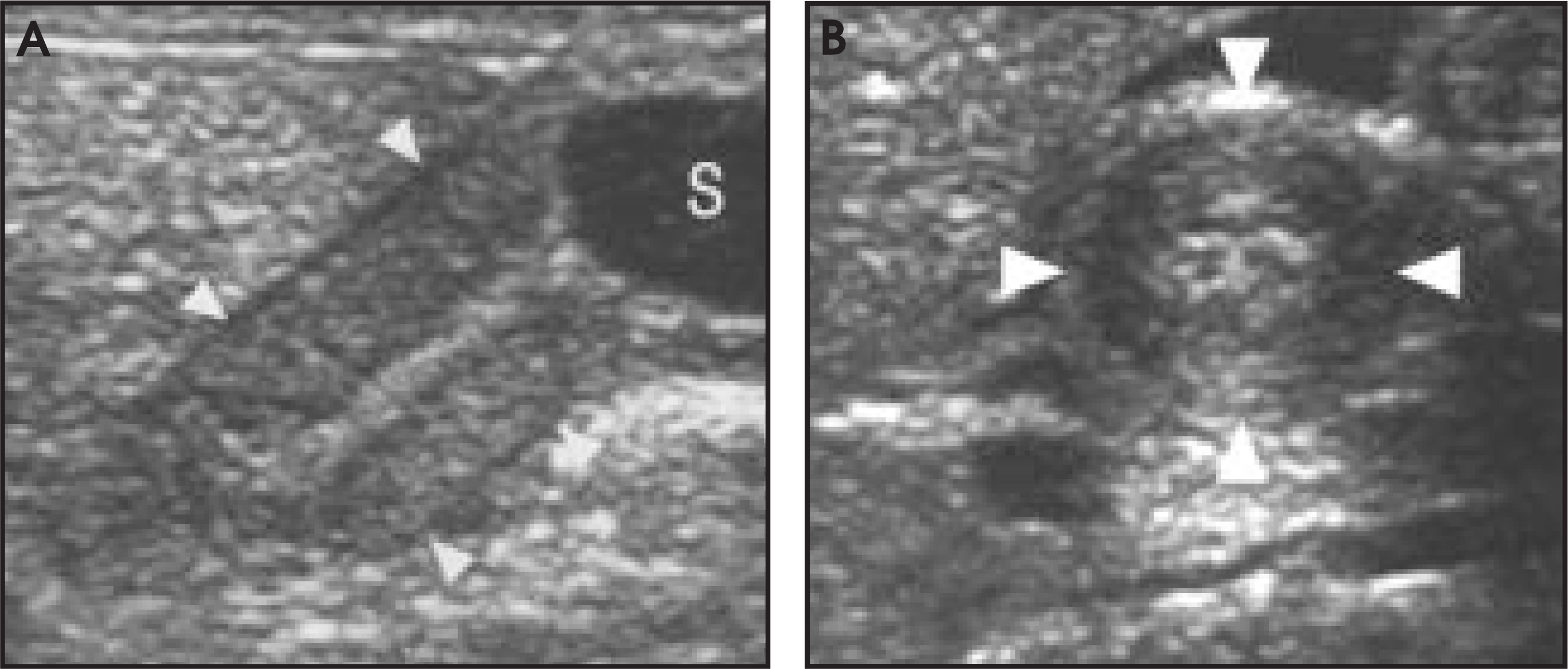
A recent report suggests that placing the infant prone to obtain posterior views may be useful in imaging the pylorus in cases in which there is an abundance of overlying bowel gas.36
The single most reliable measurement by US in the diagnosis of HPS is muscle wall thickness, measured from the base of the echogenic submucosa to the outer edge of the hypertrophic muscle layer (Figure 8). Normal infants have a pyloric muscle wall thickness ≤ 2 mm, while >4 mm is diagnostic of HPS.37 The range between 2 and 4 mm is equivocal, and these infants can be observed with follow-up examinations.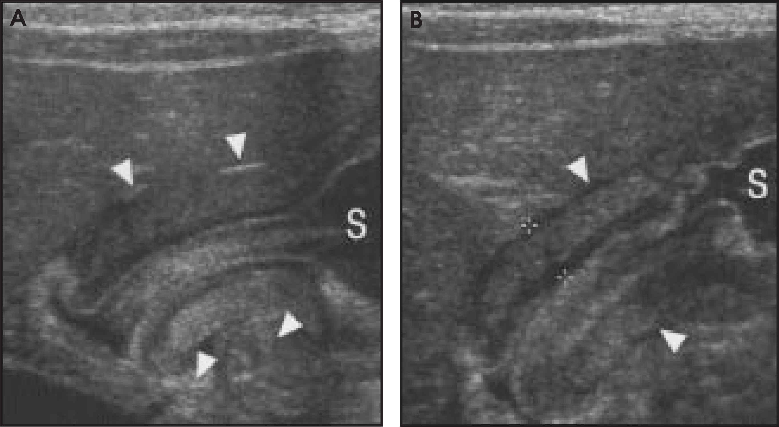
Other quantitative criteria include pyloric channel length, pyloric volume, pyloric volume to body weight ratio, pyloric muscle index, or pyloric ratio. Recently, the pyloric ratio (wall thickness to pyloric diameter) was assessed in a retrospective review of 87 children evaluated for HPS, and a ratio ≥ 0.27 yielded a sensitivity and specificity of 96% and 94%, respectively.38
In many cases, measurements and calculations are superfluous, as an experienced sonographer can establish the diagnosis reliably based on subjective evaluation. Some nonquantitative findings include the “antral nipple,” increased gastric peristalsis, prepyloric antral thickening, extension of fluid into the proximal pylorus, and lack of stomach contents traversing the pyloric channel.38
In a series of 323 examinations by Blumhagen et al,37 the sonographic diagnosis, based primarily on muscle wall thickness, was correct in 321, yielding an accuracy of 99.4%. There were two false-negative and no false-positive sonograms (sensitivity 98.2%, specificity 100%). In this study, infants with palpable pyloric masses were included, introducing a bias toward higher predictive value of a positive sonogram. A more recent study included 150 infants with normal or equivocal examinations.39 Using muscle wall thickness ≥ 3 mm as diagnostic of HPS, the sensitivity, specificity, and accuracy of sonography were found to be 100%. A similar study of 142 infants without a palpable pyloric mass or with equivocal findings at clinical examination, found an overall sensitivity of 97% and specificity of 99%.40
Other diagnoses
Ultrasound aids in the diagnosis of many other pediatric conditions that may present with acute abdominal pain. Reports have shown that many patients with acute abdominal symptoms evaluated for one suspected diagnosis were found on US evaluation to have other diagnoses. It is important to evaluate the entire abdomen and pelvis in an effort to discover alternate causes of the clinical signs and symptoms.
Conclusion
There is much evidence supporting the use of US as a key diagnostic tool in acute pediatric abdominal pain. In some cases, new observations have been shown to be useful in determination of prognosis and therapy, as well. Given all the advantages of US in the evaluation of pediatric patients, it is not surprising that ultrasonography has become an increasingly important modality in this clinical setting. AR
References
- Franken E, Kao S, Smith W, Sato Y. Imaging of the acute abdomen in infants and children. AJR Am J Roentgenol. 1989;153:921-928.
- Lowe L, Penney M, Scheker L. Appendicolith revealed on CT in children with suspected appendicitis: How specific is it in the diagnosis of appendicitis?. AJR Am J Roentgenol. 2000;175:981-984.
- Puylaert J. Acute appendicitis: US evaluation using graded compression. Radiology. 1986;158:355-360.
- Sivit C. Diagnosis of acute appendicitis in children: Spectrum of sonographic findings. AJR Am J Roentgenol. 1993;161:147-52.
- Quillin S, Siegel M. Appendicitis: Efficacy of color Doppler sonography. Radiology. 1994;191:557-560.
- Gutierrez C, Mariano M, Faddis D. Doppler ultrasound accurately screens patients with appendicitis. Am Surg. 1999;65:1015-1017.
- Lim H, Lee W, Kim T. Appendicitis: Usefulness of color Doppler US. Radiology. 1996;201:221-225.
- Hopkins K, Patrick L, Ball T. Imaging findings of perforative appendicitis: A pictorial review. Pediatr Radiol.. 2001;31:173-179.
- Rubin S, Martin D. Ultrasonography in the management of possible appendicitis in childhood. J Pediatr Surg.. 1990;25:737-740.
- Pickuth D, Heywang-Kobrunner S, Spielmann R. Suspected acute appendicitis: Is ultrasonography or computed tomography the preferred imaging technique?. Eur J Surg. 2000;166:315-319.
- Wise S, Labuski M, Kasales C. Comparative assessment of CT and sonographic techniques for appendiceal imaging. AJR Am J Roentgenol.. 2001;176:933-941.
- Karakas S, Guelfguat M, Leonidas J. Acute appendicitis in children: Comparison of clinical diagnosis with ultrasound and CT imaging. Pediatr Radiol. 2000;30:94-98.
- Dilley A, Wesson D, Munden M. The impact of ultrasound examinations on the management of children with suspected appendicitis: A 3-year analysis. J Pediatr Surg.. 2001;36:303-308.
- Sivit C, Applegate K, Stallion A. Imaging evaluation of suspected appendicitis in a pediatric population: Effectiveness of sonography versus CT. AJR Am J Roentgenol.. 2000;175:977-980.
- Lowe L, Penney M, Stein S. Unenhanced limited CT of the abdomen in the diagnosis of appendicitis in children: Comparison with sonography. AJR Am J Roentgenol.. 2001;176:31-35.
- Garcia Pena B, Mandl K, Kraus S. Ultrasonography and limited computed tomography in the diagnosis and management of appendicitis in children. JAMA.. 1999;282:1041-1046.
- Ramachandran P, Sivit C, Newman K, Schwartz M. Ultrasonography as an adjunct in the diagnosis of acute appendicitis: A 4-year experience. J Pediatr Surg.. 1996;31.
- Wong M, Casey S, Leonidas J. Sonographic diagnosis of acute appendicitis in children. J Pediatr Surg.. 1994;29:1356-1360.
- Luks F, Yazbeck S, Perreault G, Desjardins J. Changes in the presentation of intussusception. Am J Emerg Med.. 1992;10:574-576.
- Alford B, McIlhenny J. The child with acute abdominal pain and vomiting. Radiol Clin North Am.. 1992;30:441-453.
- del-Pozo G, Albillos J, Tejedor D. Intussusception in children: Current concepts in diagnosis and enema reduction. RadioGraphics.. 1999;19:299-319.
- Shanbhogue R, Hussain S, Meradji M. Ultrasonography is accurate enough for the diagnosis of intussusception. J Pediatr Surg. 1994;29.
- Rohrschneider W, Troger J. Hydrostatic reduction of intussusception under US guidance. Pediatr Radiol.. 1995;25:530-534.
- del-Pozo G, Albillos J, Tejedor D. Intussusception: US findings with pathologic correlation—The crescent-in-doughnut sign. Radiology.. 1996;199:688-692.
- Wright J, Slater S. Suspected intussusception: Is ultrasound a reliable diagnostic aid?. Aust N Z J Surg. 1996;66:686-687.
- Verschelden P, Filiatrault D, Garel L. Intussusception in children: Reliability of US in diagnosis—A prospective study. Radiology.. 1992;184:741-744.
- Harrington L, Connolly B, Hu X. Ultrasonographic and clinical predictors of intussusception. J Pediatr.. 1998;132:836-839.
- Bhisitkul D, Listernick R, Shkolnik A. Clinical application of ultrasonography in the diagnosis of intussusception. J Pediatr.. 1992;121:182-186.
- Pracros J, Tran-Minh V, Morin de Finfe C. Acute intestinal intussusception in children. Contribution of ultrasonography (145 cases). Ann Radiol (Paris).. 1987;30:525-530.
- Kong M, Wong H, Lin S. Factors related to detection of blood flow by color Doppler ultrasonography in intussusception. J Ultrasound Med. 1997;16:141-144.
- Lim H, Bae S, Lee K. Assessment of reducibility of ileocolic intussusception in children: Usefulness of color Doppler sonography. Radiology.. 1994;191:781-785.
- Lagalla R, Caruso G, Novara V. Color Doppler ultrasonography in pediatric intussusception. J Ultrasound Med.. 1994;13:171-174.
- Carter C, Evans K. Inheritance of congenital pyloric stenosis. J Med Genet. 1969;6:233-254.
- Macdessi J, Oates R. Clinical diagnosis of pyloric stenosis: A declining art. BMJ. 1993;306:553-555.
- Teele R, Smith E. Ultrasound in the diagnosis of idiopathic hypertrophic pyloric stenosis. N Engl J Med.. 1977;296:1149-1150.
- Sargent S, Foote S, Mooney D, Shorter N. The posterior approach to pyloric sonography. Pediatr Radiol.. 2000;30:256-257.
- Blumhagen J, Coombs J. Ultrasound in the diagnosis of hypertrophic pyloric stenosis. J Clin Ultrasound.. 1981;9:289-292.
- Lowe L, Banks W, Shyr Y. Pyloric ratio: Efficacy in the diagnosis of hypertrophic pyloric stenosis. J Ultrasound Med.. 1999;18:773-777.
- Hernanz-Schulman M, Sells L, Ambrosino M. Hypertrophic pyloric stenosis in the infant without a palpable olive: Accuracy of sonographic diagnosis. Radiology.. 1994;193:771-776.
- Neilson D, Hollman A. The ultrasonic diagnosis of infantile hypertrophic pyloric stenosis: Technique and accuracy. Clin Radiol.. 1994;49:246-247.
Citation
. Ultrasound of the acute pediatric abdomen. Applied Radiology. 2003;32(3):13-19. doi:10.37549/AR1171.