Urinary bladder pheochromocytoma
Applied Radiology — Vol. 38 , Issue 6 , pp. 39A -39C
DOI: 10.37549/AR1696
Published: June 1, 2009
Categories
CASE SUMMARY
A 13-year-old girl presented to the emergency room with a history of recurrent headaches in the occipital area after urination for thepast 2 years. The headaches were becoming constant after each episode of urination in the past few months prior to presentation and the headaches started approximately 1 minute after urination, and lingered for approximately 3 minutes. She reported pain of “10” on a 10-point rating scale, but indciated that the pain usually subsided or lingered at “2” out of 10.
Her medical, surgical, and family histories were significant only for a cousin with migraines and a positive history of diabetes and hypertension. Physical examination was remarkable for hypertension with a bood pressure of 132/89. The patient was started on alpha-and beta-blockers for the headache, hypertension and palpitations she experienced after voiding. During her admission, she was noticed to have systolic blood pressures of >200 mmHg right after urination and normal systolic blood pressure before urination. Ultrasound, computed tomography (CT) and magnetic resonance imaging (MRI) studies were performed (Figures 1 to 3 depict ultrasound and MRI results).
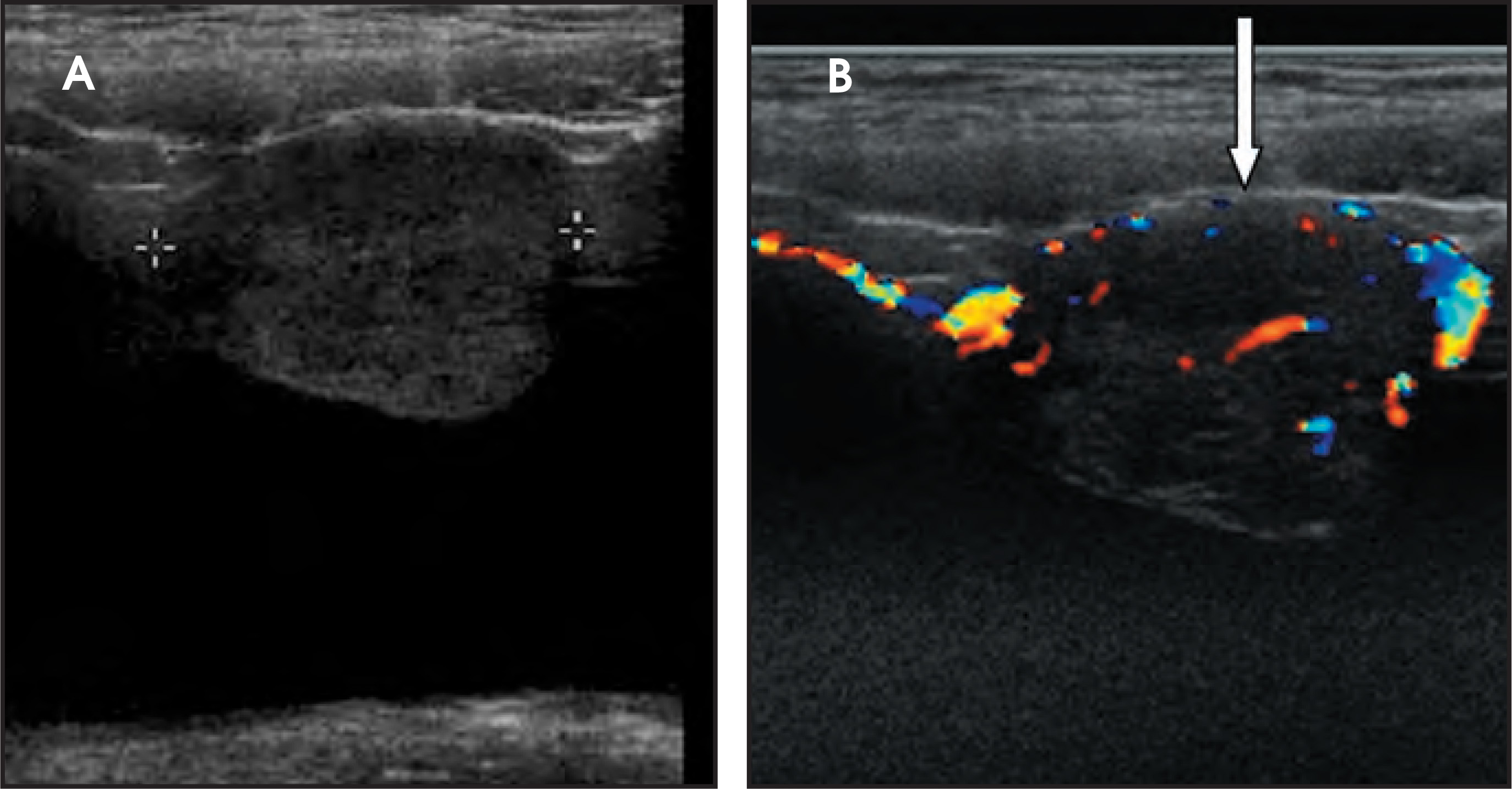
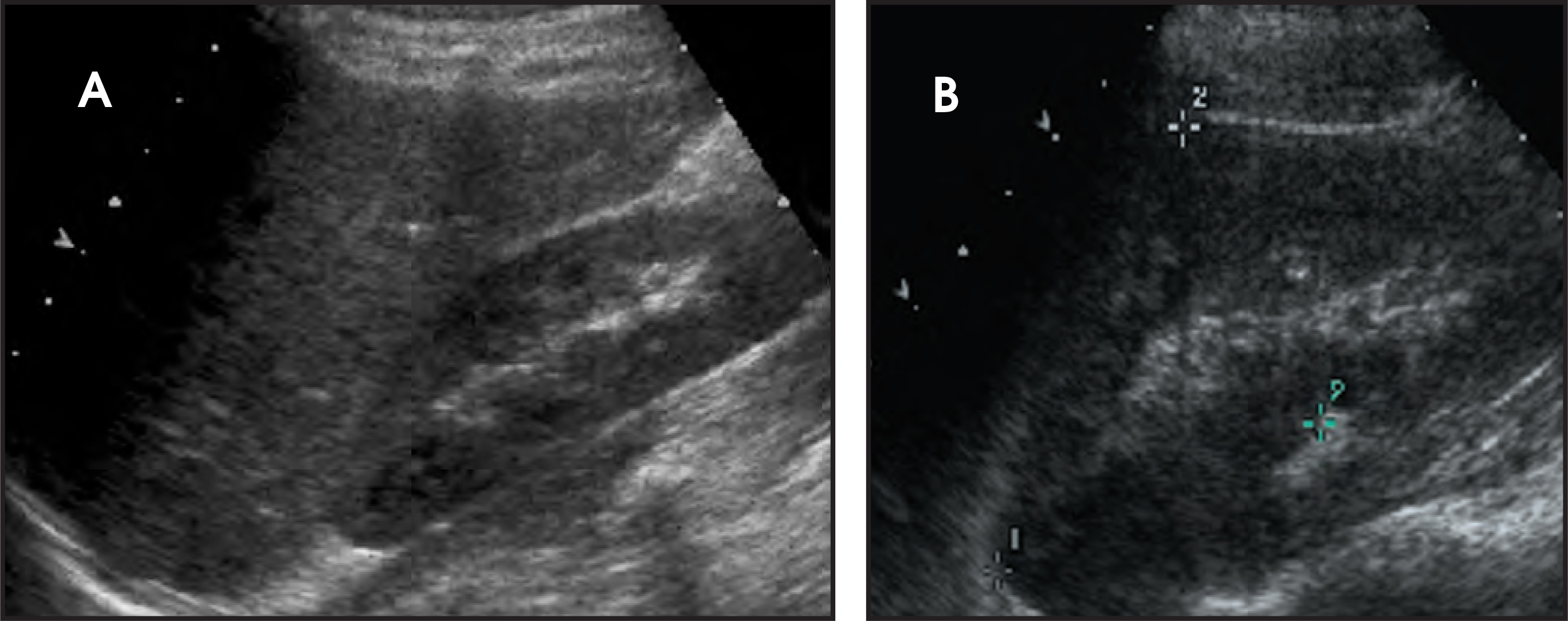
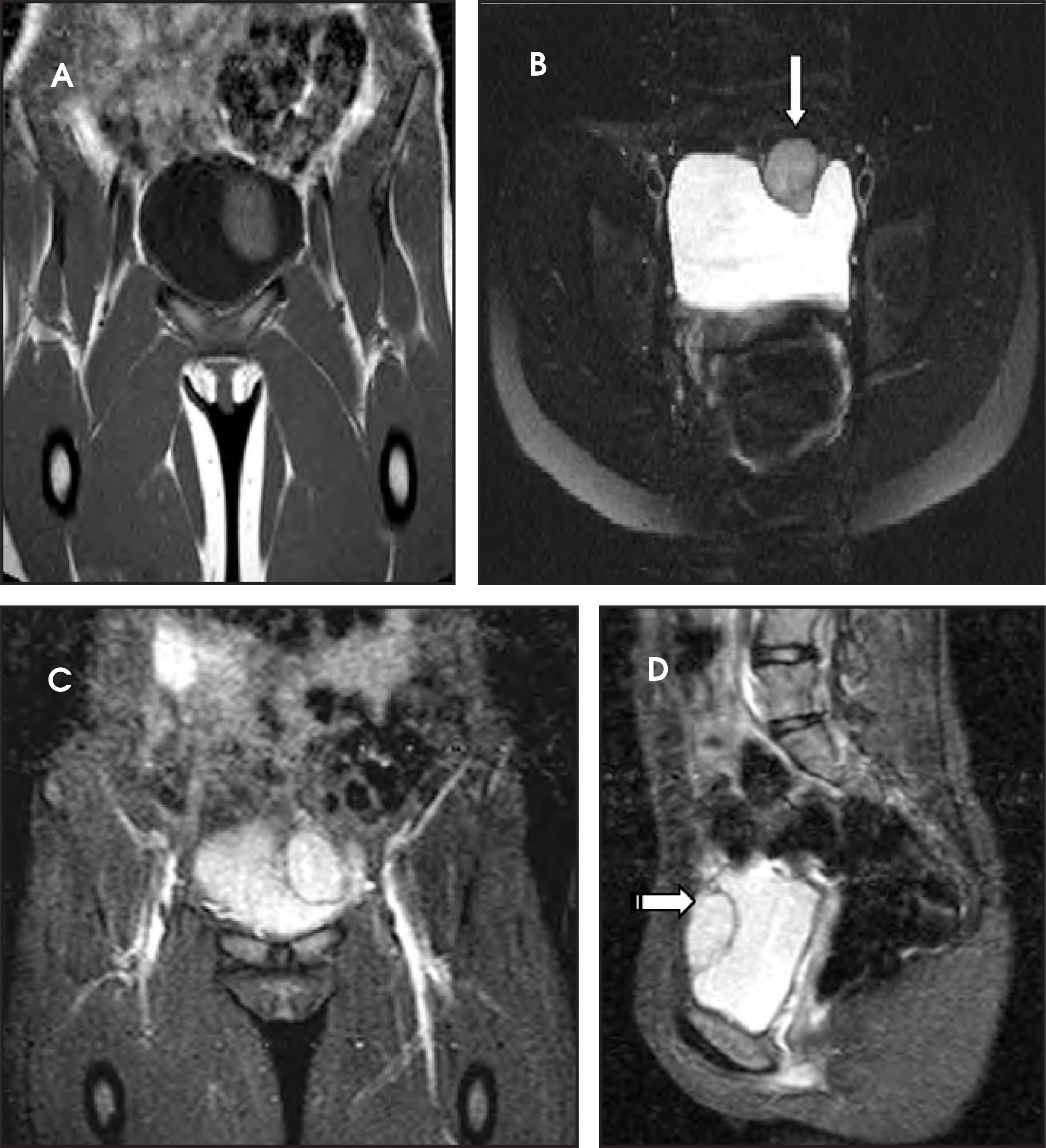
The differential diagnosis included neurofibroma, rhabdomyosarcoma, transitional cell carcinoma, leiomyosarcoma and neuroblastoma.
The urinalysis had a trace of blood and 1+ bacteria but was otherwise negative. Other findings included:
white count 8.58 with 63% segs, 28% lymphs, 5% monos, 3% eos and 1% basophil;
Hg 14.2, platelets 401;
sodium 140, potassium 3.9, chloride 106, bicarb 26, bun 10, creatinine 0.6, glucose 124;
plasma metanephrine was normal at 0.20 nmol/L (normal 0.00-0.49); and,
plasma normetanephrine was elevated at 5.36 nmol/L (normal 0.00-0.89 nmol/L).
DIAGNOSIS
Urinary bladder pheochromocytoma
IMAGING FINDINGS
The renal ultrasound (Figure 1) revealed a vascular solid mass in the anterior wall of the urinary bladder, slightly to the left of the midline, measuring 3.4 cm transverse by 2.8 cm anteroposterior by 3.9 cm craniocaudal. The abdomen and pelvis were otherwise normal(Figure 2). MRI (Figure 3) showed an abnormal soft tissue mass, measuring 3.4 x 2.8 x 3.9 cm, located anterior to the urinary bladder,just to the left of the midline, which indented the urinary bladder.
CASE FOLLOW-UP
Based on the imaging findings, the patient was started on an alpha-blocker and surgery was performed after alpha blockage was obtained. A partial cystectomy was performed and the histologic diagnosis was extra-adrenal paraganglioma (pheochromocytoma).
DISCUSSION
Pheochromocytoma of the urinary bladder is a rare entity. Patients with this lesion usually present with the triad of sustained hypertension, hematuria and postmicturition syncope. Urinary bladder pheochromocytoma has no sex predilection, but there is a slight female preponderance.1,2 Childhood pheochromocytomas are more commonly multicentric and extra adrenal and only 2% of these pheochromocytomas are malignant.2
Pheochromocytomas of the urinary bladder probably arise in the paraganglia of the visceral (autonomic) nervous system, located submucosally either in the dome or in the posterior wall, close to the trigone. These lesions can be seen in the context of familial syndromes or diseases, including neurofibromatosis, von Hippel-Lindau, Sturge-Weber, tuberous sclerosis and multiple endocrine neoplasia type II.
If pheochromocytoma is suspected on clinical and laboratory grounds, imaging studies are useful in localizing the tumor as well as determining multiple sites or metastases. Sonographically, pheochromocytoma appears as a sharply demarcated soft tissue mass. It may be purely solid or may contain foci of hemorrhage and necrosis that appear cystic. The Doppler study characteristically shows low resistance flow.3 CT has a high sensitivity for detecting adrenal (94%) and extra-adrenal (82%) pheochromocytomas.1
MR is more sensitive than all other imaging modalities. Pheochromocytomas typically display intermediate signal intensity on T1-weighted images and high signal intensity on T2-weighted images, likely because of the high intracellular water content and rich vascularity.4
In our study however, the T2-weighted images did not appear as profoundly bright as in adrenal pheochromocytomas. This may be attributed to differences in the cytologic and vascular patterns.5
Lifelong yearly follow-up with monitoring of blood pressure and signs and symptoms, as well as catecholamine assays, is recommended. Clinicians can use 131I-methyliodobenzylguanidine (MIBG) imaging when recurrence or incomplete resection is suspected. Imaging with 131I-MIBG is regarded to be 67% to 70% specific. Imaging with 18F-fluorodeoxyglucose (FDG) is regarded as being 80% to 90%sensitive.
CONCLUSION
Pheochromocytoma of the urinary bladder is a rare entity that usually presents with the triad of sustained hypertension, hematuriaand postmicturition syncope. The surgical management of urinary bladder pheochromocytoma is usually partial cystectomy. Total cystectomy with lymph node dissection is reserved for malignant lesions.
References
- Whalen R, Althausen A, Daniels G. Extra-adrenal pheochromocytoma. J. Urol. 1992;147:1-10.
- Sweetser P, Ohl D, Thompson N. Pheochromocytoma of the urinary bladder. Surgery. 1991;109:677-681.
- Cronan J, Do H, Monchik J, Stein B. Bladder pheochromocytoma: Color Doppler sonographic correlation. J Ultrasound Med. 1992;11:493-495.
- Warshawsky R, Bow S, Waldbaum R, Cintron J. Bladder pheochromocytoma with MR correlation. J Compt Assist Tomogr. 1989;13:714-716.
- Kier R, McCarthy S. MR characterization of adrenal masses: Field strength and pulse sequence considerations. Radiology. 1989;171:671-674.
Citation
. Urinary bladder pheochromocytoma. Applied Radiology. 2009;38(6):39A-39C. doi:10.37549/AR1696.