Use of combined PET/CT imaging in evaluation of the solitary pulmonary nodule: Principles, techniques, and pitfalls
Applied Radiology — Vol. 35 , Issue 4 , pp. 24 -43
DOI: 10.37549/AR1417
Published: April 1, 2006
Categories
Solitary pulmonary nodule (SPN) is typically defined as an intraparenchymal focal, round or oval area of increased opacity <3 cm in diameter.1,2 Nearly 1 in every 500 chest radiographs taken reveals a newly diagnosed SPN. More than 150,000 SPNs are detected annually in the United States alone.3 This estimate is mainly based on chest radiographs. Now, with increasing use of computed tomography (CT) of the chest for screening of lung cancer and chest CT angiography (CTA) for diagnosing pulmonary embolus and for cardiac evaluation, this number is rapidly increasing.
Data on the use of CT screening in smokers from the Early Lung Cancer Action Project showed noncalcified nodules in 233 out of 1000 participants.4 The Mayo Clinic study found 2244 noncalcified nodules in 1000 of 1520 participants.5 It is important to note that although overall survival in lung cancer is poor, patients with stage IA (T1 N0 M0) cancer have a reported 5-year survival of 61% to 85%,6 and, thus, early resection of a malignant nodule can make a large difference in patient outcome. However, with <30% of SPNs representing a primary or metastatic malignancy, determining the appropriate course of action is a challenge.
Growth rate assessment is a good differentiating factor between a benign and malignant process, but given that the typical doubling time of a malignant nodule is between 30 and 400 days, most commonly proposed follow-up protocols are at 3-month intervals for up to 1 year, and then every 6 months for another year. Some people advocate considering an even longer follow-up.7 Many patients, however, believe that waiting 3 months, not to mention up to 2 years, without at least a preliminary diagnosis is an unacceptable option. Additionally, stability over a short interval cannot exclude malignancy, yet delay can worsen the prognosis (Figure 1).
Evaluation of growth in a small nodule is often quite imprecise as well. Morphologic characteristics of an SPN are often helpful in determining its etiology. Thin-section CT can improve detection of calcification within an SPN. However, the majority of pulmonary nodules will still remain indeterminate following thin-section CT imaging. It has also been shown that despite all the accumulated data on morphologic characteristics of benign and malignant nodules, simple Bayesian analysis of patient characteristics and selected radiologic features is superior to evaluation by experienced radiologists in the stratification of benign and malignant nodules.8
Surgical resection of the nodule is definitely diagnostic and may be curative. However, the morbidity and the expense associated with resection of every incidentally found nodule make this approach simply impractical. Biopsy has not been proven to be a viable option either. Transthoracic needle aspiration yields a positive tissue diagnosis in approximately 60% of lesions <2 cm,9 with pneumothorax rates as high as 64.2%.10 Bronchoscopy has a lower complication rate, yet its diagnostic yield for nodules <2 cm is approximatedly 10% and is only 40% to 60% for nodules 2 to 4 cm in diameter (Figure 2).7 Use of contrast enhancement with CT is reported to have high sensitivity (98%) for malignant nodules; however, specificity and overall accuracy are much lower.11
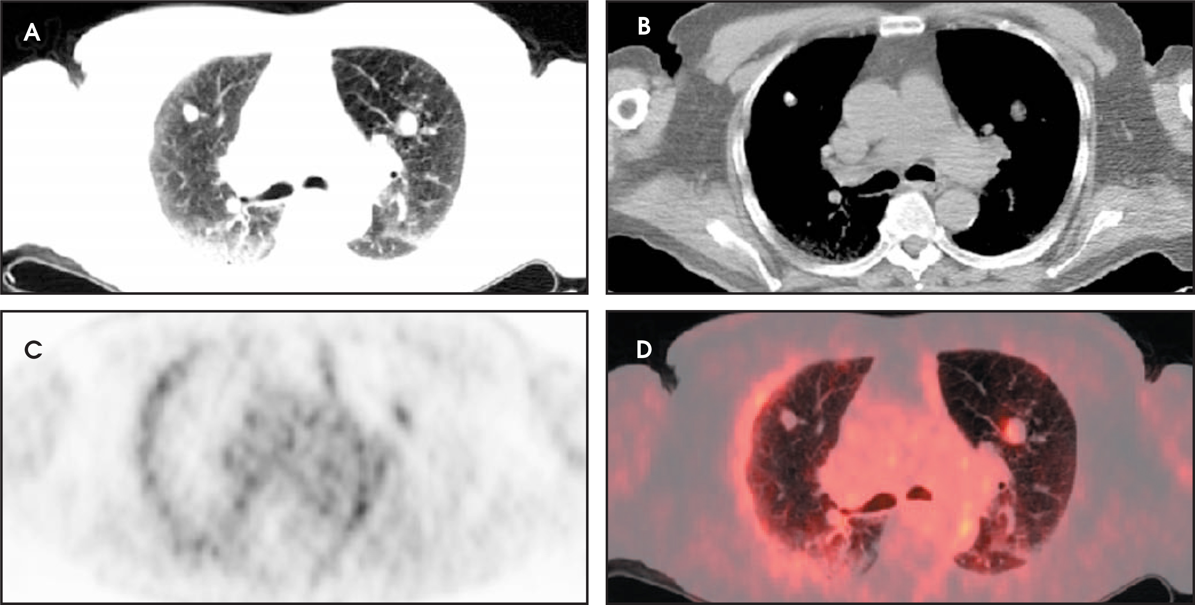
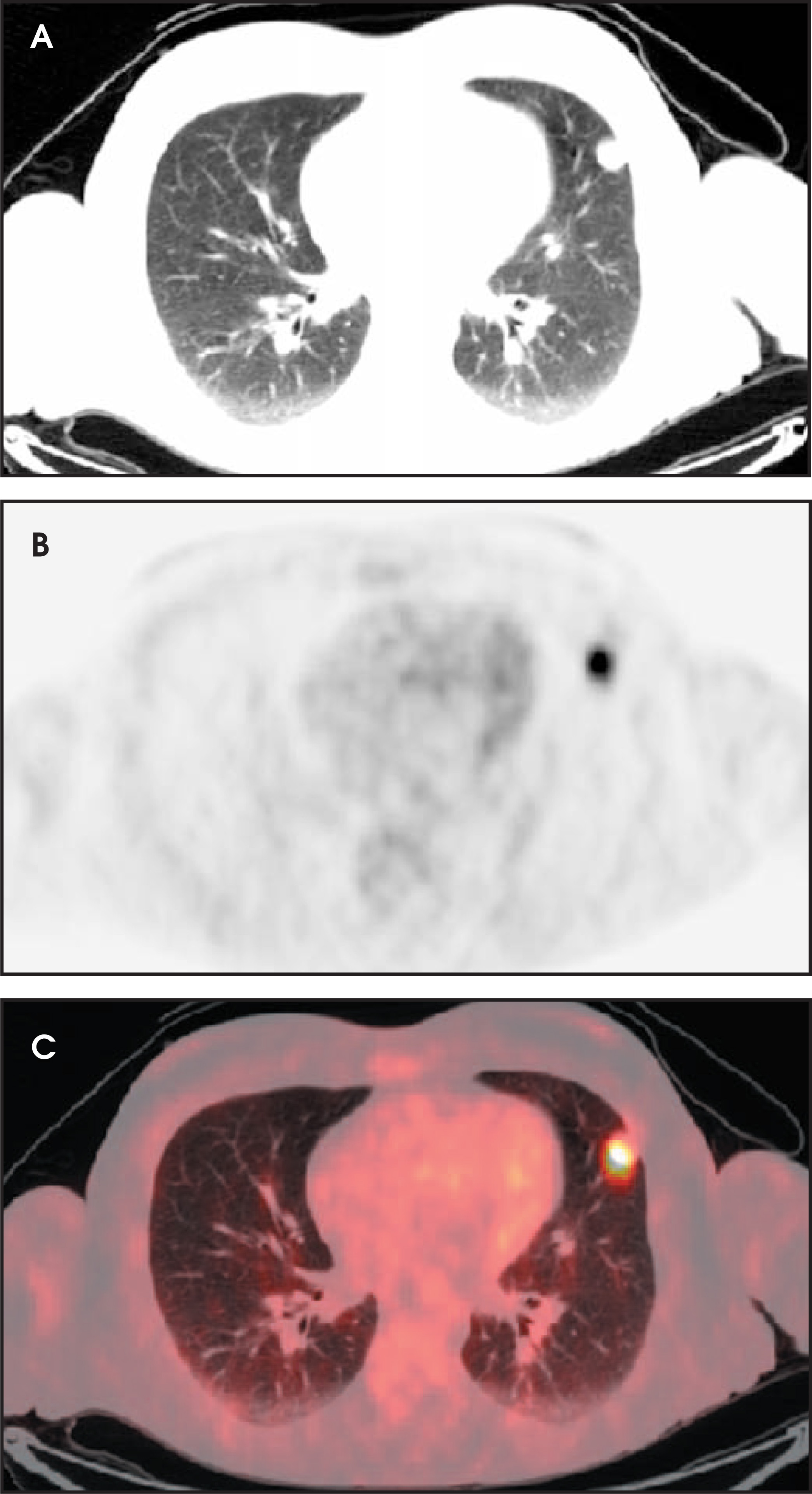
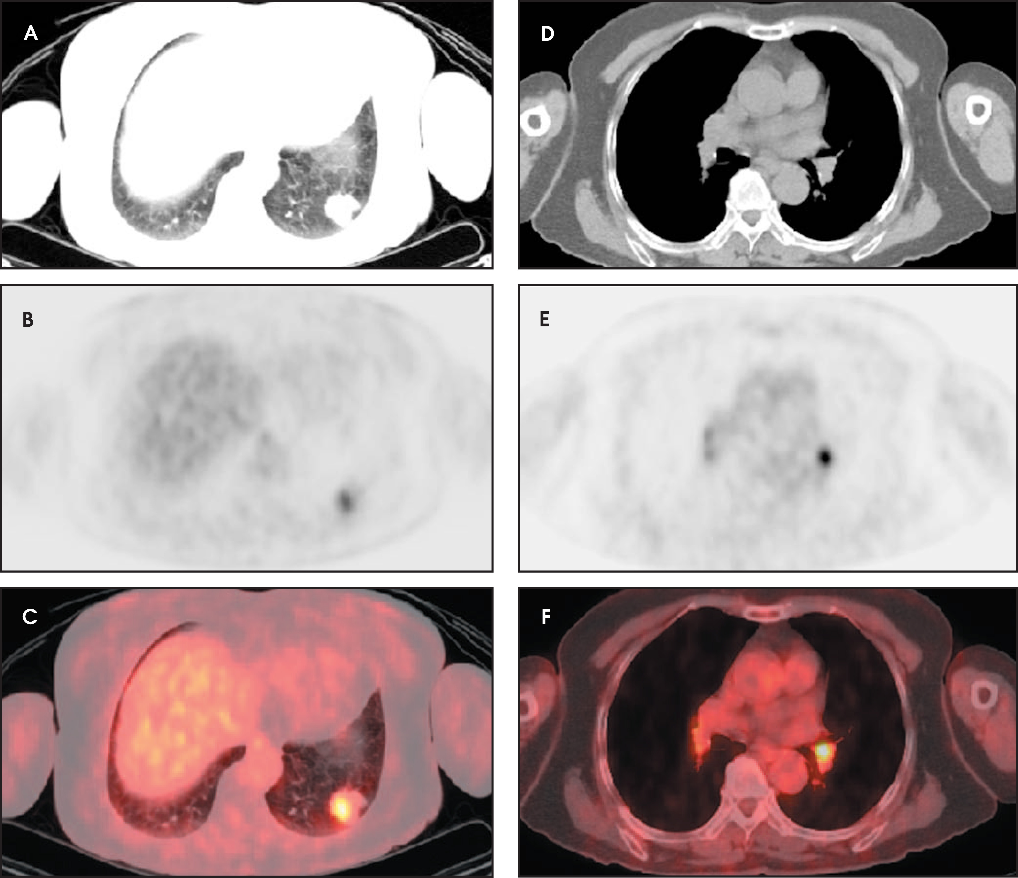
The problem facing a clinician is even greater if >1 nodule is present, which is not that uncommon, especially after a CT of a patient initially presenting with an SPN on chest radiography. Granulomatous disease, lung cancer with metastases, and metastases from a remote primary cancer all can present with multiple nodules. In patients without known malignancy, there is no clear algorithm to follow, especially since resection of all of the nodules is quite impractical. A different problem along the same lines is an SPN in a patient with a recent or remote history of malignancy, but without known metastatic disease (Figures 3 through 5).
PET scanning
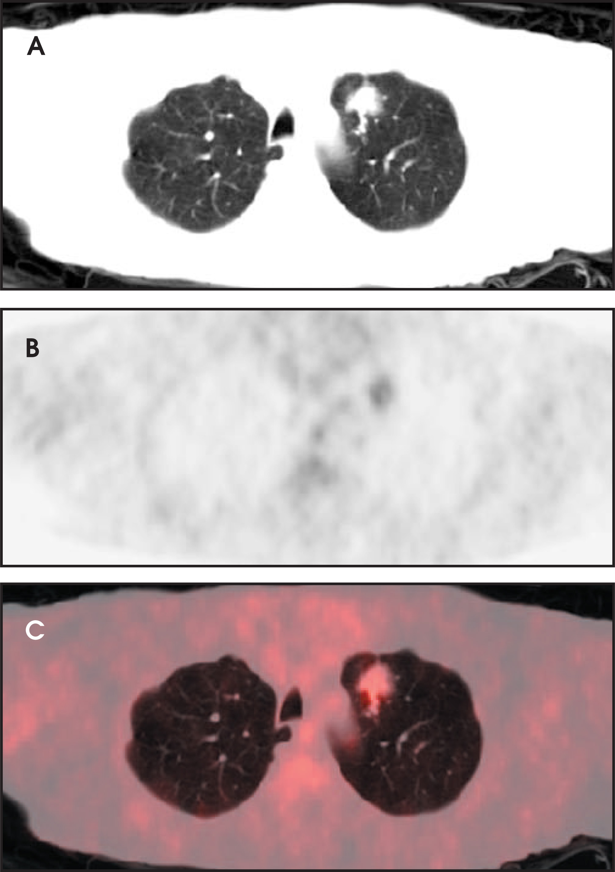
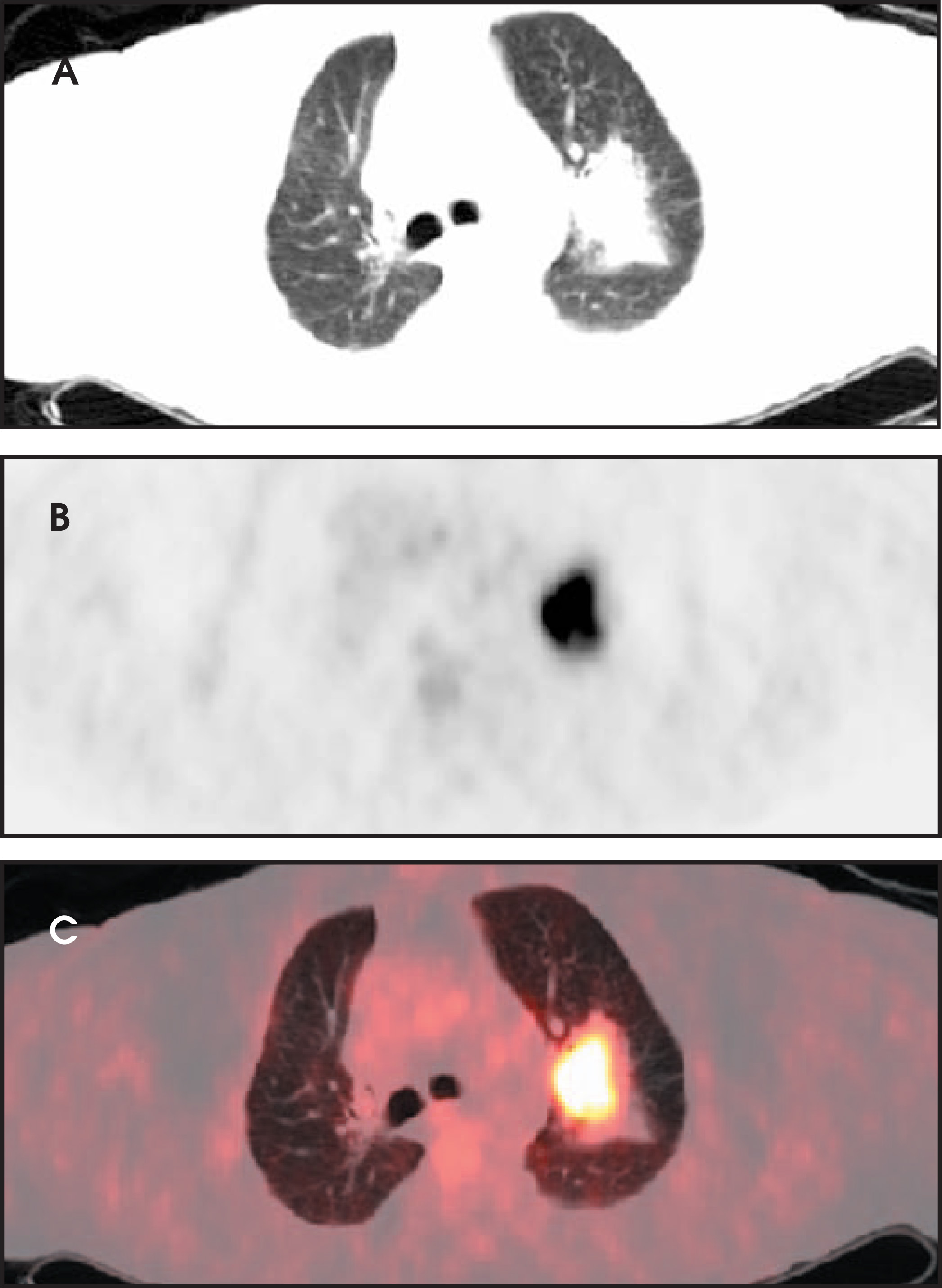
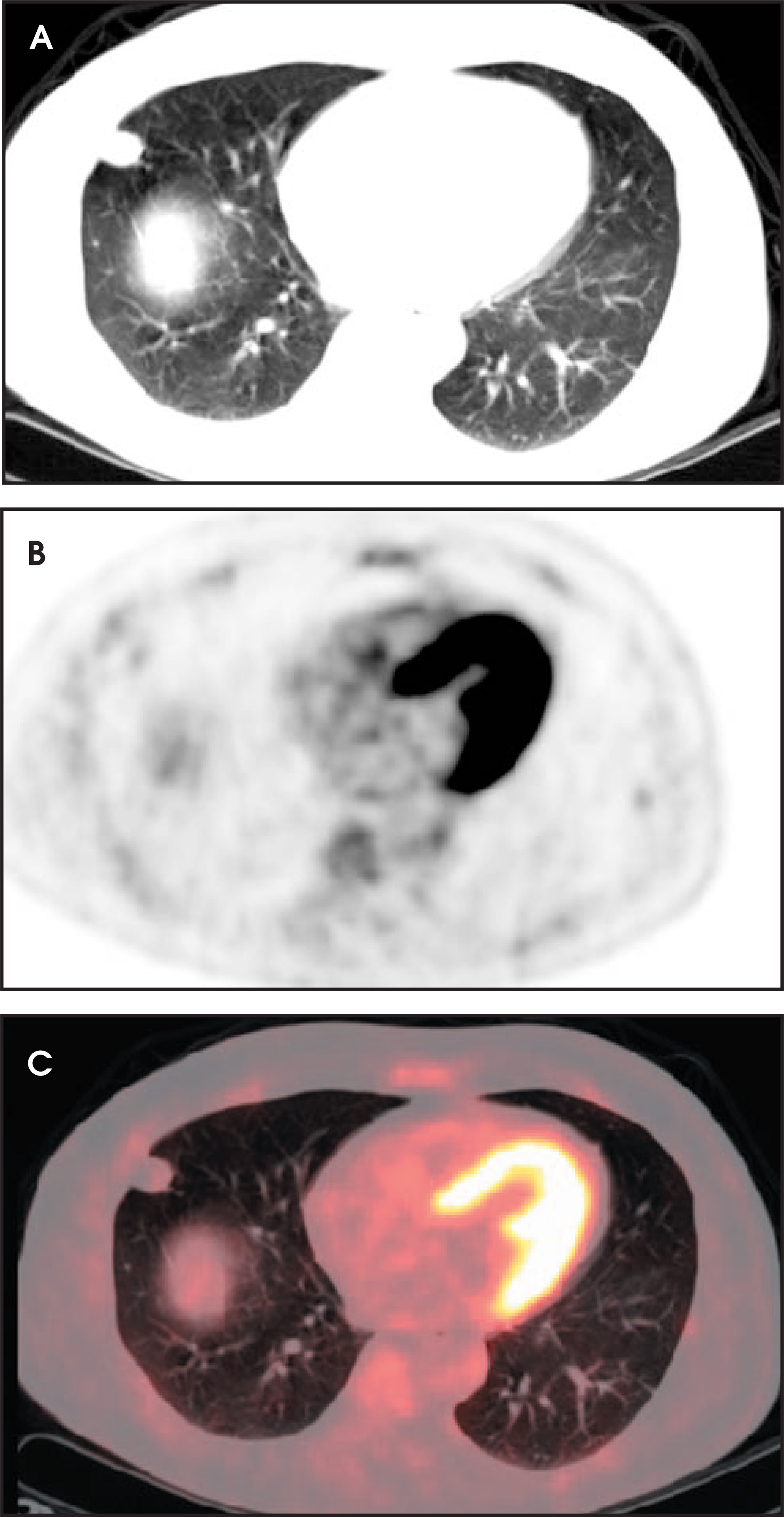
Recently, positron emission tomography (PET) has come to the aid of clinicians. PET uses a radioactive glucose analogue, fluorine-18–labeled fluoro-2-deoxyglucose (FDG), to map metabolic activity of tissues based on their use of blood glucose. Uptake in the lesion in question is then assessed. Quite often, tumor/blood ratios are used for qualitative interpretation. Lesions with uptake higher than that of blood pool often are malignant, while those with less intense uptake are most typically benign. To quantify FDG distribution, the amount of FDG uptake in a particular lesion is compared with the total body administered dose. A standardized uptake value (SUV)— which is the ratio of FDG concentration in the lesion to the average FDG concentration in the body—of >2.5 has been shown to be very sensitive and reasonably specific for malignant lesions,12 although the use of an SUV of 2 or 3 has been proposed by some (Figures 5 through 10).13 Benign lesions typically show FDG uptake in the 0.4 to 2 SUV range (Figures 11 and 12).
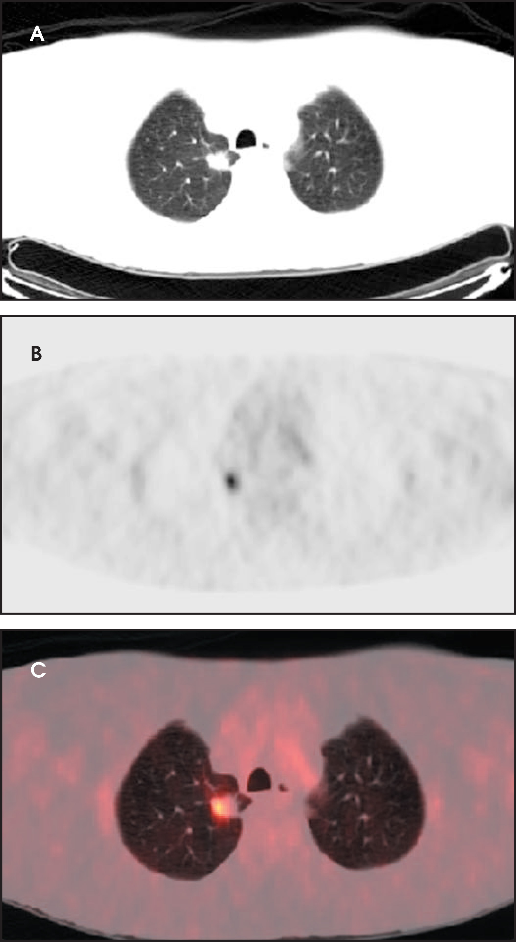
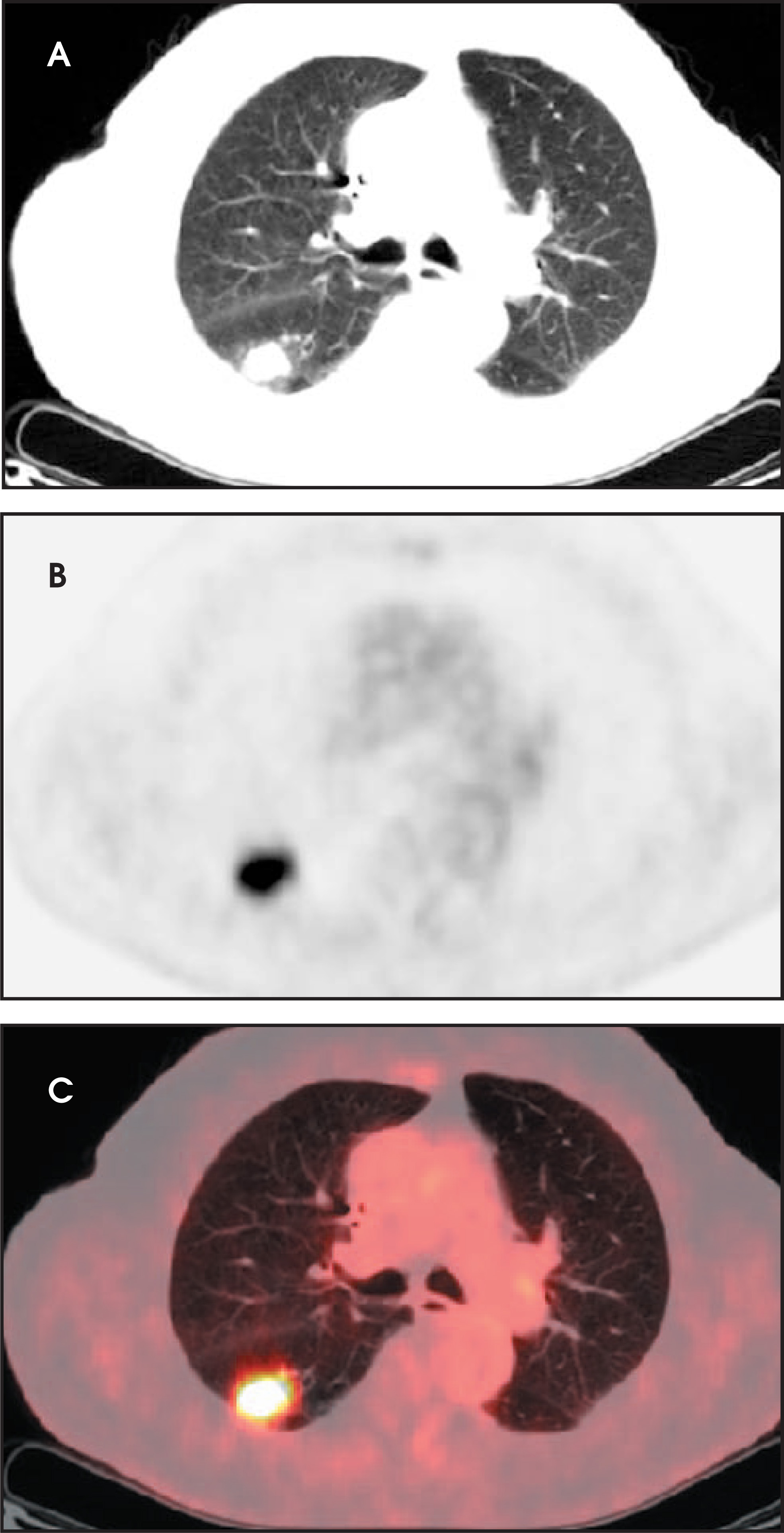
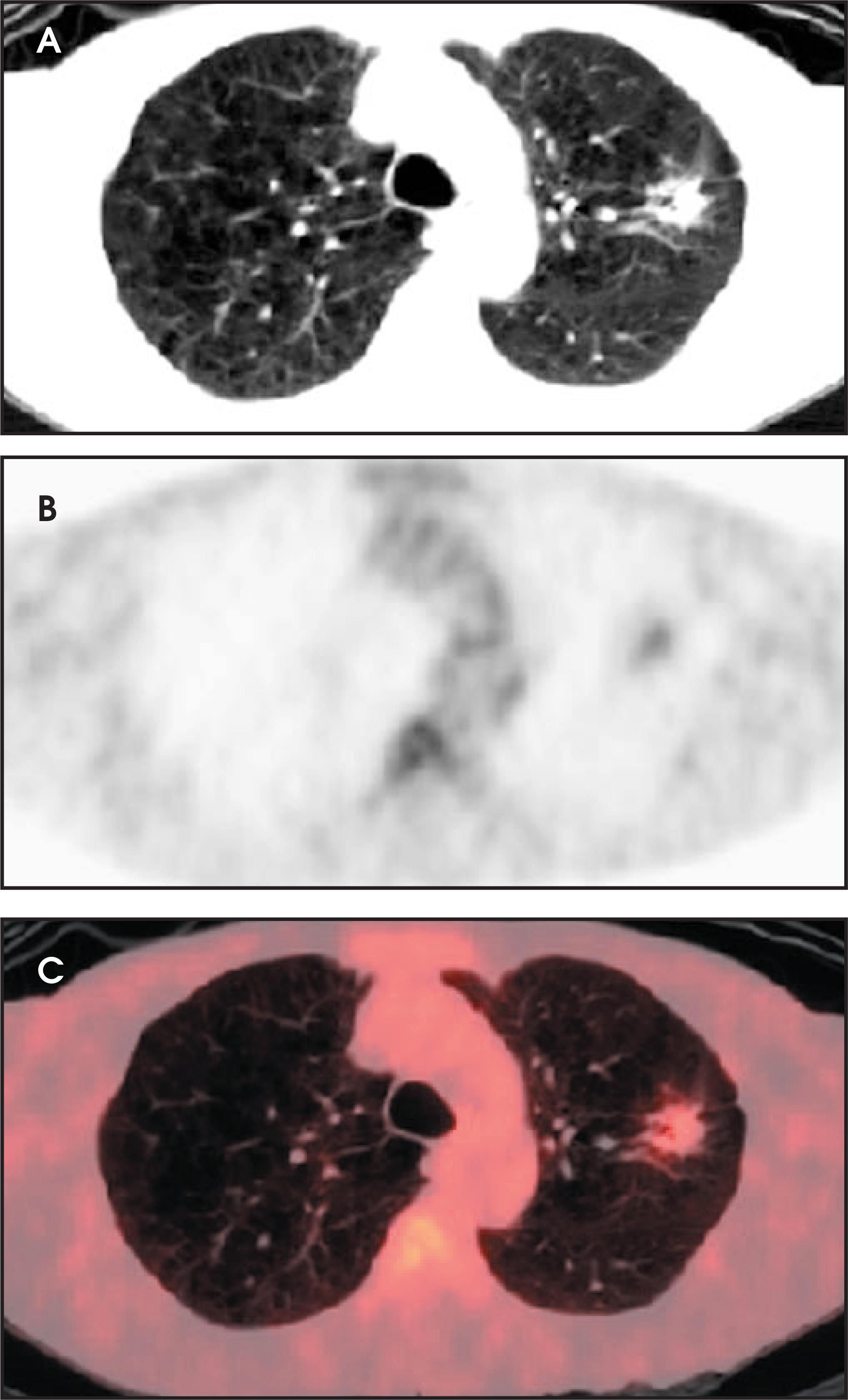
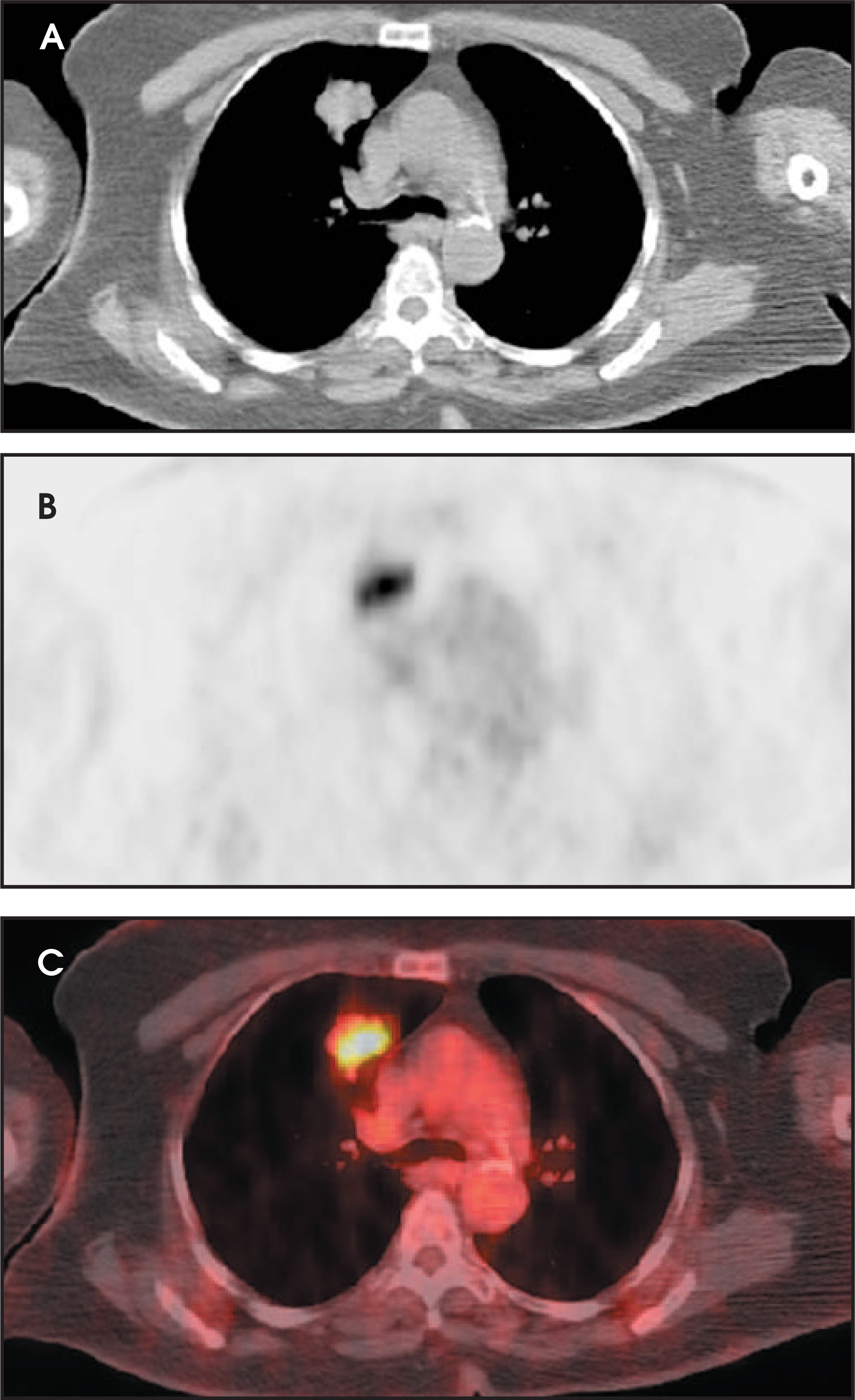
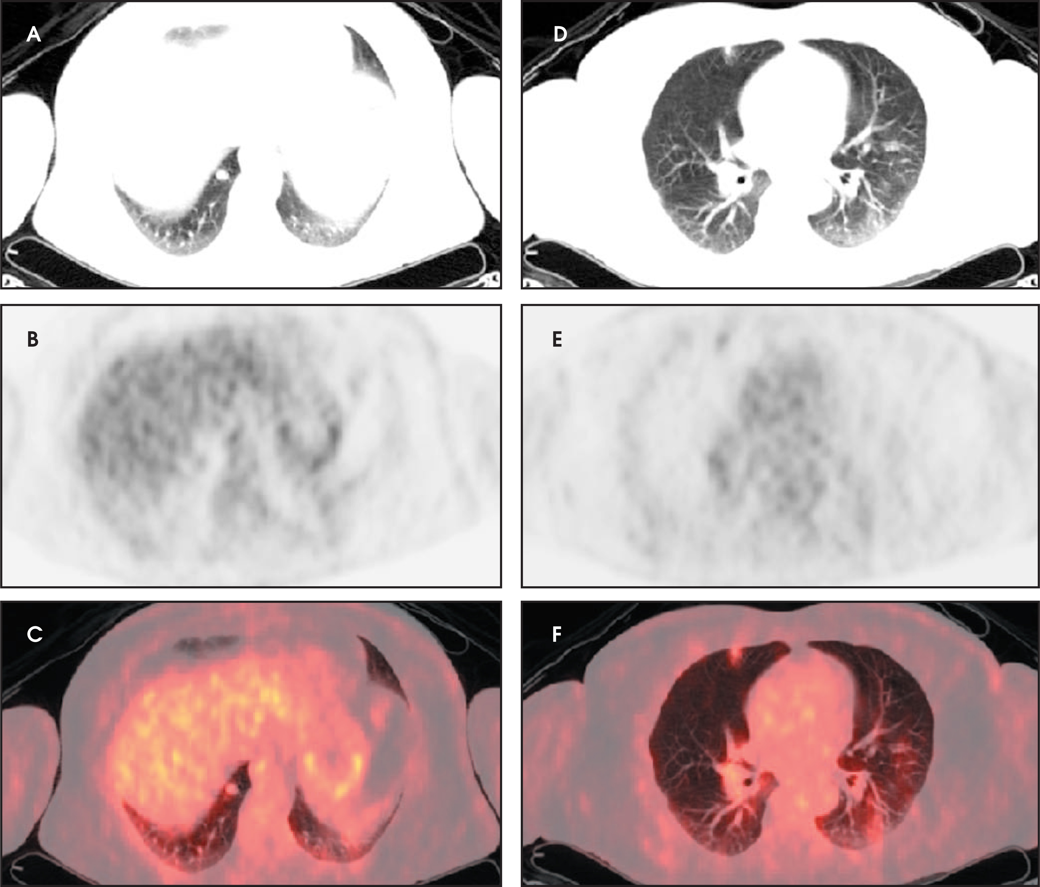
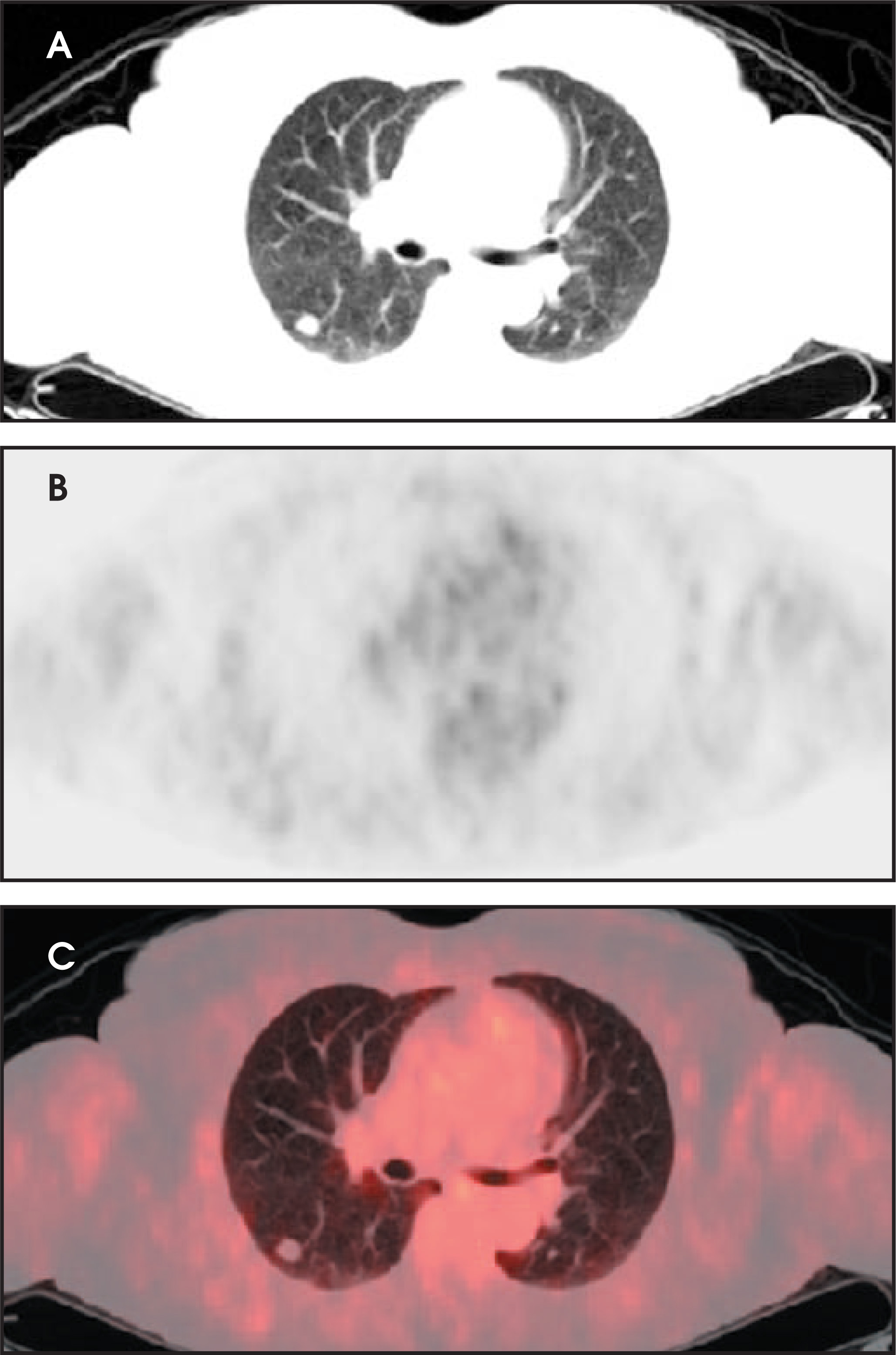
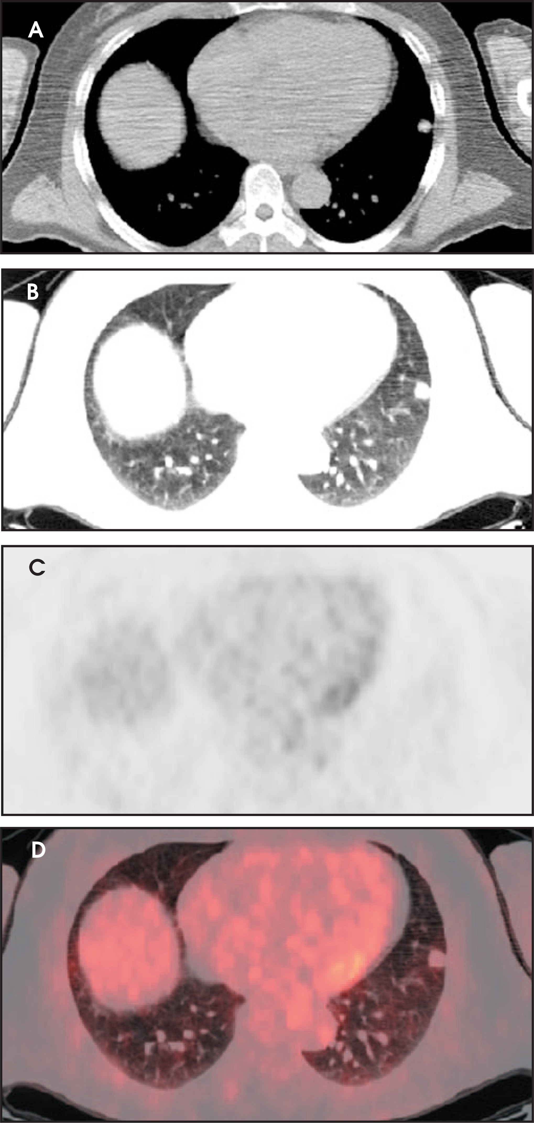
Characterization of SPNs by FDG-PET has been reported to have a sensitivity of 97% and a specificity of 78% in a recent meta-analysis.14 For the detection of malignant nodules, FDG-PET has an overall sensitivity of 95% to 100% and a specificity of 80% to 89%. However, these numbers may be somewhat lower for smaller (<1.5 cm) nodules. The association between FDG uptake and cell differentiation and, in turn, prognosis, has been also suggested by available data.9 The Centers for Medicare and Medicaid Services has approved the use of FDG-PET for evaluation of an SPN.
In addition to characterizing a nodule, FDG-PET has been shown to be more sensitive and more specific than CT in identifying mediastinal involvement and in detecting distant metastases.15 Given that 26% of newly diagnosed non–small-cell lung cancer (NSCLC) patients present with mediastinal lymph node disease and 49% present with extrathoracic metastases, this is a very significant benefit of PET.16 PET has produced better results in nodal staging of bronchogenic carcinoma than CT, MRI, endoscopic ultrasound (EUS), or, because of limited accessibility, even mediastinoscopy. The diagnostic accuracy of FDG-PET is 92% compared with 75% for CT.17 The reported positive predictive value for PET is 79% compared with 56% for CT and 75% for EUS, and negative predictive value is 93% compared with 83% for CT and 79% for EUS (Figure 13).15 In some studies, negative predictive value of PET was as high as 98.4%.18 In a recent study, PET staging of NSCLC was highly correlated with survival, while CT alone could not accurately predict survival.19
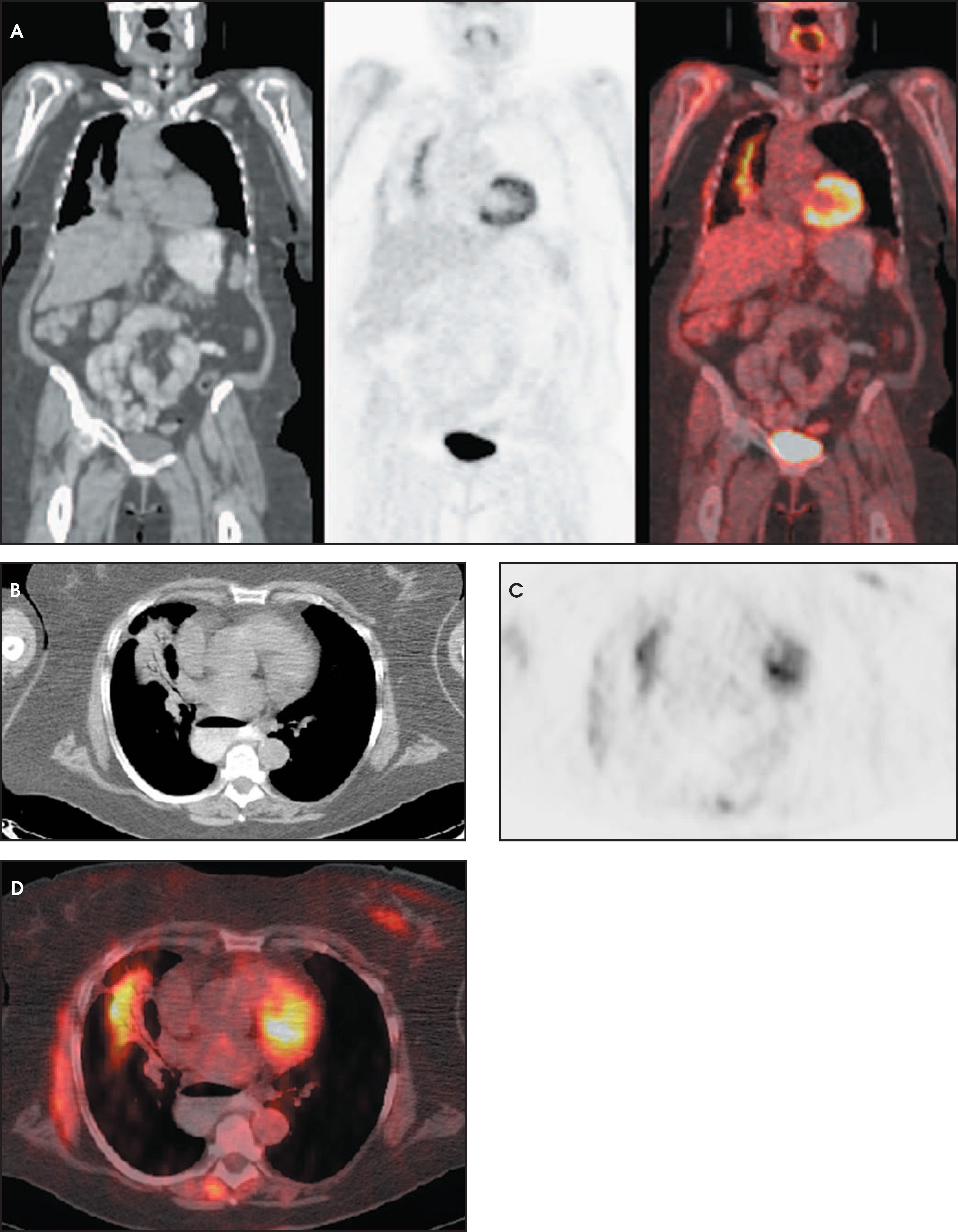
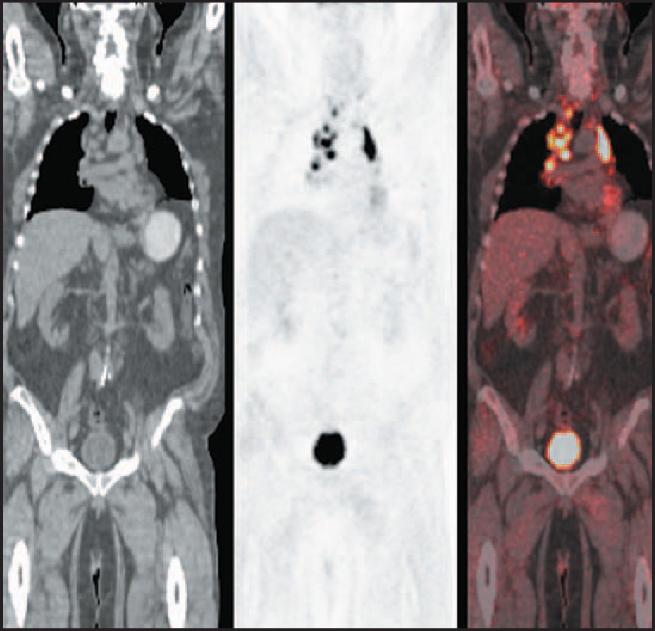
In cases of metastatic disease, PET can be helpful in finding a primary malignancy, or conversely, in excluding one, since the whole body is imaged without additional radiation exposure (Figures 14 and 15). PET finds metastatic disease in approximately 10% of patients that were metastasis-free on a routine CT scan. PET can improve the diagnostic yield of a biopsy, by guiding intervention to the metabolically active lesions.9 Using PET in the management algorithm of the SPN and in potentially operable NSCLC evaluation has been shown to be a cost-effective approach.20 It is very helpful to clinicians to have a single study that not only characterizes the nodule as benign or malignant but also stages the malignant cases.
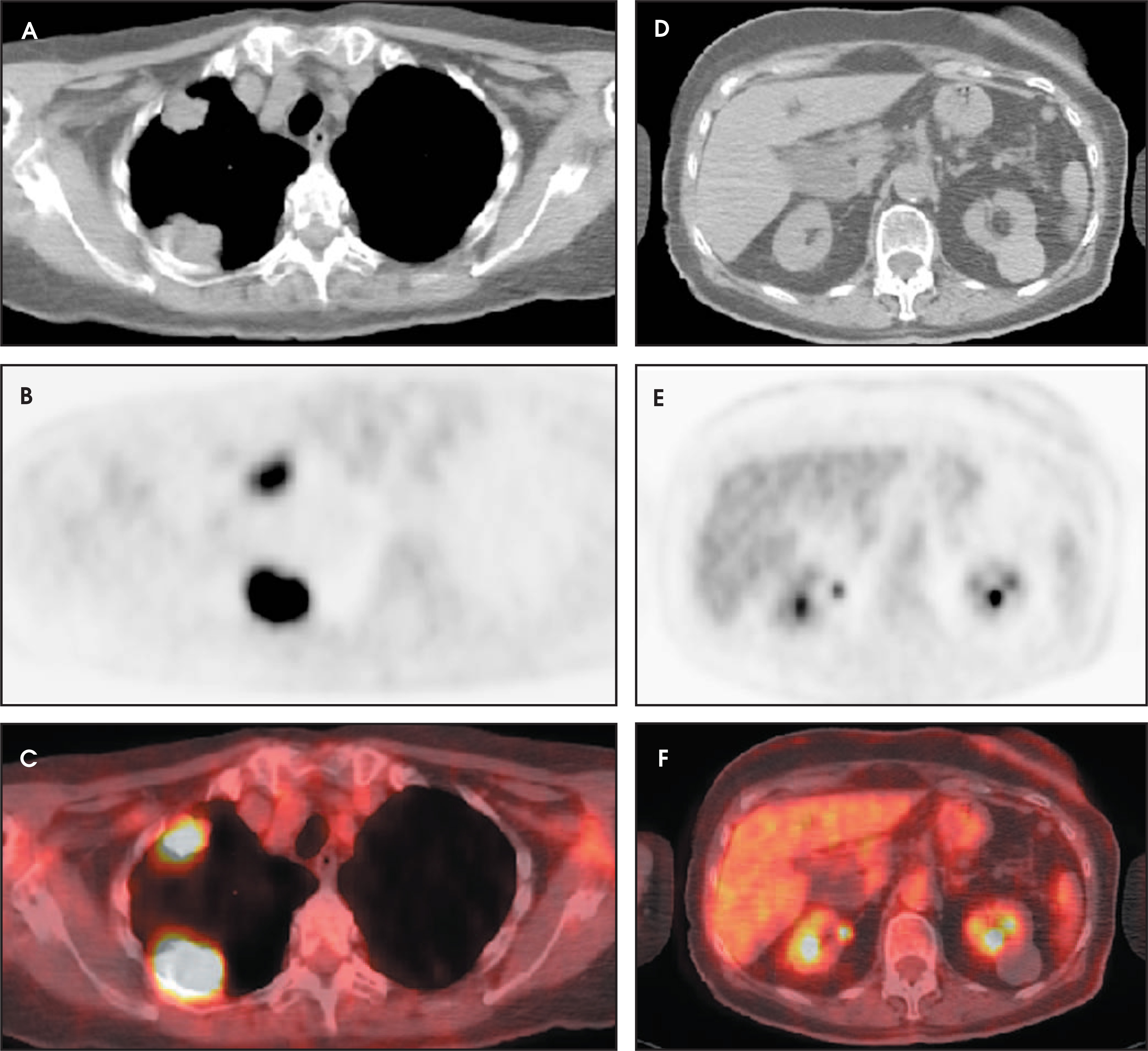
PET, unfortunately, has some shortcomings and blind spots. Inflammatory and granulomatous processes (such as tuberculosis, histoplasmosis, aspergillosis, coccidiomycosis, sarcoid, Wegener’s, and even pneumonia) can produce false-positive results, especially in cases of a fulminate process (Figures 16 and 17). Yet in the majority of cases, PET is still able to provide an accurate diagnosis. If postobstructive pneumonia is suspected, or if infiltrate does not resolve in a short time interval with treatment, PET can be of significant diagnostic value, despite decreased specificity in the setting of an infectious process (Figure 18).
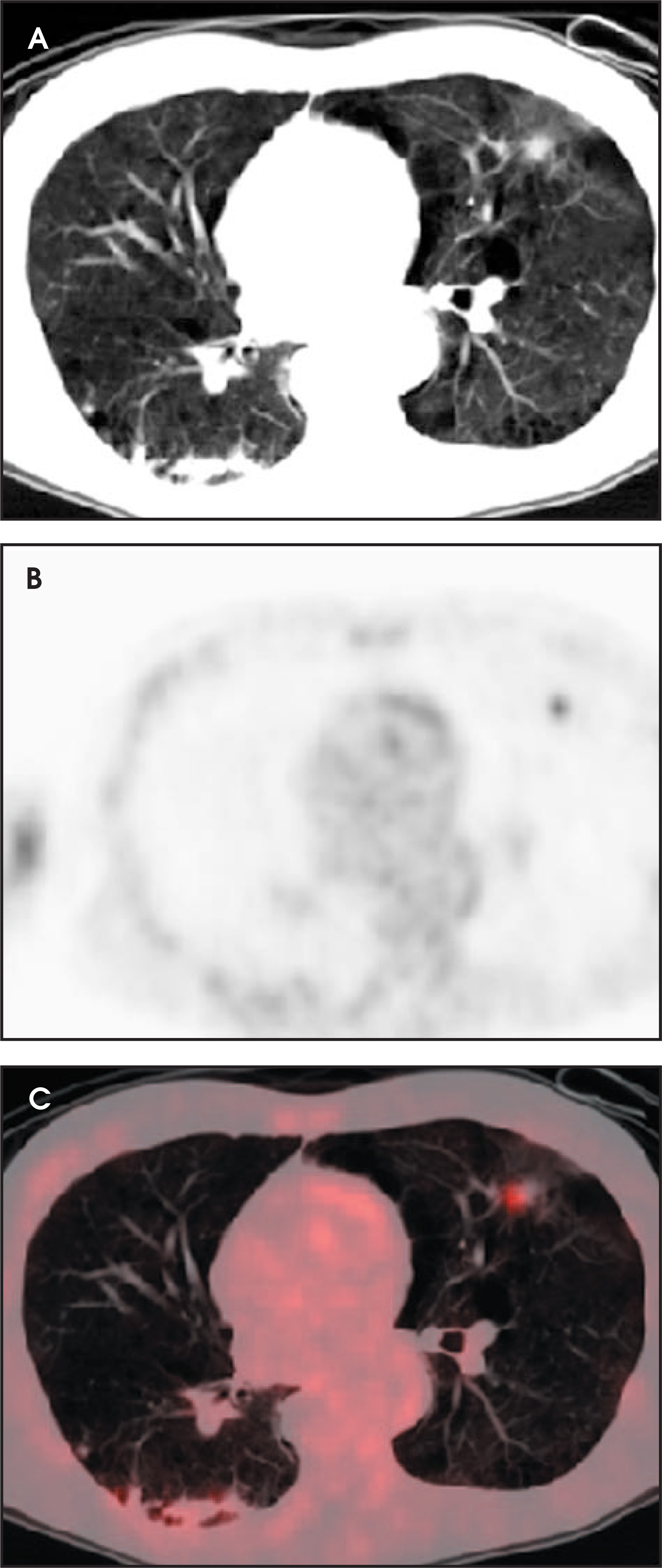
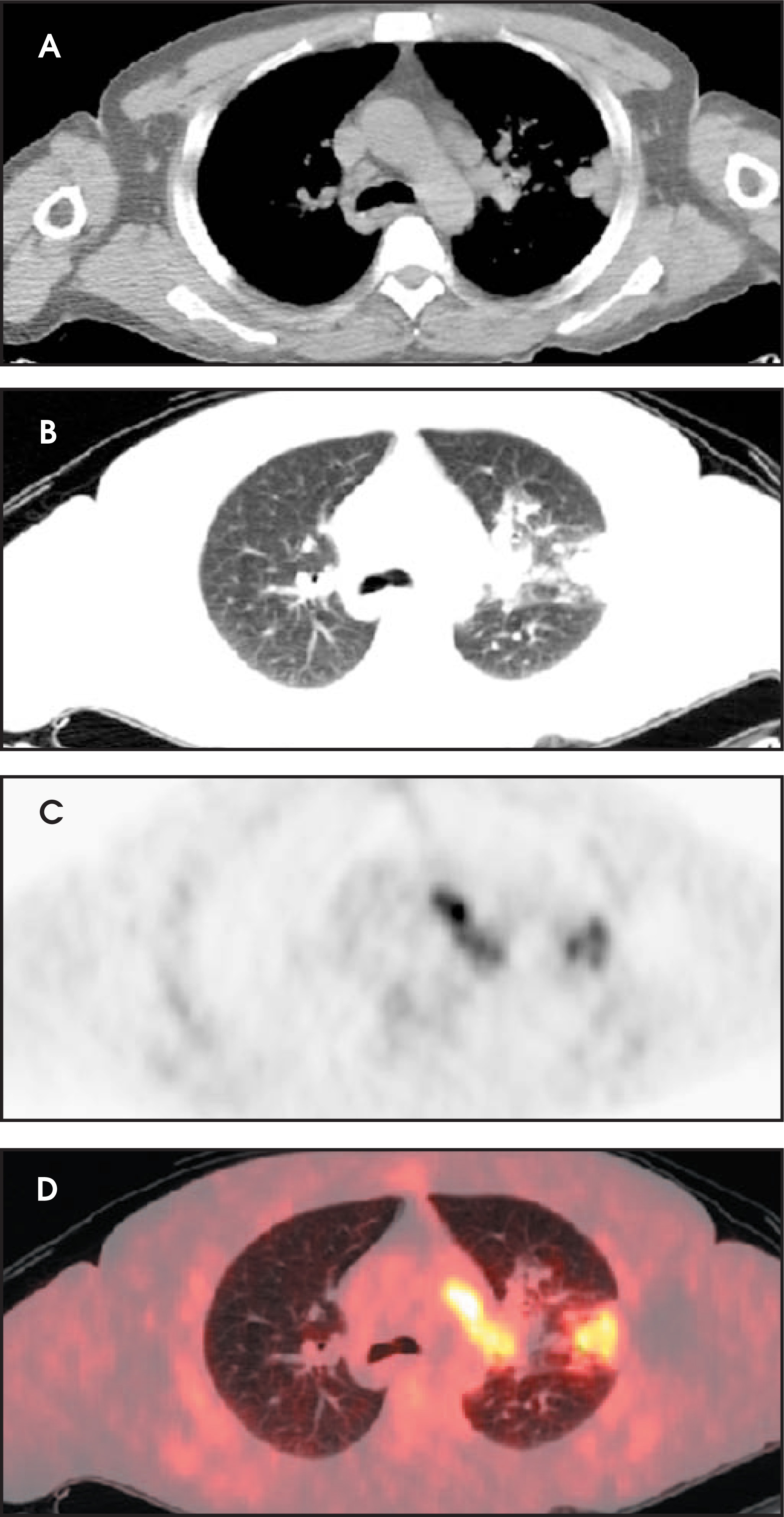
Neoplasms with low metabolic activity, such as bronchoalveolar cell carcinoma and carcinoid, can sometimes give a borderline or a false-negative result (Figures 19 and 20). Two other causes of false-negative results are related to the technique itself. FDG tumor uptake can be competitively inhibited by blood glucose in patients with high blood glucose levels (>250 mg/dL) during the study. Thus, it is very important to check the patient’s glucose level before injecting FDG. The second PET shortcoming is low spatial resolution, resulting in false-negative results in lesions <7 mm in size or even up to 1 cm. Also, SUV calculations can be affected by changes in body weight; so, if PET is used to assess treatment response and there has been a significant weight change, it is helpful to correct for the lean body mass.
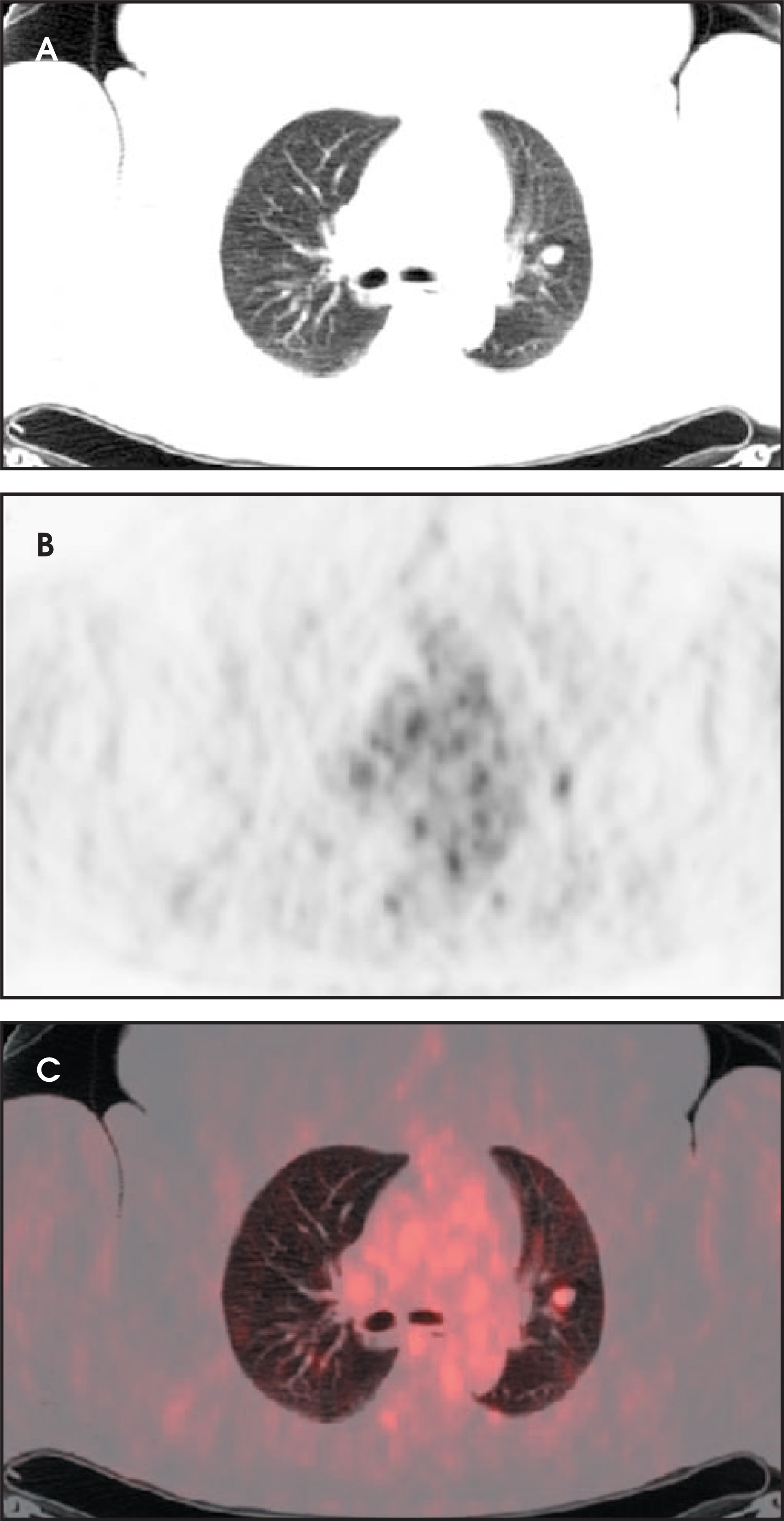
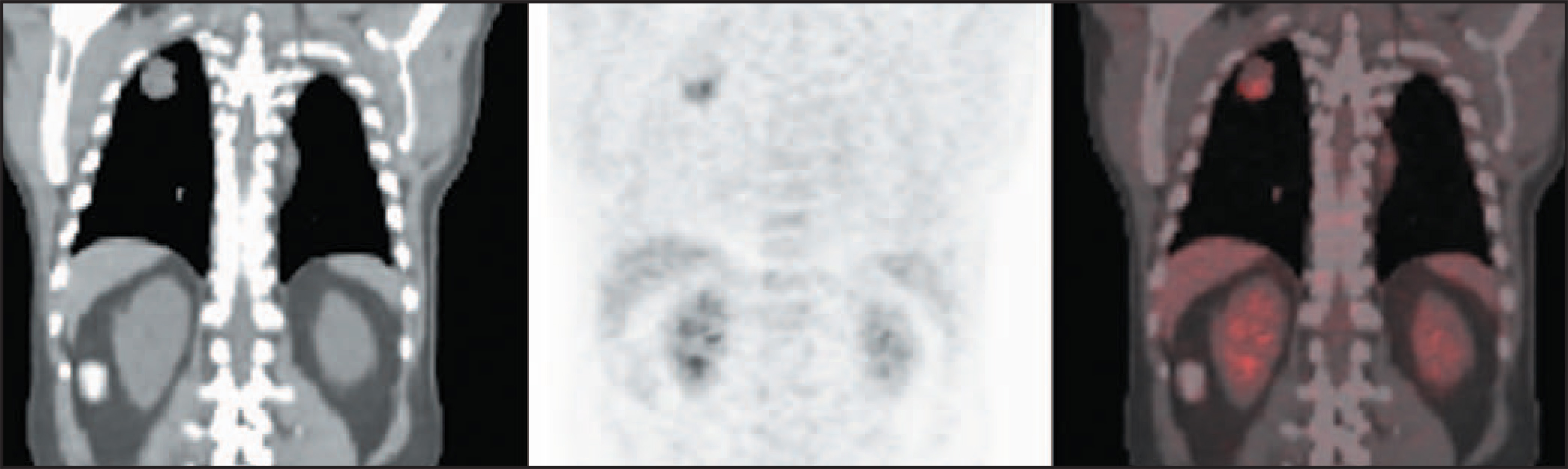
Before interpreting an FDG-PET scan, it is very important to be familiar with normal distribution of FDG in the chest. In some patients, increased FDG uptake can be seen in the myocardium (Figures 5 and 14), depending on the balance and availability of free fatty acids, typical myocardial energy substrate, and glucose. After physical activity, increased uptake can be seen in participating muscles. Also, if the patient is uncomfortable on the table, increased FDG uptake can be seen in cervical, paraspinal, and trapezius muscles due to muscle tension.21 Increased uptake can be seen in brown fat of the upper mediastinum/ chest if the room temperature is too low for the patient.22
PET/CT scanning
Given the success of PET and a large volume of data on lesion morphologic characteristics on CT, it is only logical to combine physiologic and anatomic imaging into a single study—PET/CT. While having the benefits of both studies, PET/CT offers additional available synergies. CT allows acquisition of attenuation correction data in <1 minute, compared with roughly 12 minutes for a regular PET study.23 The decrease in examination time increases the number of studies that can be performed in a day, thus improving patient access to this new and exciting—but not yet readily available—technology. Given that the patient must lie still for the entire examination and that motion degrades the quality of the data, the decrease in study time results in improved data, especially in mildly claustrophobic or anxious patients. The improvement is especially noticeable for small lesions. Fusion with high–spatial-resolution CT images is also most helpful in small lesions. Coregistering thoracic CT and PET data sets has been reported to significantly improve tumor staging and localization, nodal staging, and interpretation of metastatic involvement.24-26 PET/CT images have been shown to significantly increase diagnostic accuracy regarding lesion type and location compared with PET alone.23 Coregistered PET and CT images can be used for treatment guidance in addition to diagnosis. Significant changes in radiation treatment plans were made in a majority of cases after coregistering planning CT and PET images in a recent prospective study.27 When lesions have areas of different metabolic activity, such as a cystic area or a region of necrosis, PET/CT is invaluable in guiding intervention toward metabolically active portions of the lesion, thus improving the diagnostic yield of biopsy (Figure 20). When only 1 of ≥2 small lesions, located in close proximity to each other, is metabolically active, PET/CT is again an irreplaceable tool to guide biopsy (Figure 21).
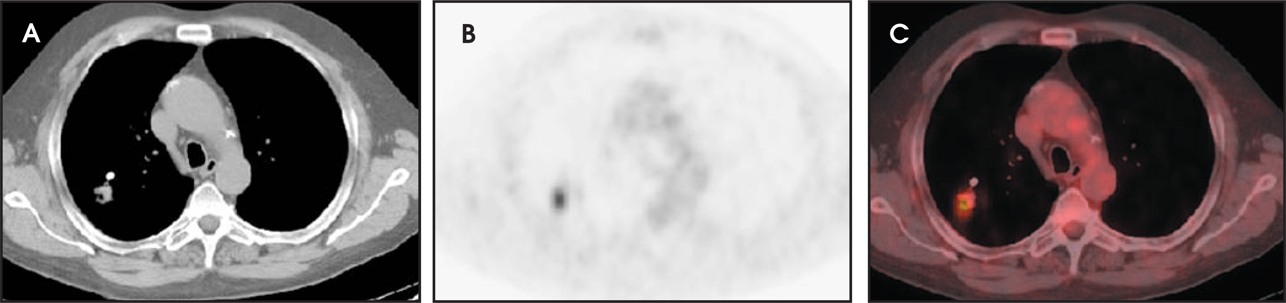
Another benefit of simultaneous acquisition is in patient positioning. Although the vast majority of patients can lie supine for a 15to 45-second CT scan, back pain or other problems make it difficult for some patients to lie flat for 45 minutes, which is the typical acquisition time of a PET scan. Patients may be accommodated in a semidecubitus position for the PET/CT study, still allowing excellent coregistration images (Figure 22).
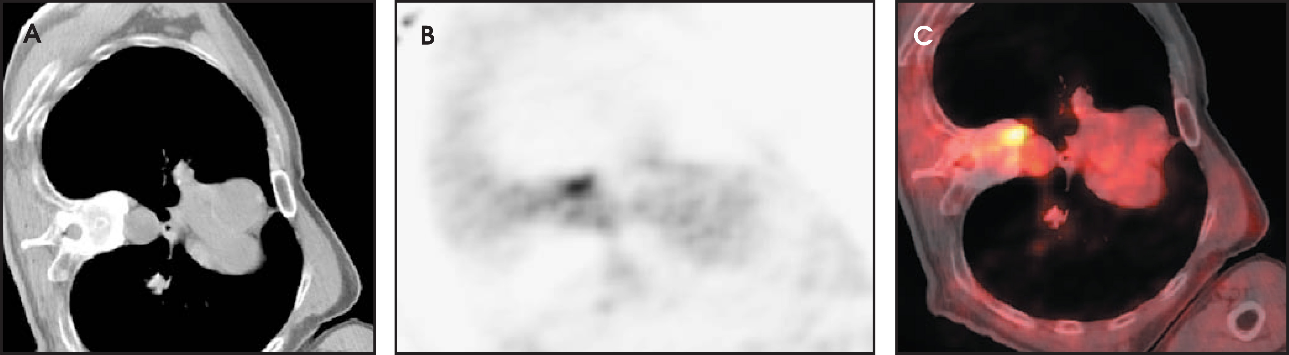
PET/CT localizes with greater precision compared with a low–spatial-resolution PET, easily distinguishing an osteophyte in the thoracic spine or recent rib trauma from a peripheral lung lesion (Figure 22). While coregistration in PET/CT is vastly better than fusion of images done days or even weeks apart on different scanners, one still needs to be aware of some pitfalls. Patient motion can degrade the quality of the data. In evaluation of lesions, especially near the diaphragm, one needs to be aware that breathing can sometimes result in misregistration (Figure 23).
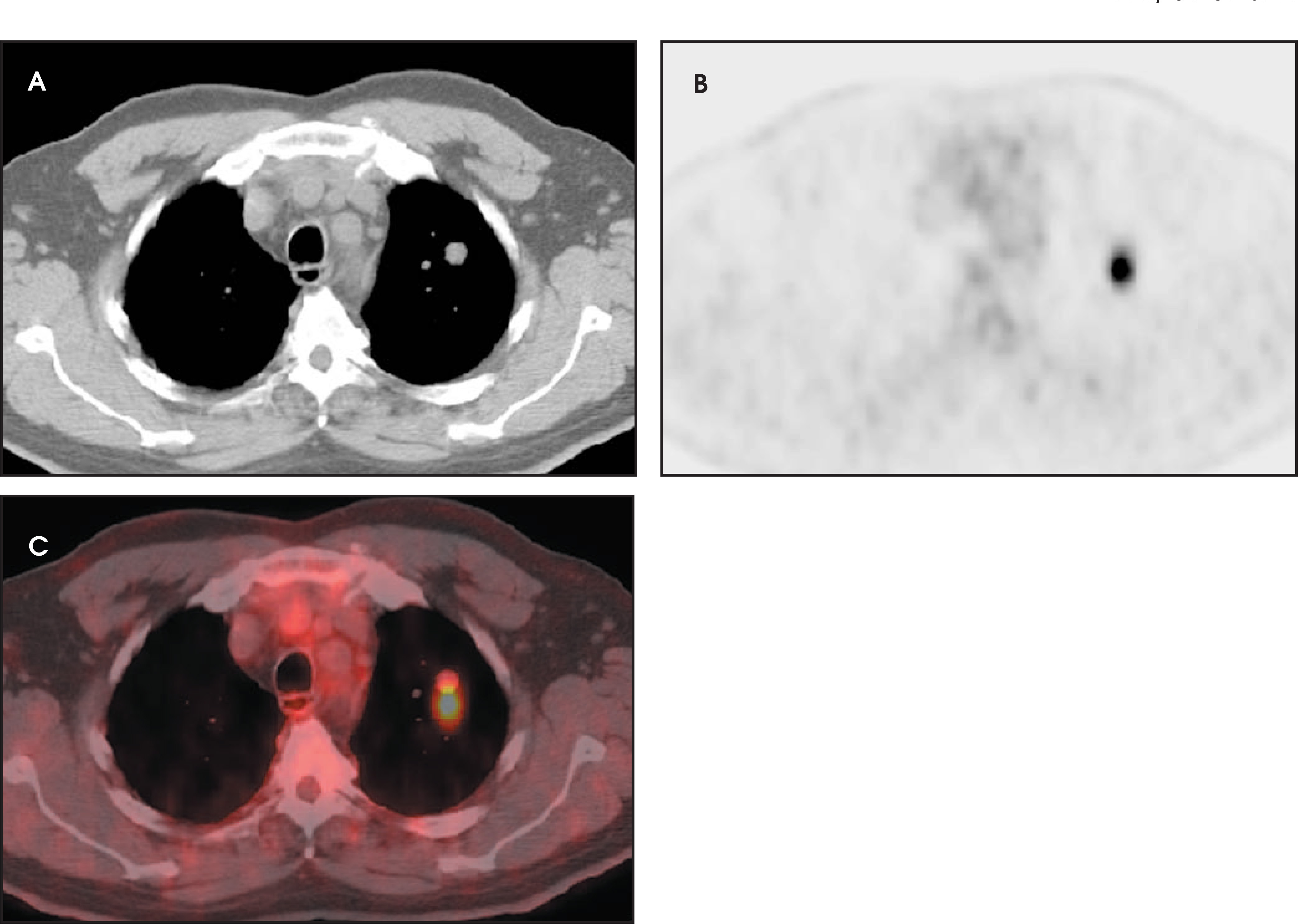
Data suggests that CT images acquired with a breath-hold after a normal expiration are best to match with PET images. The difference in the level of the diaphragmatic dome between CT and PET is only 4 mm on average with this technique, which is consistent with the spatial resolution of PET in general. Misregistration using other breathing techniques is much higher, in the range of 9.5 to 44.4 mm. Patients were also more attentive and were more compliant with the breath-hold after a normal expiration approach.28,29
It has been shown recently that the CT portion of a PET/CT can be acquired with a tube current as low as 10 mA and still yield good diagnostic information.23 There appears to be no benefit in CT scanning with tube current above 80 mA.23 Thus, the increase in radiation dose to the patient with PET/CT is very minimal when compared with PET alone. If a separate CT can be avoided after the PET/CT, the total exposure for patient evaluation actually becomes lower. Appropriate training in CT interpretation is essential for the person interpreting the study.
Overall, PET/CT appears to be a major step forward from the use of CT or PET alone. In our experience, it is more accepted than PETby both patients and referring physicians.
In spite of the emergence of PET/CT, there is still no clear “ideal” algorithm for the evaluation of an SPN. However, new options allow for more patient-friendly and cost-effective protocols. It has already been shown that, given a low prevalence of malignant SPNs, either PET with radiologic follow-up for 2 years, or PET and CT with follow-up produced a net cost reduction of $1600 per patient versus CT alone with follow-up. The majority of savings came from reducing the number of invasive procedures, which also means decreased patient morbidity.13 Given the availability of PET/CT at our institution, we often use it as the most appropriate next step in a workup of any SPN that is indeterminate or suggestive of malignancy that was initially diagnosed by chest X-ray or CT. Nodules that are clearly benign based on CT appearance or that show no FDG uptake at all can be followed at 3- to 6-month intervals for 24 months. Nodules that show intermediate FDG uptake (>1, but < 2.5 SUV) deserve a close follow-up with CT or PET/CT or a biopsy, depending on the likelihood of an inflammatory process based on clinical symptoms and history, and the patient’s level of comfort with a follow-up approach. A similar conservative strategy using chest CT and PET has shown potential cost savings of $1150 per patient, with no decrease in life expectancy. With this strategy, all positive sites of abnormal tracer accumulation found on CT and PET that are consistent with unresectable disease are biopsied to confirm the finding, so that 100% of surgical candidates are definitively identified. The largest component of the cost savings arises from avoiding unnecessary mediastinoscopy and thoracotomy.13
Conclusion
PET/CT is a safe, noninvasive technique that frequently allows fast and accurate diagnosis of an SPN and should be part of a routine algorithm for SPN evaluation. Its value is especially evident in patients with SPNs that are indeterminate by CT criteria, in patients with heterogeneous masses, or in patients whose clinical history is complicated by previous malignancy.
References
- Khouri N, Meziane M, Zerhouni E. The solitary pulmonary nodule: Assessment, diagnosis, and management. Chest. 1987;91:128-33.
- Viggiano R, Swensen S, Rosenow E. Evaluation and management of solitary and multiple pulmonary nodules. Clin Chest Med. 1992;13:83-95.
- Liptay M. Solitary pulmonary nodule: Treatment options. Chest. 1999;116(suppl 6):517S-518S.
- Henschke C, Naidich D, Yankelevitz D. Early Lung Cancer Action Project: Initial findings on repeat screenings. Cancer. 2001;92:153-159.
- Swenson S, Jett J, Sloan J. Screening for lung cancer with low-dose spiral computed tomography. Am J Respir Crit Care Med. 2002;165:508-513.
- Mountain C. Revisions in the International System for Staging Lung Cancer. Chest. 1997;111:1710-1717.
- Ost D, Fein A, Feinsilver S. Clinical practice: The solitary pulmonary nodule. N Engl J Med. 2003;348:2535-2542.
- Erasmus J, McAdams H, Connolly J. Solitary pulmonary nodules: Part II. Evaluation of the indeterminate nodule. RadioGraphics. 2000;20:59-66.
- Line B, Maragh M, Ahamed T. PET imaging of lung and esophageal cancer. Appl Radiol. 2002;31(6):9-17.
- Cox J, Chiles C, McManus C. Transthoracic needle aspiration biopsy: Variables that affect risk of pneumothorax. Radiology. 1999;212:165-168.
- Swensen S, Brown L, Colby T. Lung nodule enhancement at CT: Prospective findings. Radiology. 1996;201:447-455.
- Knight S, Delbeke D, Stewart J, Sandler M. Evaluation of pulmonary lesions with FDG-PET: Comparison of findings in patients with and without a history of prior malignancy. Chest. 1996;109:982-988.
- Goldsmith S, Kostakoglu L. Nuclear medicine imaging of lung cancer. Radiol Clin North Am. 2000;38:511-524.
- Gould M, Maclean C, Kuschner W. Accuracy of positron emission tomography for diagnosis of pulmonary nodules and mass lesions: A meta-analysis. JAMA. 2001;285:914-924.
- Toloza E, Harpole L, McCrory D. Noninvasive staging of non-small cell lung cancer: A review of current evidence. Chest. 2003;123(suppl 1):137S-146S.
- Jemal A, Thomas A, Murray T, Thun M. Cancer statistics, 2002. CA Cancer J Clin CA Cancer J Clin CA Cancer J Clin.
- Wahl R. Targeting glucose transporters for tumor imaging: “Sweet” idea, “sour” result. J Nucl Med. 1996;37:1038-1041.
- Graeter T, Hellwig D, Hoffmann K. Mediastinal lymph node staging in suspected lung cancer: Comparison of positron emission tomography with F-18-fluorodeoxyglu-cose and mediastinoscopy. Ann Thorac Surg. 2003;75:231-235.
- Dunagan D, Chin R, McCain T. Staging by positron emission tomography predicts survival in patients with non-small cell lung cancer. Chest. 2001;119:333-339.
- Dietlein M, Moka D, Weber K. [Cost-effectiveness of PET in the management algorithms of lung tumors: Comparison of health economic data.]. Nuklearmedizin. 2001;40:122-128.
- Kostakoglu L, Agress H, Goldsmith S. Clinical role of FDG PET in evaluation of cancer patients. RadioGraphics. 2003;23:315-340.
- Cohade C, Osman M, Pannu H, Wahl R. Uptake in supraclavicular area fat (“USA-Fat”): Description on 18F-FDG PET/CT. J Nucl Med. 2003;44:170-176.
- Hany T, Steinert H, Goerres G. PET diagnostic accuracy. Improvement with in-line PET-CT system: Initial results. Radiology. 2002;225:575-581.
- Lardinois D, Weder W, Hany T. Staging of non-small-cell lung cancer with integrated positron-emission tomography and computed tomography. N Engl J Med. 2003;348:2500-2507.
- Aquino S, Asmuth J, Moore R. Improved image interpretation with registered thoracic CT and positron emission tomography data sets. AJR Am J Roentgenol. 2002;178:939-944.
- Antoch G, Stattaus J, Nemat A. Non-small cell lung cancer: Dual-modality PET/CT in preoperative staging. Radiology. 2003;229:526-533.
- Mah K, Caldwell C, Ung Y. The impact of (18)FDG-PET on target and critical organs in CT-based treatment planning of patients with poorly defined non-small-cell lung carcinoma: A prospective study. Int J Radiat Oncol Biol Phys(52):339-350.
- Goerres G, Kamel E, Heidelberg T. PET-CT image co-registration in the thorax: Influence of respiration. Eur J Nucl Med Mol Imaging. 2002;29:351-360.
- Goerres G, Kamel E, Seifert B. Accuracy of image coregistration of pulmonary lesions in patients with non-small cell lung cancer using an integrated PET/CT system. J Nucl Med. 2002;43:1469-1475.
Citation
. Use of combined PET/CT imaging in evaluation of the solitary pulmonary nodule: Principles, techniques, and pitfalls. Applied Radiology. 2006;35(4):24-43. doi:10.37549/AR1417.