Uterine artery embolization: Where does it stand in the management of uterine leiomyomas? Part 1
Applied Radiology — Vol. 33 , Issue 9 , pp. 22 -29
DOI: 10.37549/AR1282
Published: September 1, 2004
Categories
Authors’ note: In this two-part series on the management of uterine fibroids, we will discuss some of the most commonly employed treatment options for women with symptomatic uterine fibroids. The first part, which is presented in this issue, will be dedicated to uterine artery embolization (UAE): when it is applicable, how to perform it, and what to expect from the procedure. The second part, which will be published in the October issue of this journal, will discuss more traditional treatments and outcomes for fibroids, which will help the reader understand how UAE fares against these alternatives. Finally, we will touch on some of the newest cutting-edge therapies to watch for.
Interventional radiologists have made great advances in the treatment of symptomatic uterine fibroids. Uterine artery embolization (UAE) eliminates the need for surgery, general anesthesia, and prolonged recovery time. During this 1- to 2-hour procedure, the uterine arteries are blocked with particles, resulting in infarction of the fibroids with subsequent hyalinization and fibrosis. The fibroids and the overall volume of the uterus shrink over the next several months. Nine of 10 women get complete symptomatic relief from their fibroid-related symptoms. Uterine artery embolization is a minimally invasive technique that is gaining popularity as a safe and effective alternative to myomectomy and hysterectomy.
Leiomyomas are the most common benign pelvic tumor in women of reproductive age, affecting approximately 20% to 40% of women >35 years of age.1 Reproductive characteristics associated with the development of fibroids include early menarche, low parity, late age at first birth, infertility, longer interval since last childbirth, and early use of oral contraceptives.2 Not all women with fibroids are symptomatic. Approximately 10% to 20% of women with fibroids have symptoms,3 including: menorrhagia, pain, bloating/pressure, urinary frequency, constipation, sciatica, and anemia. These symptoms have a significant negative impact on patients’ quality of life.
Since the 1970s, UAE has been a recognized nonsurgical treatment for acute pelvic hemorrhage. In 1989, Ravina et al 4 began performing preoperative UAE to control intraoperative blood loss during myomectomy. They discovered that the fibroids actually shrank during the time interval between the transcatheter procedure and the myomectomy. They subsequently published the first series of bilateral UAE for the primary treatment of fibroids.4 Since this time, multiple series have been published indicating that UAE is effective in controlling fibroid-related symptoms in 80% to 95% of women.6
Patient selection for UAE
Fibroid patients have revolutionized the way in which interventional procedures have been adopted. Many well-informed women whose quality of life is significantly impaired by fibroid-related symptoms have circumvented normal referral patterns and have done extensive research on the Internet prior to referring themselves for this procedure. Gynecologists’ opinion of UAE is mixed. While some gynecologists will refer patients for UAE if this treatment option best meets the patient’s preferences and future reproductive goals, others still consider UAE an experimental procedure. Regardless of the referral pattern in your practice, a close relationship between the gynecologist and interventional radiologist is helpful for optimal patient care.
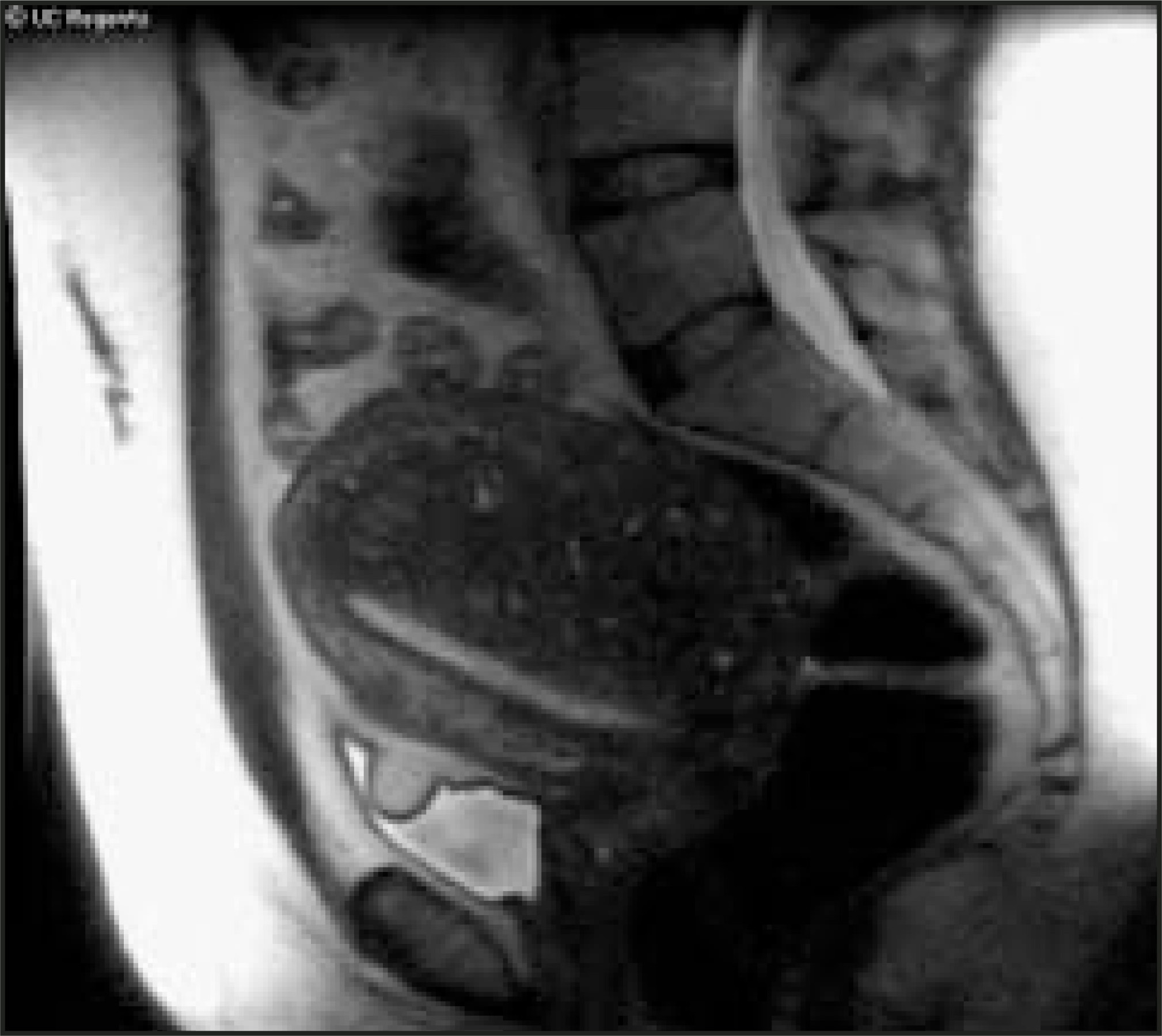
A thorough workup of the patient must be performed before considering UAE (see “UAE technique,” below). During this process, other causes of the patients’ symptoms must be excluded. If cancer or adenomyosis are diagnosed, the patient should be triaged to the appropriate treatment path. At the present time, the treatment of adenomyosis with UAE is controversial (Figure 1).
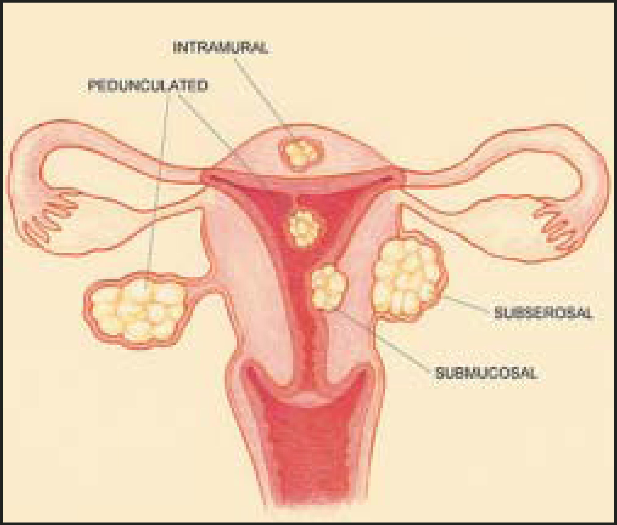
The Society of Interventional Radiology (SIR) believes that UAE is most appropriate for premenopausal patients with no desire for future fertility. A patient’s fibroids must be documented with an imaging study, and symptoms should correlate with the size and location of the fibroid(s) within the uterus13(Figure 2). Differing opinions exist regarding offering UAE to postmenopausal women and women who plan to have children in the future.
In many postmenopausal women, uterine fibroids will shrink without treatment after hormonal stimulation decreases. In patients who continue to have symptoms, it is critical to exclude cancer. Postmenopausal women with abnormal bleeding patterns should first have a thorough workup performed by their gynecologist, including a complete history and physical examination. Additional studies such as ultrasound, magnetic resonance imaging (MRI), or endometrial biopsy may also be of value in determining the cause of abnormal bleeding and excluding diagnoses other than fibroids. If the workup remains negative for an alternate diagnosis and the patient does not desire a hysterectomy, UAE could be considered.
Similarly, controversy exists regarding UAE in women who have future childbearing plans. Some physicians believe that no patient who wishes to preserve future fertility should have UAE, because of concerns about postprocedural amenorrhea or the side effects of embolization on the uterus. The most commonly reported rates of permanent amenorrhea post-UAE range from 2% to 7%7,8,10,11,14 and the incidence may be significantly higher in women >45 years of age.15,16 The etiology of post-UAE amenorrhea is not yet clearly defined. Several possibilities include: nontarget embolization, decreased vascular supply to the ovaries, or alteration of the uterine-ovarian hormonal balance.10,15,16 Further studies are necessary to understand the cause of earlier menopause in some women post-UAE and to determine the effect of UAE on future pregnancies.
If a premenopausal woman wishes to preserve her fertility and has fibroid symptoms severe enough to warrant a hysterectomy or multiple difficult myomectomies, UAE should be considered. The Agency for Healthcare Research and Quality (AHRQ) Evidence Report on the Management of Uterine Fibroids17 showed that increasing the number of fibroids removed was a significant predictor of complications and transfusions. In addition, as compared with those who have a single fibroid, women who have multiple fibroids removed have poorer long-term results in terms of pregnancy rate, risk of recurrence, and need for subsequent surgeries.17 Prospective controlled studies comparing UAE with myomectomy will need to be completed to better guide patient care.
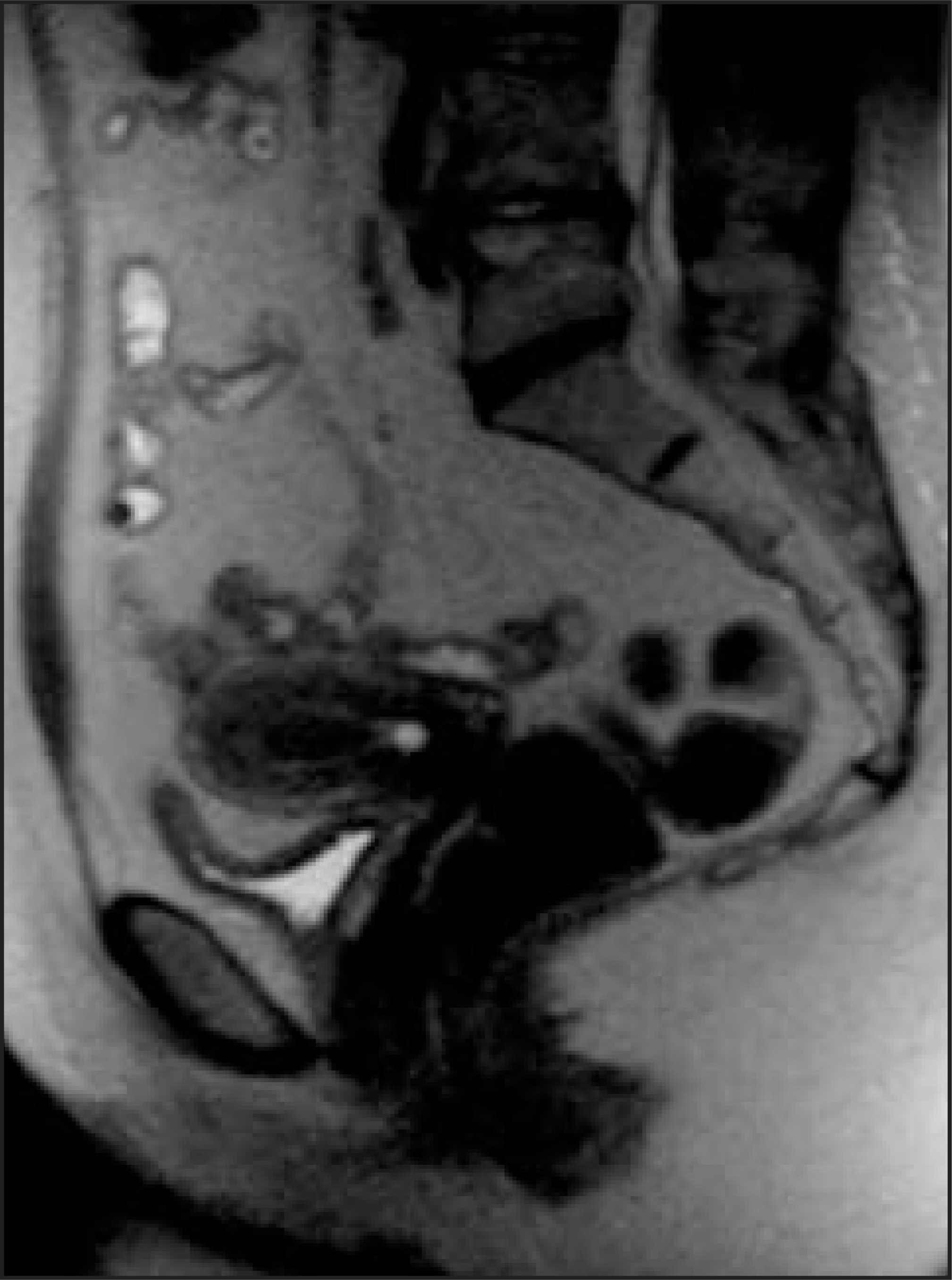
Other factors, which should be considered in patient selection, are fibroid location and size. Fibroids that protrude into the uterine cavity and have a thin stalk are better removed hysteroscopically (Figure 3). If embolized, these fibroids have a tendency to slough off and could result in infection, bleeding, and significant pain during passage.18 All fibroids with a submucosal component, submucosal fibroids, or intramural fibroids with a submucosal component may have a similar risk of leiomyoma tissue passage with associated symptoms of fever, pain, and bleeding (Figure 4). This complication of tissue passage can happen as long as 1 year postprocedure and is the most common post-UAE complication requiring rehospital-ization.14 In a recently published study of 400 consecutive patients who underwent UAE, Spies et al14 reported 17 rehospitalizations, 9 of which were secondary to leiomyoma passage. Treatment after rehospitalization consists of some combination of intravenous (IV) antibiotics, IV narcotics, transfusion, hysteroscopy, and possible dilation and curettage and/or hysterectomy. Patients must, therefore, be appropriately consulted before the procedure regarding these risks and must be educated about worrisome postprocedure symptoms, so that they will seek early medical treatment. Alternatively, hysteroscopic myomectomy may be a better option.
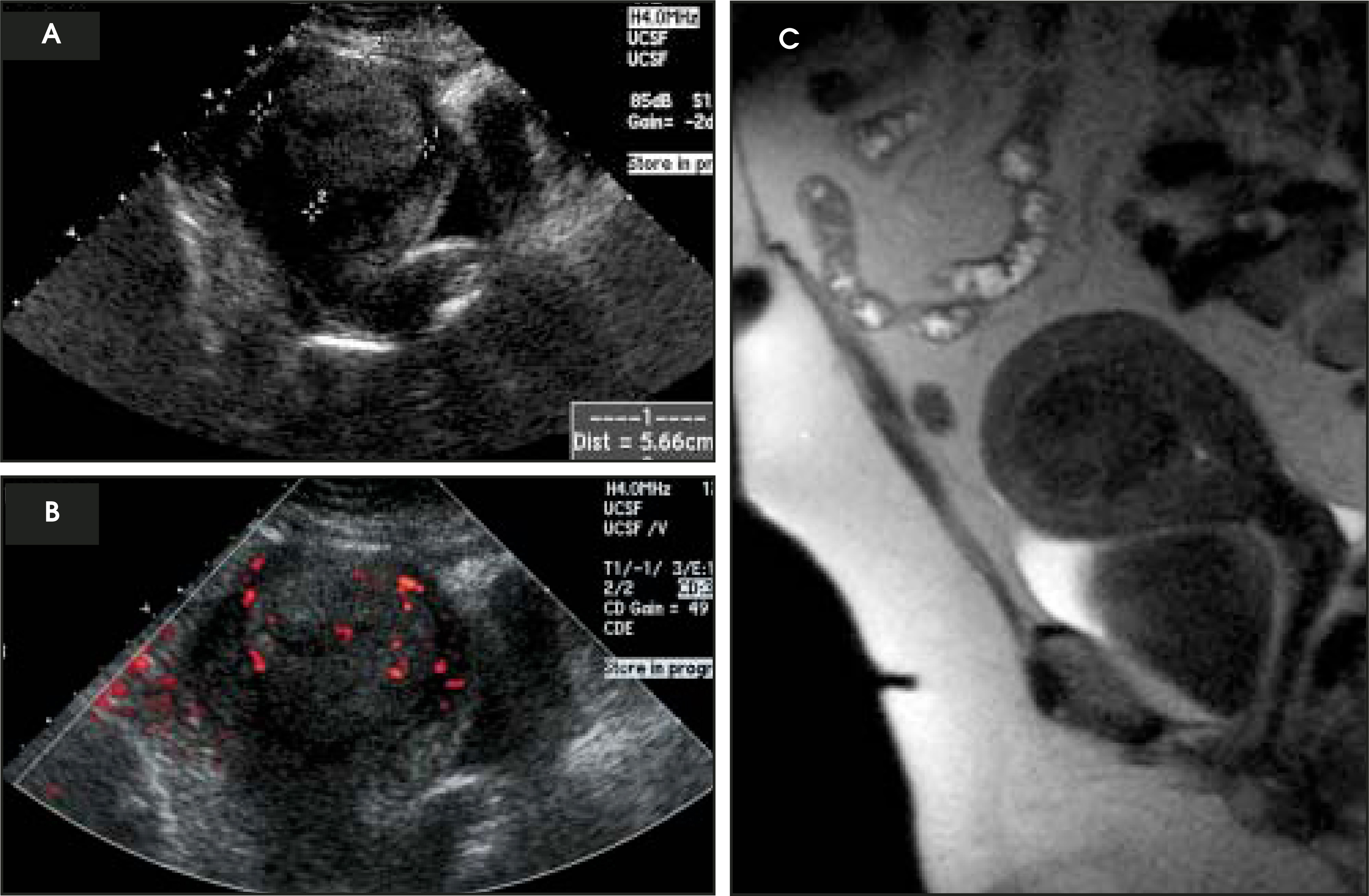
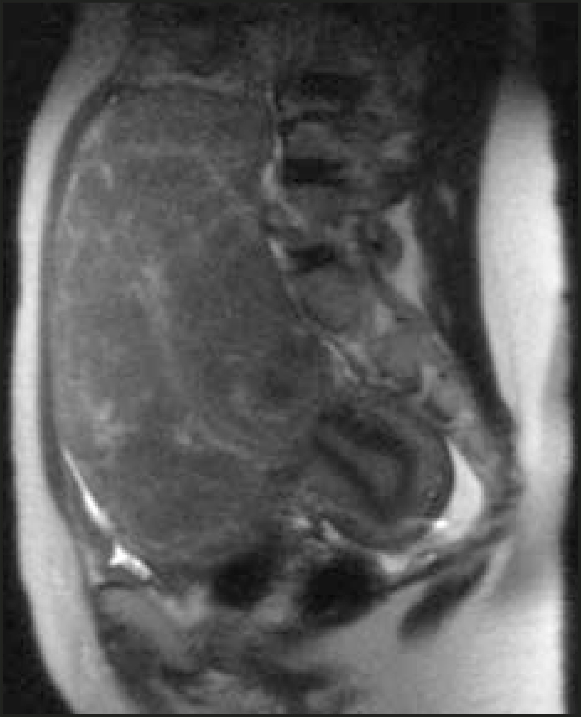
Finally, size of the dominant fibroid must be considered in correlation with the patient’s symptoms. Some studies have reported treatment failure to be more likely with fibroids >8 cm.19 Although these large fibroids usually undergo an appropriate degree of volume reduction, the residual fibroid may still be large enough in size to result in persistent bulky symptoms (Figure 5). This could, therefore, result in “perceived” treatment failure. In addition, serious complications, although rare, may also be more likely with large myomas.10,18 Patients with very large fibroids will require counseling before UAE to determine if the patient’s symptoms and expectations from the procedure are realistic. Alternatively, surgical treatment may be more appropriate.
UAE technique
The technique used to perform UAE varies among institutions and individual physicians. The method described here is the standard procedure used at University of California San Francisco, which is tailored when necessary on a patient-by-patient basis.
Preprocedural workup and discussions are emphasized as critical to a successful procedure. We employ a multidisciplinary approach to fibroid therapy and each of our patients sees both a gynecologist and an interventional radiologist before the procedure. A thorough history and physical examination are performed; a complete blood count, creatinine, and coagulation panel is obtained; and a pelvic MRI is acquired. All of this data is reviewed carefully to exclude other diagnoses and to determine if the patient is a good candidate for UAE.
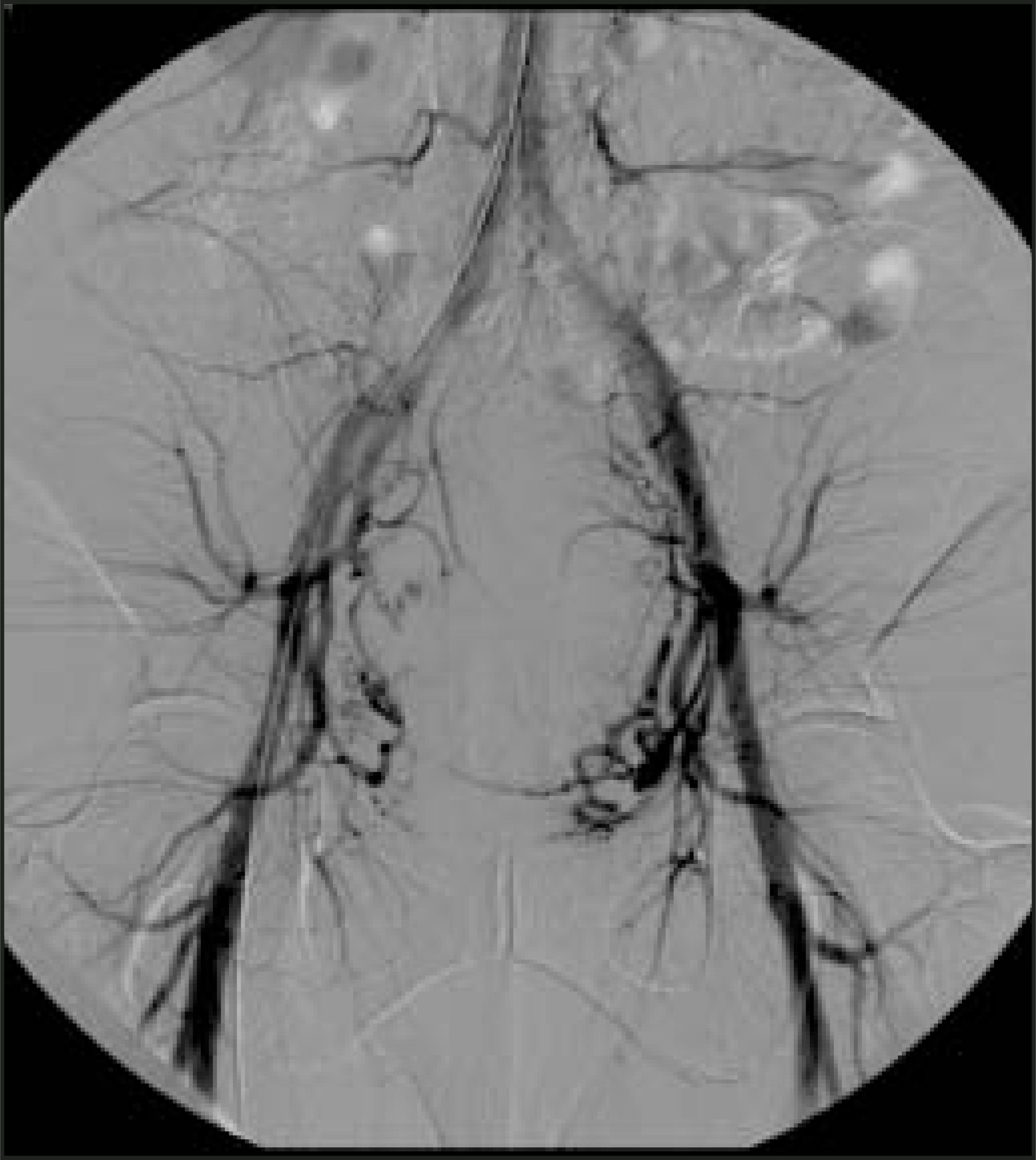
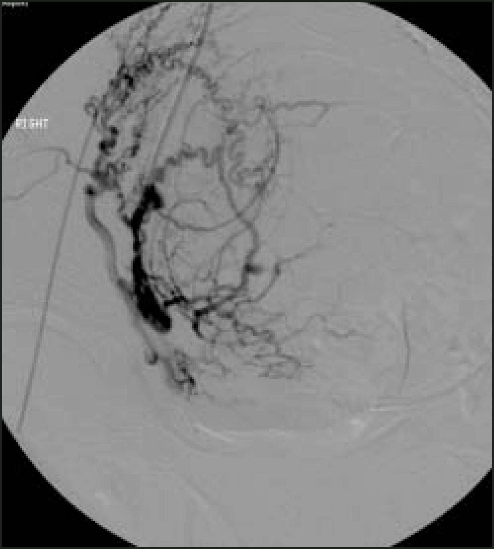
In our practice, a unifemoral approach is typically used. A 5F sheath (Cordis, Miami Lakes, FL) is placed into the right common femoral artery. A pigtail catheter (Cook, Bloomington, IN) is then advanced over a 15J wire and positioned at the level of the renal arteries. A preprocedural abdominal aortogram is then performed (Figure 6). At the present time, we are still performing a preprocedural aortogram, although this has changed our treatment approach in only a few cases. A study by Binkert et al20 supports this, reporting that the preprocedural aortogram altered treatment in only 6% of cases. It is possible that in the future we will eliminate this step, decreasing both radiation and contrast load. A 5F Cobra2 (C2) catheter (Cook, Bloomington, IN) is then used in combination with a Terumo wire (Terumo Medical Corp., Somerset, NJ) to select the anterior division of the left internal iliac artery. An arteriogram is performed to guide selection of the left uterine artery (UA). Once the 5F catheter is engaged in the origin, a microcatheter (Mass Transit, Cordis, or Renegade Hi Flow, Boston Scientific, Natick, MA) is then advanced coaxially through the 5F catheter and positioned at the horizontal portion of the UA, beyond the cervicovulvar branches. A microcatheter system is used to decrease the probability of spasm during catheter/wire manipulation. An arteriogram is done to confirm position (Figure 7) and an embolization is performed using 500µ to 700µ polyvinyl alcohol (PVA) particles (Cook, Bloomington, IN) suspended in a mixture of approximately 50% sterile saline and 50% Omnipaque (GE Healthcare, Princeton, NJ) contrast material. Embolization is considered complete when flow is near stasis (Figure 8).
The right UA is catheterized in a similar fashion using a Waltman loop formed from the C2catheter and the microcatheter. A uterine arteriogram is performed to verify optimal position, and embolization is performed to near stasis. A complete abdominal aortogram with a pigtail catheter (Cook) positioned at the level of the renal arteries is repeated to help detect persistent collateral flow to the fibroid(s).
Collateral supply
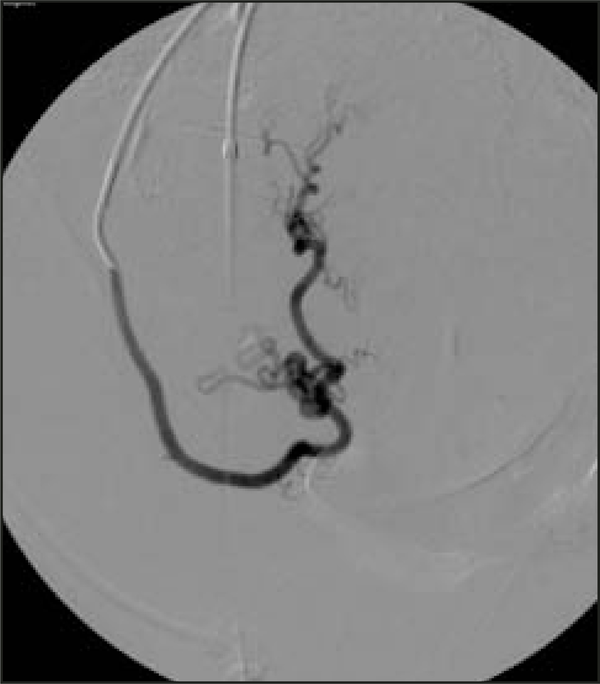
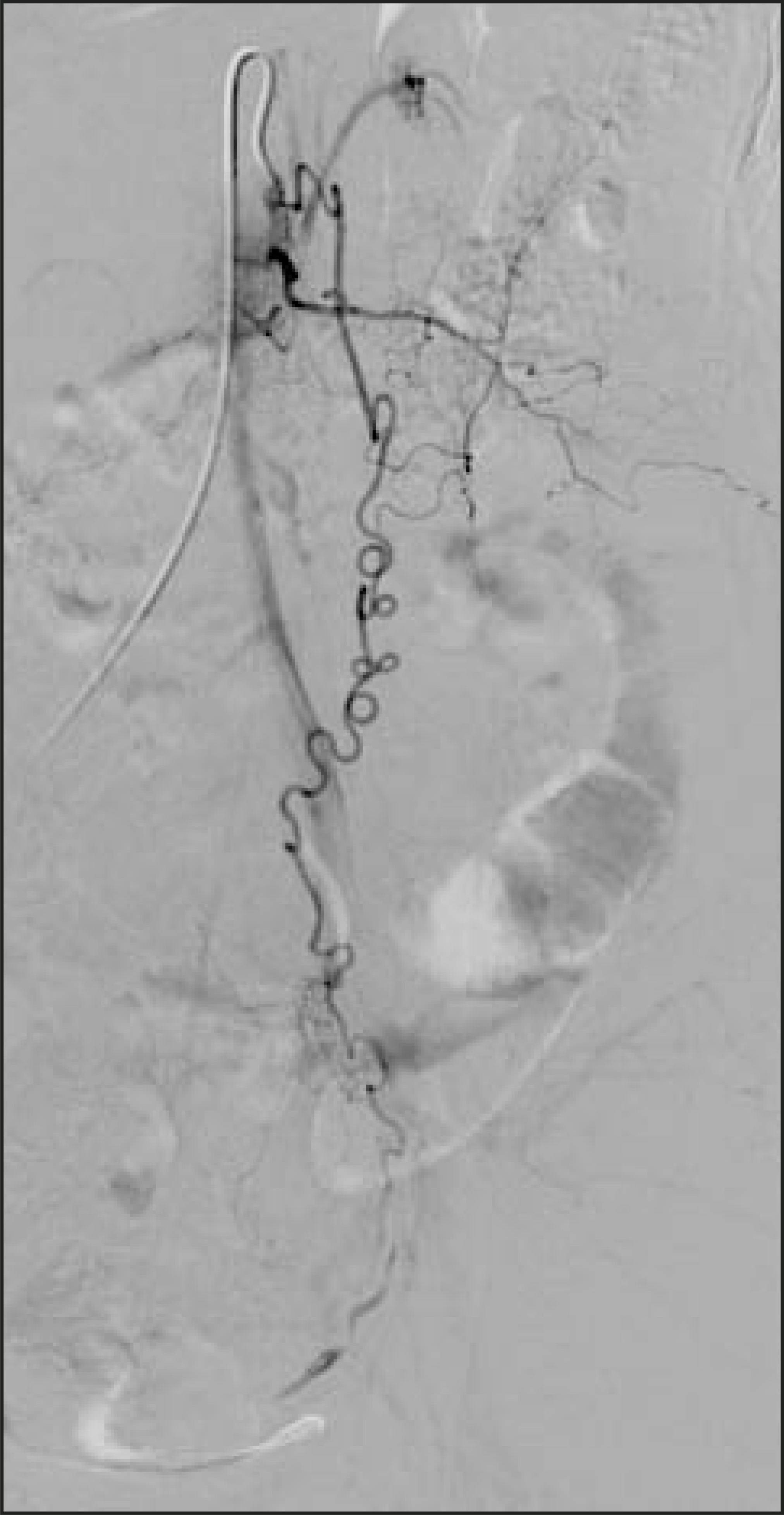
If collateral supply to the fibroid is identified, several options are available. Many interventionalists will take a “wait-and-see” approach. If the patient’s symptoms do not improve within 3 months, ovarian artery embolization could be considered. Several reports in the literature describe successful embolization of communications between the uterine artery and the ovarian artery.21,22 The ovarian arteries are usually very tortuous (Figure 9), and embolization distal to the ovary can be technically difficult. A full discussion of alternatives, potential risks, and benefits must occur before embarking on this treatment option.
End point of embolization
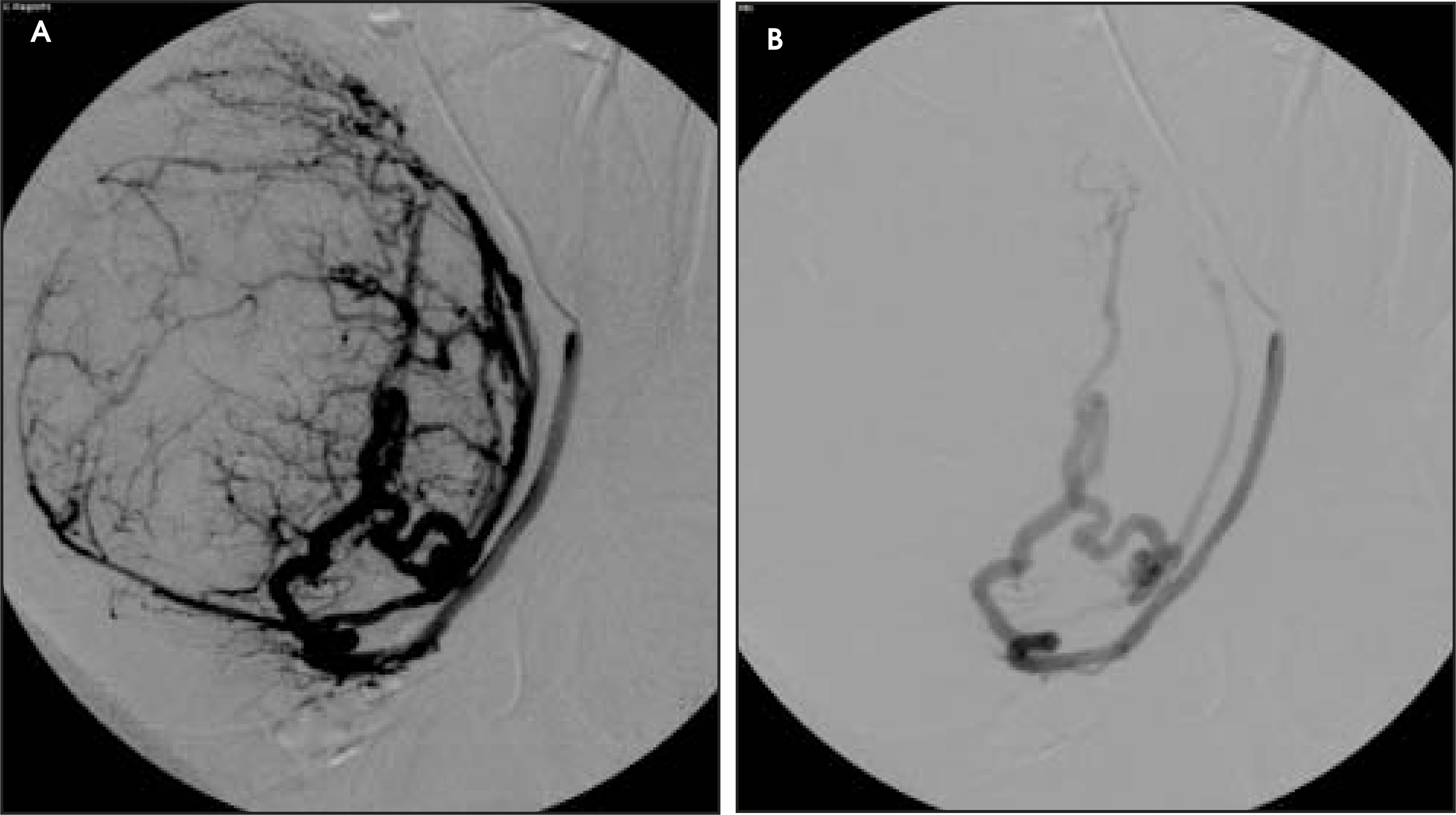
Several technical points regarding UAE are worthy of comment. The end point of embolization continues to evolve as interventionalists gain experience. Initially, embolization was performed to stasis. At completion, there was a static column of contrast in the uterine artery. Now, as techniques and embolic materials change, several other end points are considered appropriate indications of adequate inter-ruption of flow to the fibroids. These include: 1) the appearance of new collateral-reflux into the ovarian artery or cross-uterine branch opacification that were not seen on the initial uterine arteriogram; 2) increased resistance in the arteries of the uterine body manifested by reflux back along the angiographic catheter; 3) a “pruned-tree” appearance (Figure 10); and 4) the Shlansky- Goldberg method: sluggish forward flow in the main uterine artery with contrast still visible for 5 cardiac beats.23 Spies et al23 recommend waiting at least 5 minutes after em- bolization to re-evaluate flow and have found that further embolization is needed in 10% to 20% of cases.
Particle selection
A second area of discussion is the type of particles to be used: PVA versus calibrated microspheres. There is no consensus at the present time regarding the agent of choice for UAE. A majority of the published studies reporting the success of UAE have used PVA for embolization. Since calibrated microspheres have en-tered the market, a few published reports have supported their use in UAE.4-26Recently, the U.S. Food and Drug Administration (FDA) approved the use of microspheres for UAE.
Several properties inherent to an agent may affect the embolization process. Polyvinyl alcohol clumps after intravascular administration and may, therefore, form larger units that result in a more proximal occlusion. Dilution and slow injection may help to optimize delivery more distally.
Spherical agents are reported to clump less, which, in theory, could allow more accurate size definition and enhance delivery to the perifibroid plexus. Pelage et al27 compared the behavior of PVA with calibrated microspheres in sheep and found that the calibrated microspheres were associated with less uterine necrosis and allowed for a segmental arterial occlusion that correlated with the size of the particles. Further studies are needed to define the optimal embolant and size. Currently, many interventionalists continue to use PVA particles in either the 355 to 500µ or 500 to 700µ ranges because of familiarity with this agent.
Results
The first case of UAE for the treatment of symptomatic fibroids was performed in France in 1989. Since that time, more than 10,000 cases have been performed.23 Several large series published within the last year report that 91% to 93% of patients are satisfied with the procedure and would recommend it to others.11,28 Current technical success rates are 98% to 100% in both large and small series.5-11 Overall, clinical success rates range from 80% to 95%.5-12,28 In the largest prospective multicenter trial published to date, the Ontario Uterine Fibroid Embolization Trial,12 reported a study of 555 women who were followed for 3 months following UAE. Significant improvements were reported for menorrhagia (83%), dysmenorrhea (77%), and urinary frequency (86%). In this study, uterine volumes decreased by 35% at 3-month follow-up, with dominant fibroid reduction of 42%.12
Spies et al28 prospectively evaluated symptom improvement and volume changes in 200 patients over a longer postprocedure follow-up, mean follow-up of 21 months. They similarly found symptomatic improvement in heavy bleeding (90%) and bulky symptoms (91%) at 1 year after UAE.28 Patient symptoms tended to improve over a 3-month period, after which they remained stable despite continued fibroid volume reduction on imaging studies.28 Mean fibroid volume reduction was 58% after 12 months, with corresponding uterine volume decreases of 38%.28 In experienced hands, UAE seems to be effective at alleviating symptoms and improving quality of life in the majority of patients.
Complications
In reviewing complications, it is difficult to compare results of different studies because of variations in reporting and follow-up periods. Using established standardized definitions of postoperative morbidity from the obstetrics and gynecology literature may be helpful in evaluating the safety of this procedure.
Georgetown University recently published a prospective study of 400 consecutive UAE patients, specifically examining the frequency and severity of short-term complications after UAE. The overall perioperative complication rate in this study was 8.5%14; many of the complications were minor.
One fifth of all adverse events were caused by allergic reactions or rashes, which were treated pharmacologically.14 Using the American College of Obstetrics and Gynecology (ACOG) definitions of perioperative complications, the overall morbidity after UAE was 5%.14 Serious complications were uncommon, occurring in only 5 (1.25%) of 400 pa-tients.4 Others have reported serious complications in approximately 1% to 4% of patients.,10 These complications include uterine infection (often precipitated by leiomyoma passage), pulmonary embolism, arterial thrombosis, prolonged pain, and, very rarely, death.
In the study by Spies et al,143.5 % of patients were readmitted after discharge. Passing of a leiomyoma was the most common complication that resulted in rehospitalization (Figure 11). Emergent hysterectomy is required to treat complications in approximately 1% of patients.10,11 Spies and coworkers14 reported that only 1 patient in 400 required hysterectomy, although several other patients required lesser procedures of dilation and curettage or hysteroscopy.
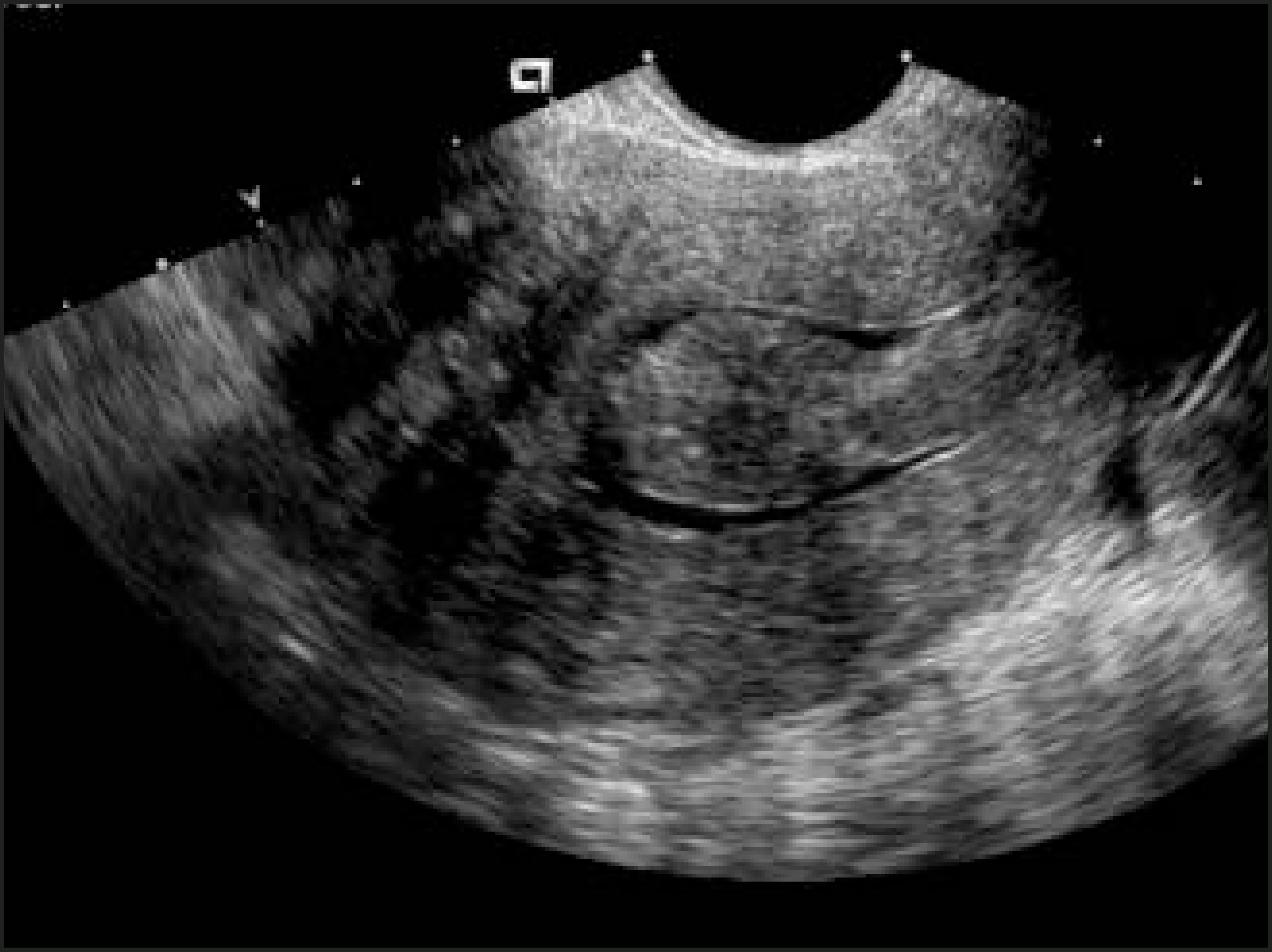
At the present time, no data are available regarding long-term complications. Although it is difficult to compare complication rates of hysterectomy, myomectomy, and UAE, recent publications suggest that UAE is at least as safe as the other procedures.14,29 Prospective Phase-II comparative FDA trials are under way that will more accurately compare the relative safety of UAE versus myomectomy and UAE versus hysterectomy. Results from these trials are eagerly awaited.
Conclusion
The treatment of women with symptomatic uterine fibroids continues to evolve. Uterine artery embolization is an effective, less invasive option for some women. To understand which patients will receive the greatest benefit from UAE, it is necessary to be familiar with and have knowledge of other fibroid treatment options and their strengths. In the October issue, Part 2 of this article will address other fibroid management strategies that will help select the best treatments for your patients.
References
- Wallach E, Thompson J, Rock J. Te Linde’s Operative Gynecology. 1992:47-62.
- Marshall L, Spiegelman D, Goldman M. A prospective study of reproductive factors and oral contraceptive use in relation to the risk of uterine leiomyomata. Fertil Steril. 1998;70:432-439.
- Wilcox L, Koonin L, Pokras R. Hysterectomy in the United States, 1988-1990. Obstet Gynecol. 1994;83:549-555.
- Ravina J, Herbreteau D, Ciraru-Vigneron N. Arterial embolisation to treat uterine myomata. Lancet. 1995;346:671-672.
- Bradley E, Reidy J, Forman R. Transcatheter uterine artery embolisation to treat large uterine fibroids. Br J Obstet Gynaecol. 1998;105:235-240.
- Worthington-Kirsch R, Popky G, Hutchins F. Uterine arterial embolization for the management of leiomyomas: Quality-of-life assessment and clinical response. Radiology. 1998;208:625-629.
- Goodwin S, McLucas B, Lee M. Uterine artery embolization for the treatment of uterine leiomyomata midterm results. J Vasc Interv Radiol. 1999;10:1159-1165.
- Spies J, Scialli A, Jha R. Initial results from uterine fibroid embolization for symptomatic leiomyomata. J Vasc Interv Radiol. 1999;10:1149-1157.
- Hutchins F, Worthington-Kirsch R, Berkowitz R. Selective uterine artery embolization as primary treatment for symptomatic leiomyomata uteri. J Am Assoc Gynecol Laparosc. 1999;6:279-284.
- Pelage J, Le Dref O, Soyer P. Fibroid-related menorrhagia: Treatment with superselective embolization of the uterine arteries and midterm follow-up. Radiology. 2000;215:428-431.
- Walker W, Pelage J. Uterine artery embolisation for symptomatic fibroids: Clinical results in 400 women with imaging follow up. BJOG. 2002;109:1262-1272.
- Pron G, Bennett J, Common A. The Ontario Uterine Fibroid Embolization Trial. Part 2. Uterine fibroid reduction and symptom relief after uterine artery embolization for fibroids. Fertil Steril. 2003;79:120-127.
- . 2003:193-199.
- Spies J, Spector A, Roth A. Complications after uterine artery embolization for leiomyomas. Obstet Gynecol. 2002;100(5 Pt 1):873-880.
- Chrisman H, Saker M, Ryu R. The impact of uterine fibroid embolization on resumption of menses and ovarian function. J Vasc Interv Radiol. 2000;11:699-703.
- Spies J, Roth A, Gonsalves S, Murphy-Skrzyniarz K. Ovarian function after uterine artery embolization for leiomyomata: Assessment with use of serum follicle stimulating hormone assay. J Vasc Interv Radiol. 2001;12:437-442.
- Management of uterine fibroids. Evidence Report. Technol Assess. 2001:1-173.
- Goodwin S, Walker W. Uterine artery embolization for the treatment of uterine fibroids. Curr Opin Obstet Gynecol. 1998;10:315-320.
- Al-Fozan H, Tulandi T. Factors affecting early surgical intervention after uterine artery embolization. Obstet Gynecol Surv. 2002;57:810-815.
- Binkert C, Andrews R, Kaufman J. Utility of nonselective abdominal aortography in demonstrating ovarian artery collaterals in patients undergoing uterine artery embolization for fibroids. J Vasc Interv Radiol. 2001;12:841-845.
- Razavi M, Wolanske K, Hwang G. Angiographic classification of ovarian artery-to-uterine artery anastomoses: Initial observations in uterine fibroid embolization. Radiology. 2002;224:707-712.
- Andrews R, Bromley P, Pfister M. Successful embolization of collaterals from the ovarian artery during uterine artery embolization for fibroids: A case report. J Vasc Interv Radiol. 2000;11:607-610.
- . 2003:62-73.
- Pelage J, Le Dref O, Beregi J. Limited uterine artery embolization with tris-acryl gelatin microspheres for uterine fibroids. J Vasc Interv Radiol. 2003;14:15-20.
- Banovac F, Ascher S, Jones D. Magnetic resonance imaging outcome after uterine artery embolization for leiomyomata with use of tris-acryl gelatin microspheres. J Vasc Intervent Radiol. 2002;13:681-688.
- Spies J, Benenati J, Worthington-Kirsch R, Pelage J. Initial experience with use of tris-acryl gelatin microspheres for uterine artery embolization for leiomyomata. J Vasc Interv Radiol. 2001;12:1059-1063.
- Pelage J, Laurent A, Wassef M. Uterine artery embolization in sheep: Comparison of acute effects with polyvinyl alcohol particles and calibrated microspheres. Radiology. 2002;224:436-445.
- Spies J, Ascher S, Roth A. Uterine artery embolization for leiomyomata. Obstet Gynecol. 2001;98:29-34.
- Razavi M, Hwang G, Jahed A. Abdominal myomectomy versus uterine fibroid embolization in the treatment of symptomatic uterine leiomyomas. AJR Am J Roentgenol. 2003;180:1571-1575.
Citation
. Uterine artery embolization: Where does it stand in the management of uterine leiomyomas? Part 1. Applied Radiology. 2004;33(9):22-29. doi:10.37549/AR1282.