Uterine artery embolization: Where does it stand in the management of uterine leiomyomas? Part 2
Applied Radiology — Vol. 33 , Issue 10 , pp. 11 -11
DOI: 10.37549/AR1289
Published: October 1, 2004
Categories
Authors’ note: The first article in this two-part series on the management of uterine fibroids was published in the September issue and was dedicated to uterine artery embolization (UAE): when it is applicable, how to perform it, and what to expect from the procedure. This second part will discuss more traditional treatments and outcomes for fibroids, which will help the reader understand how UAE fares against these alternatives. Finally, we will address some of the newest cutting-edge therapies to watch for.
Uterine artery embolization (UAE) is an effective alternative to the more conventional approaches to the treatment of uterine fibroids, including medical therapies, myomectomy, and hysterectomy. In this article, we will review some of these more traditional gynecologic options to help the reader better understand which therapy meets a pa-tient’s needs and future reproductive goals. We will also offer a glimpse into the future of fibroid therapy.
Other therapeutic options
Before the advent of UAE, fibroids were primarily treated with close monitoring, oral contraceptives, gonadotropin-releasing hormone agonists (GnRH-a), myomectomy, and hysterectomy. Each of these treatments has a unique set of limitations, risks, and complications.
Monitoring
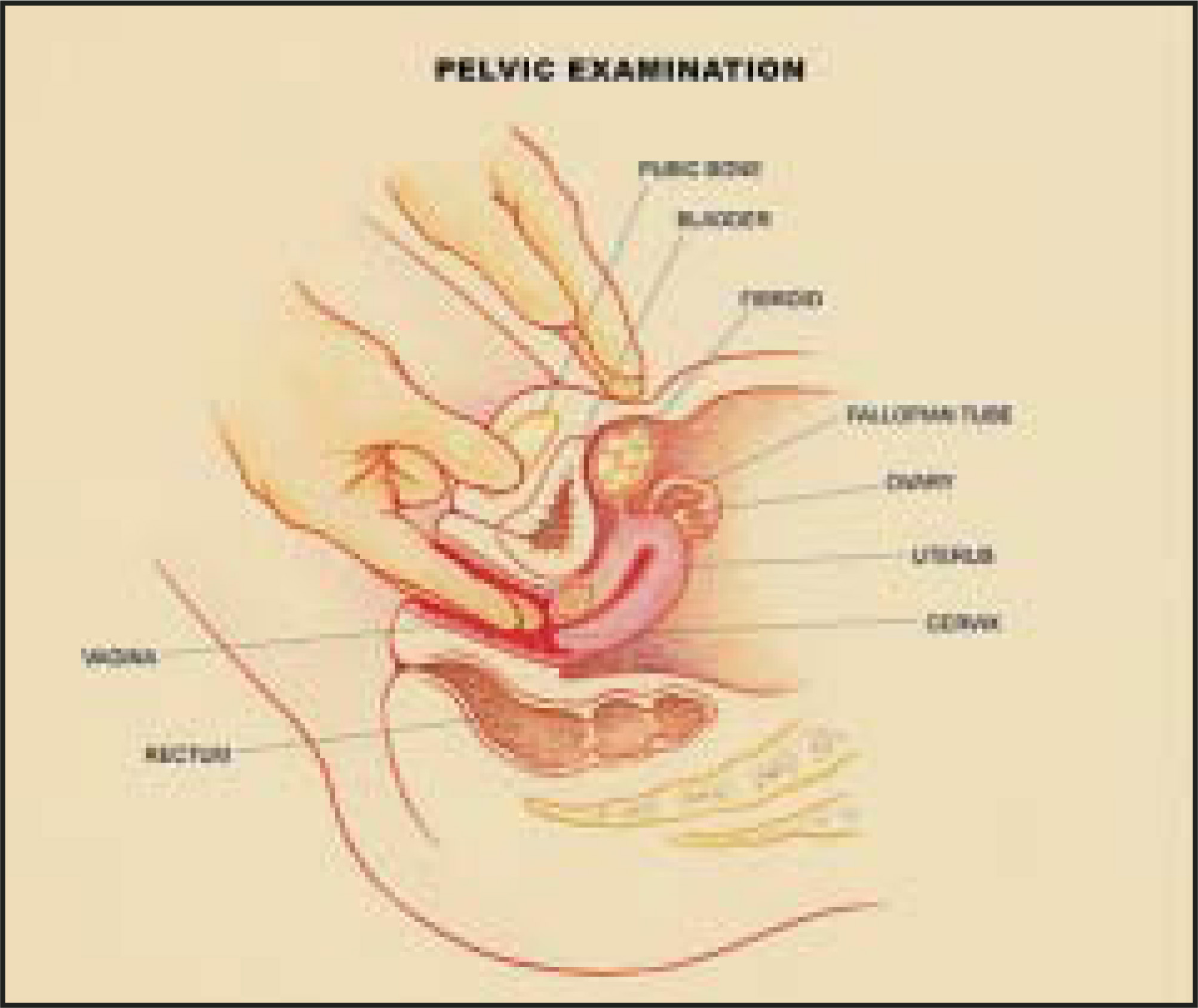
Many patients are asymptomatic and do not require any therapy. These patients are monitored with annual bimanual examinations (Figure 1) to evaluate changes in the size of the uterus. Oral birth control pills can be useful in some patients to help with symptoms of menorraghia. Oral contraceptives and progestational agents can help regulate the menstrual cycle and reduce bleeding, but they will not shrink the fibroids.
GnRH-a
Gonadotropin-releasing hormone agonists are medical treatments that work by inhibiting the release of gonadotropins by down-regulating pituitary GnRH receptors, thereby ultimately decreasing estrogen production. Deprived of estrogen, fibroids shrink up to one third to one half of their original size after 2 to 3 months of therapy. This treatment, however, is only temporary; once the medication is stopped, the fibroids usually re-grow. Gynecologists typically recommend taking GnRH-a for <6 months because of the risk of osteoporosis.1,2
Hysterectomy
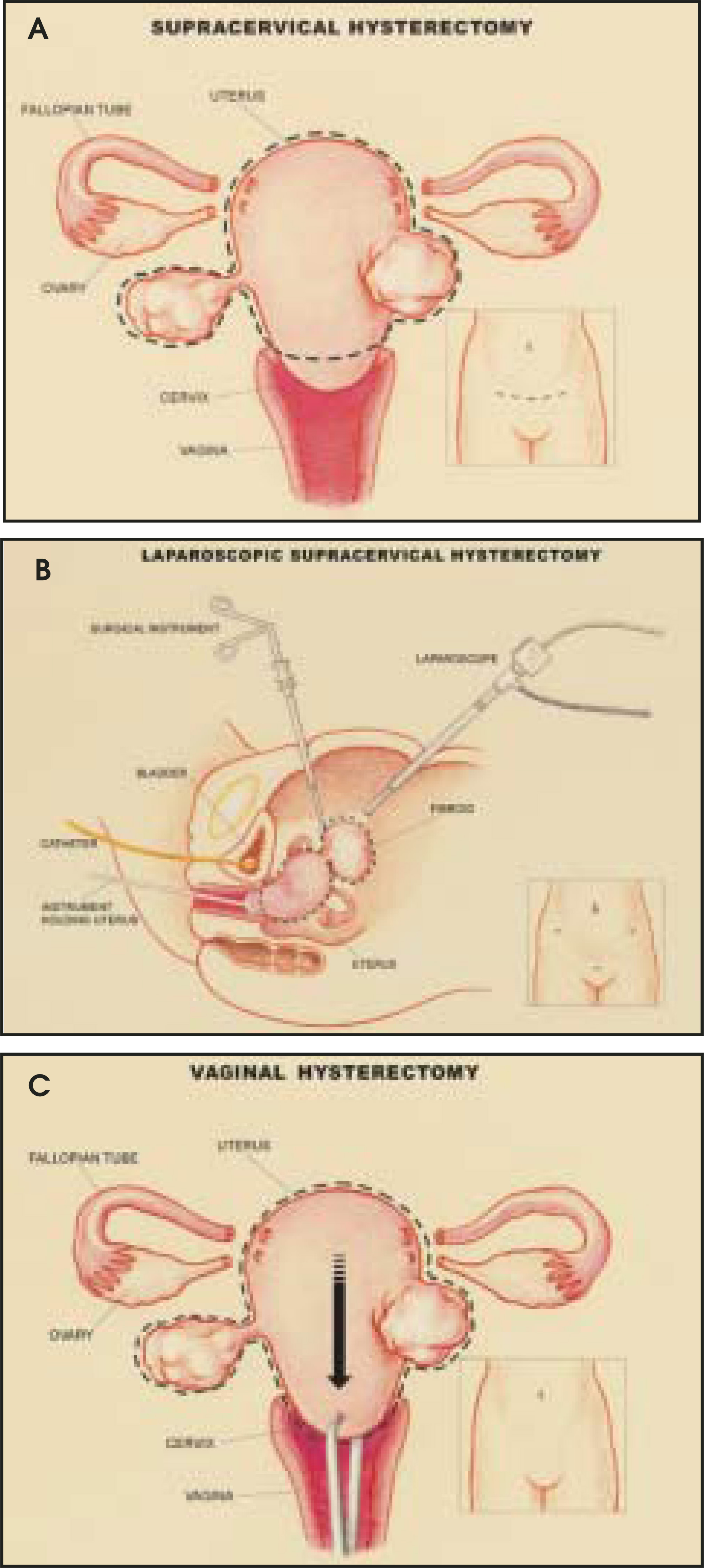
Hysterectomy is the surgical resection of the uterus. More than 600,000 hysterectomies are performed each year in the United States with symptomatic leiomyomas being the most common indication, accounting for approximately 30% of hysterectomies performed in Caucasians and 60% in African-Americans.3,4 Hysterectomy is presently the only permanent cure for fibroids, eliminating both symptoms and the chance of recurrence. The procedure can be undertaken abdominally, vaginally, or laparoscopically (Figure 2). Each hysterectomy method has specific risks, and the appropriate surgical route for each patient must be decided based on the patient’s history, the size of the uterus, and the surgeon’s technical expertise. The only formal guideline established by the American College of Obstetricians and Gynecologists suggests that vaginal hysterectomy is most appropriate for women with benign disease and mobile uteri that are smaller then 12 weeks’ gestational size or approximately 280 grams.5 Some patients with enlarged fibroid uteri will therefore not be able to have a vaginal resection and will be required to have an abdominal hysterectomy or possibly a laparoscopically assisted procedure.
Hysterectomy is a major surgical procedure that requires general anesthesia. The surgery takes approximately 1.5 to 2 hours.6 Patients typically stay in the hospital for approximately 3 to 5 days after an abdominal hysterectomy and for 1 to 3 days after a vaginal or laparoscopic hysterectomy.7-9 The postoperative recovery period is 6 to 8 weeks for an abdominal hysterectomy and somewhat less for the other approaches. The convalescence period is the shortest for laparoscopic hysterectomies, averaging 21.5 ± 8.8 days.6
Based on the Agency for Healthcare Research and Quality (AHRQ), which reviewed hysterectomy outcome studies between the years 1975 and 2000,10 there are 3 prospective studies performed in the United States that have reported both short- and long-term outcomes, using standardized measures. Each of these studies showed significant improvement in symptoms for a majority of women at 1 or 2 years after hysterectomy.11-13 In one of these studies, 98% of women responded that they “would make the decision to have the hysterectomy again, if the conditions were the same.”13 Satisfaction post-hysterectomy is high, but it is difficult to determine accurate short- and long-term complication risks, based on the literature, because of the lack of controlled trials and variable definitions of morbidity.
A landmark paper by Dicker et al14 assessed the comparative risks of complications in women undergoing hysterectomy via abdominal and vaginal approaches. This study used data from the Collaborative Review of Sterilization (CREST), a prospective multicenter observational study organized by the Center of Disease Control. The CREST study established standard definitions for morbidity after hysterectomy and set rates of expected short-term complications for abdominal and vaginal hysterectomies. Despite some changes since 1982 in surgical technique and practice, these short-term complication rates have remained quite durable over time.15 The results of the CREST study (Table 1) documented that women who had vaginal hysterectomies had fewer complications than those who had abdominal hysterectomies.14
Few studies have prospectively compared complication rates between abdominal, vaginal, and laparoscopic hysterectomies. A large prospective nationwide study done in Finland in 1996 evaluated operative-related morbidity and postoperative complications in all hysterectomies performed for benign diseases.6 A total of 10,110 hysterectomies were performed with overall complication rates of 17.2% for abdominal, 23.3% for vaginal, and 19% for laparoscopic hysterectomies6 (Table 2). The overall complication rate for abdominal hysterectomy is significantly less in this study (17.2%) as compared with the CREST trial (42.8%). This is thought to be largely due to fewer infections, secondary to increased use of prophylactic antibiotics. In addition, the apparent difference in bleeding complications between this study and the CREST study may be because of varying definitions. Ureteral and bladder injuries were significantly more common in the laparoscopic hysterectomy group when compared with both the abdominal and vaginal hysterectomy groups. Bowel complications, however, occured more frequently in vaginal hysterectomy patients. It was also noted in this study that the surgeon’s experience is a significant factor in complication rates.6
Regardless of the method of hysterectomy, all hysterectomies have the common result of permanent loss of fertility and are, therefore, considered primarily for women who do not desire future fertility.
Myomectomy
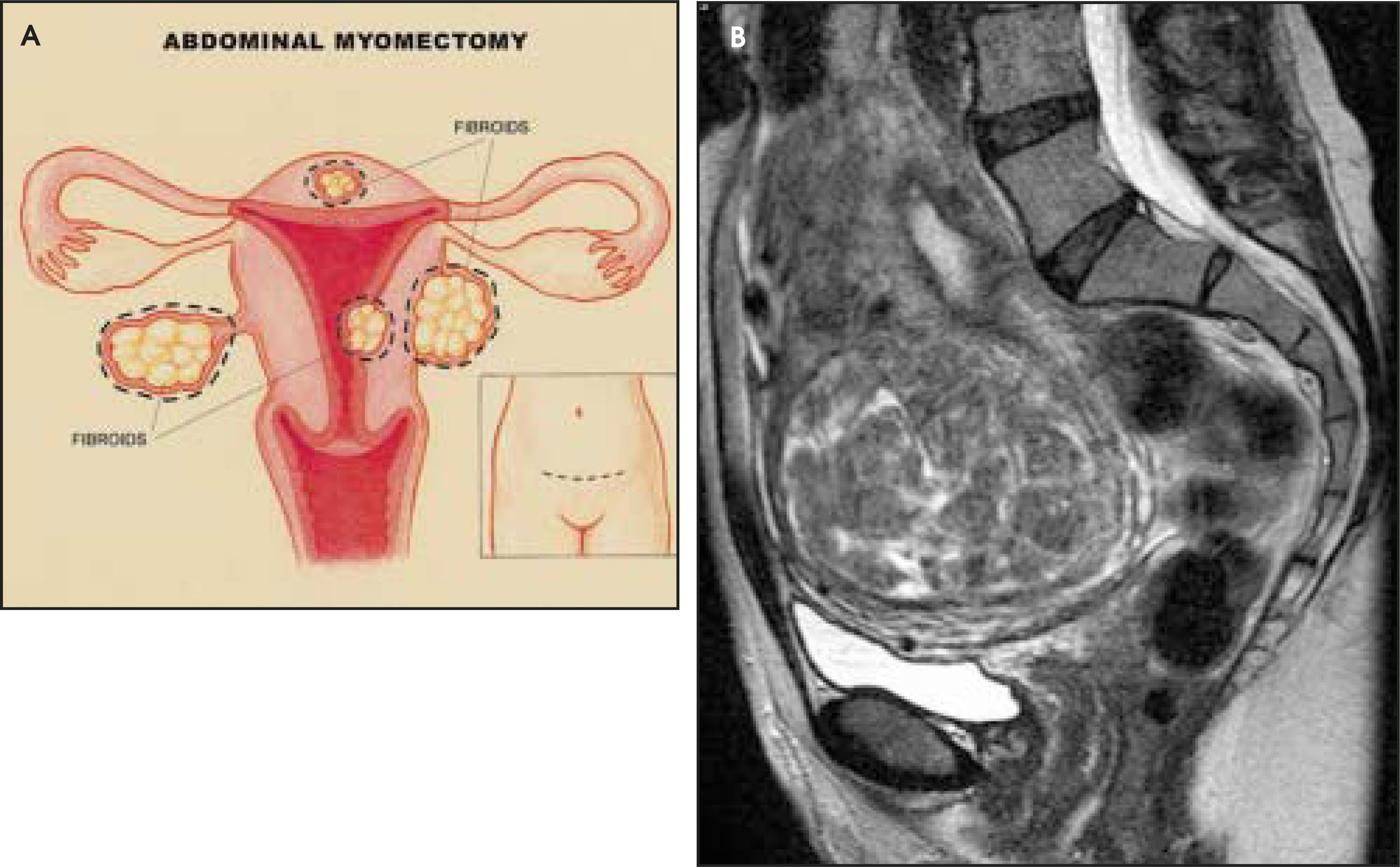
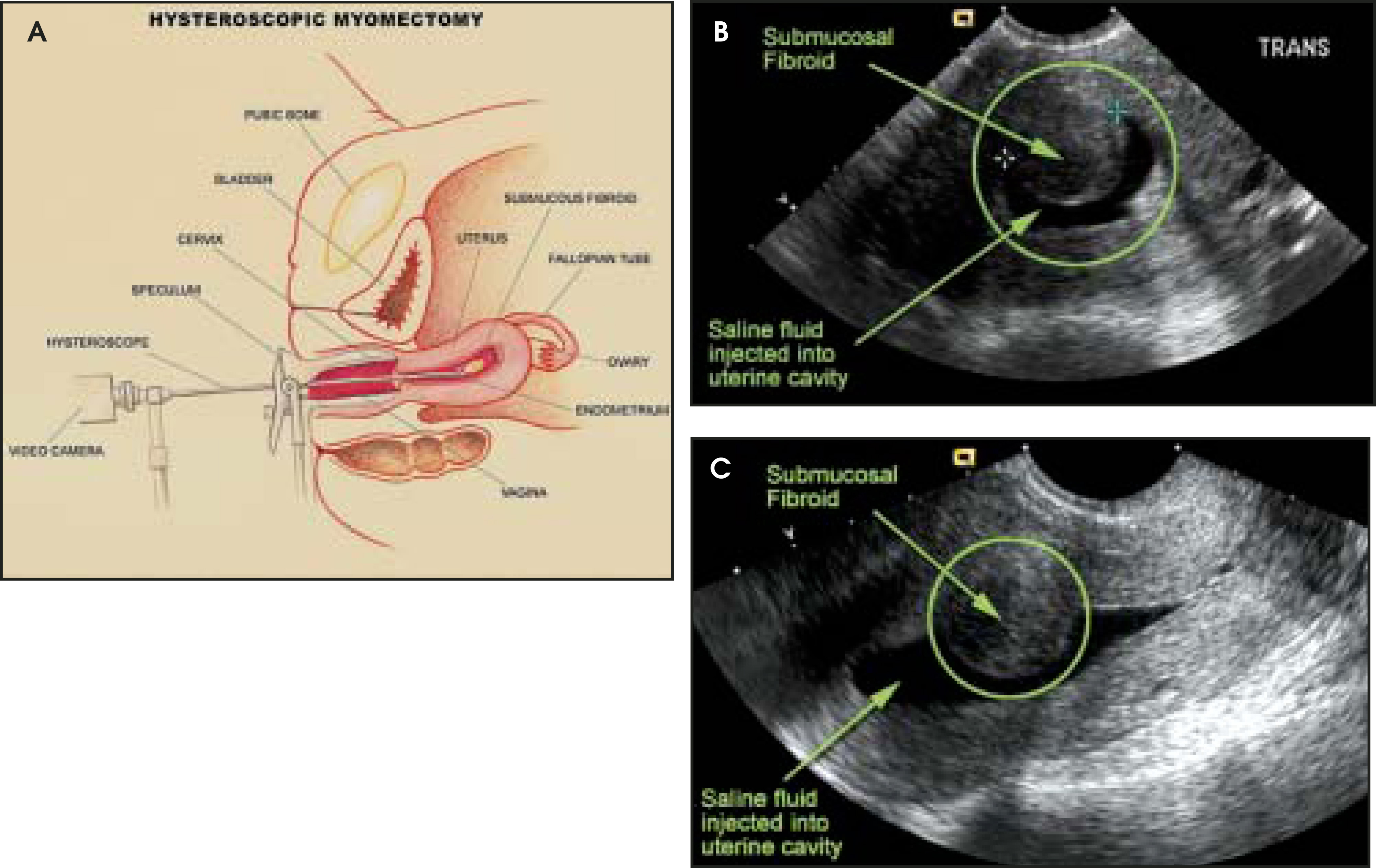
Myomectomy is a procedure in which uterine fibroids are surgically removed. It is the surgical alternative for women who wish to preserve their fertility. One tenth as many myomectomies as hysterectomies are performed annually.16 Myomectomies can be performed transabdominally, hysteroscopically, or laparoscopically. The choice of method is based on the size of the uterus and the number, size, and location of the fibroids. If the uterus is significantly enlarged (<17 weeks gestational size), if there are multiple fibroids, if the fibroids are <8 cm, or if they are deep in the myometrium, an abdominal approach is often chosen as the optimal route of resection (Figure 3). Submucosal fibroids are usually addressed from a hysteroscopic approach (Figure 4). Smaller fibroids and serosa-based pedunculated fibroids are amenable to laparoscopic removal17-20 (Figure 5).
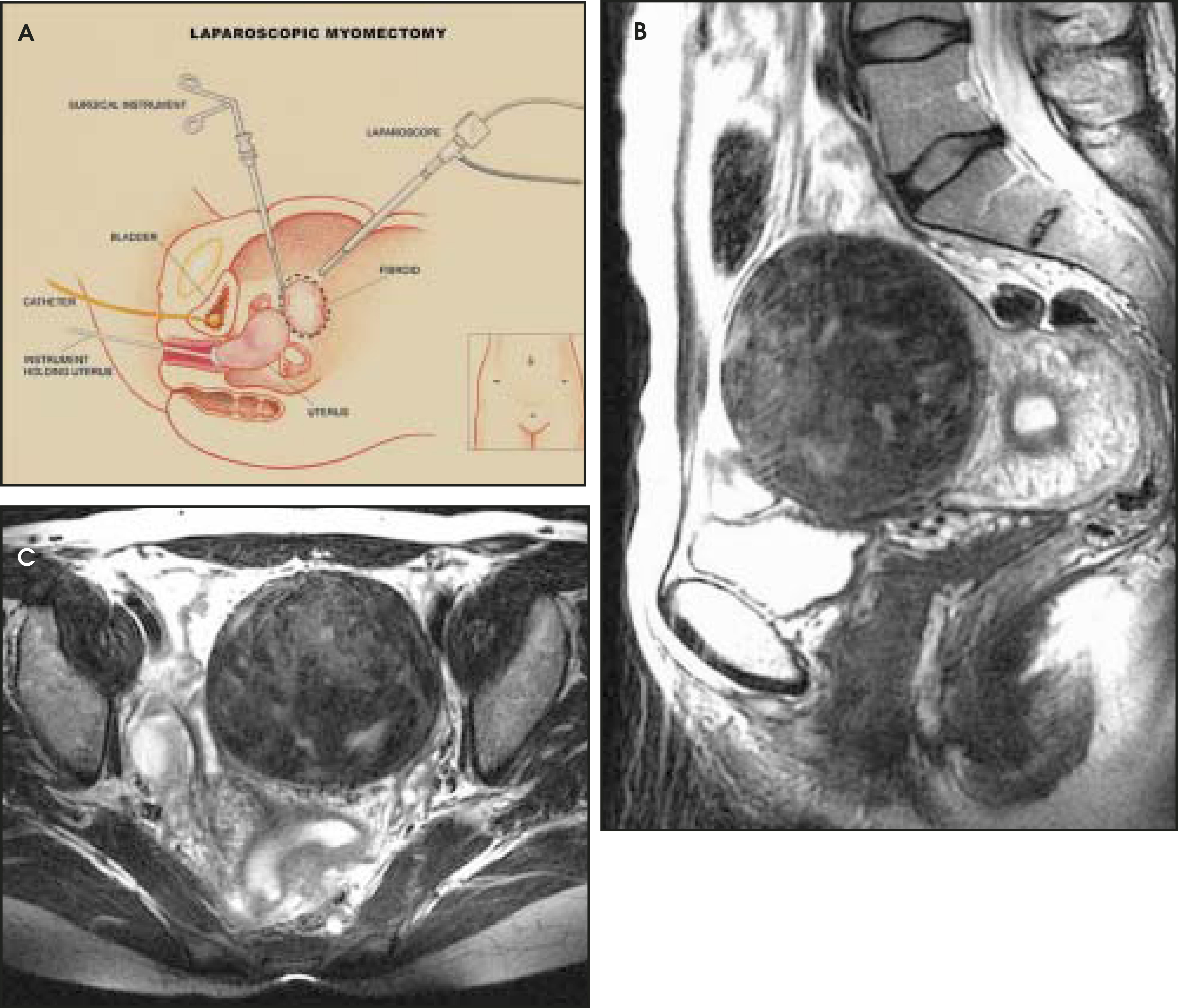
Myomectomies are often performed under general anesthesia, although some patients receive regional anesthesia with an epidural. The surgery generally takes between 1 and 3 hours, depending on the approach, size, and number of fibroids removed.21,22 Postoperative hospital stays vary but are typically 2 to 4 days for abdominal myomectomies22,23 and approximately 2 to 3 days for hysteroscopic or laparoscopic myomectomies.24 The convalescence period is 4 to 8 weeks for abdominal myomectomies and approximately 15 to 20 days for hysteroscopic or laparoscopic myomectomies.21,24
The majority of studies examining the outcomes of myomectomies are case series with variable definitions, follow-up, and reporting. The AHRQ Evidence Report on the management of fibroids reviewed all publications on myomectomy outcomes in women with symptomatic fibroids between 1975 and 2000.10 They found that a majority of patients had resolution or significant improvement in symptoms postmyomectomy with a range from 67% to 100% across all resection methods.10
Blood loss is the greatest risk of myomectomy. Transfusion is required in 1.2% to 18% of cases.10 Preoperative use of GnRH-a25 or intraoperative use of vasopressin26 results in statistically decreased estimated blood loss. Difficulty in achieving hemostasis during myomectomy can result in the formation of adhesions that may impair future fertility or lead to postoperative ileus or bowel obstruction. Certain surgical techniques (including avoiding a posterior uterine incision, proper irrigation, maintaining tissue moisture, minimizing tissue abrasion, using small-diameter sutures on serosa surfaces, and eliminating postoperative bleeding) will help decrease the postoperative risk of adhesions.17 Occasionally, bleeding can be so severe as to require conversion to a hysterectomy (≤1%).27,28
For years, myomectomy has been perceived to be associated with a longer surgical time and greater operative and postoperative complications than hysterectomy.29 A recent large retrospective outcomes study from the University of Pennsylvania compared perioperative morbidity from abdominal myomectomy with that of abdominal hysterectomy (Table 3).22 They found that after controlling for patient variables, there was no significant difference in perioperative morbidity and concluded that myomectomy should be considered a safe alternative to hysterectomy.22 Only uterine size and the presence of adhesions at surgery were associated with a significant increase in perioperative morbidity.22
There are several points unique to lap-aroscopic myomectomy that should be addressed. Laparoscopic myomectomies often take longer than abdominal myomectomies, and there is a 2% to 11% conversion rate to an open procedure.18-20 Despite multiple techniques available for laparoscopic suturing, repair of the uterus by laparoscopic means is controversial, and closures may not compare to open repairs. Both the formation of uteroperitoneal fistulas and a risk of uterine rupture during future pregnancies have been reported.18-20 Another problem is the fibroid recurrence rate following laparoscopic myomectomy. Approximately one third of patients have a recurrence after laparoscopic myomectomy. This may be higher than after other forms of myomectomy.30 This increased rate of recurrence could be related to an inability to manually palpate the uterus and detect small or deep fibroids or may be a spurious finding related to improved detection of fibroids over recent years.
The major drawback of myomectomy is the risk of developing new symptomatic fibroids. Women who have children after a myomectomy have a decreased risk of recurrence.31 As our ability to detect smaller fibroids improves with the use of transvaginal ultrasound (TVUS) and magnetic resonance imaging (MRI), the reported recurrence rates have also increased. The highest estimates of recurrence detected by TVUS were 51% at 5 years.31 Fortunately, however, not all detected fibroids cause symptoms. Approximately 10% to 26% of patients require a second major surgery after the first myomectomy.32-34
Minimally invasive ablative therapy
Patients who wish to avoid major surgery have driven the medical field to develop less invasive means of treating fibroids. Endometrial ablation, with or without hysteroscopic myomectomy, is an alternative for women who have symptomatic fibroids and do not want future fertility. There are multiple variants of this technique with a common end point of destroying the uterine lining.
Myolysis is the destruction of uterine tissue by thermo/cryoablation. This procedure is currently recommended only to women who do not want future fertility because of the risk of adhesion formation or rupture during pregnancy.35 Myolysis began in the late 1980s and was first performed by laparoscopic means using the neodymium-yttrium-aluminum-garnet laser. A significant problem with this technique was the formation of bowel adhesions.36 Since this time, multiple other ablative devices used laparoscopically or hysteroscopically have been tried, including bipolar needles, cryoprobes, and myoma interstitial thermotherapy (MITT) using diode lasers. The search continues for the optimal ablation device.
What’s new in fibroid treatment?
Multidisciplinary teams continue to develop innovative ways to treat fibroids. Medical therapies to date have primarily focused on estrogen’s role in fibroid formation and growth. Progesterone, however, is clearly important in fibroid tumor cell proliferation and growth,37 and fibroid tissue has been found to express both estrogen receptors and progesterone receptors.38 A small study of the effects of RU486 (an antiprogesterone compound) on fibroids found a significant decrease in fibroid size and blood fiow to the myomas.39 Based on this knowledge, a new class of progesterone ligands are under development, although they are not yet approved for clinical use. These selective progesterone receptor modulators (SPRMs) have both progestogenic and antiprogestogenic activities, depending on dose and the presence or absence of progesterone. Selective progesterone receptor modulators could potentially decrease fibroid size and blood fiow to the myomas without systemic hypoestrogenicity.40 In addition, SPRMs affect the spiral arteries, inhibiting proliferation of the endometrium, which could potentially control abnormal uterine bleeding. These are an exciting set of compounds that have potential in multiple aspects of female reproductive health. Large clinical trials will be necessary to evaluate their future role in fibroid treatment.
Another class of compounds, which is under investigation for fibroid therapy, is aromatase inhibitors. They directly inhibit estrogen synthesis in the ovary, which results in a rapid decrease in estrogen without the initial fiare-up period that can occur with GnRH-a.41
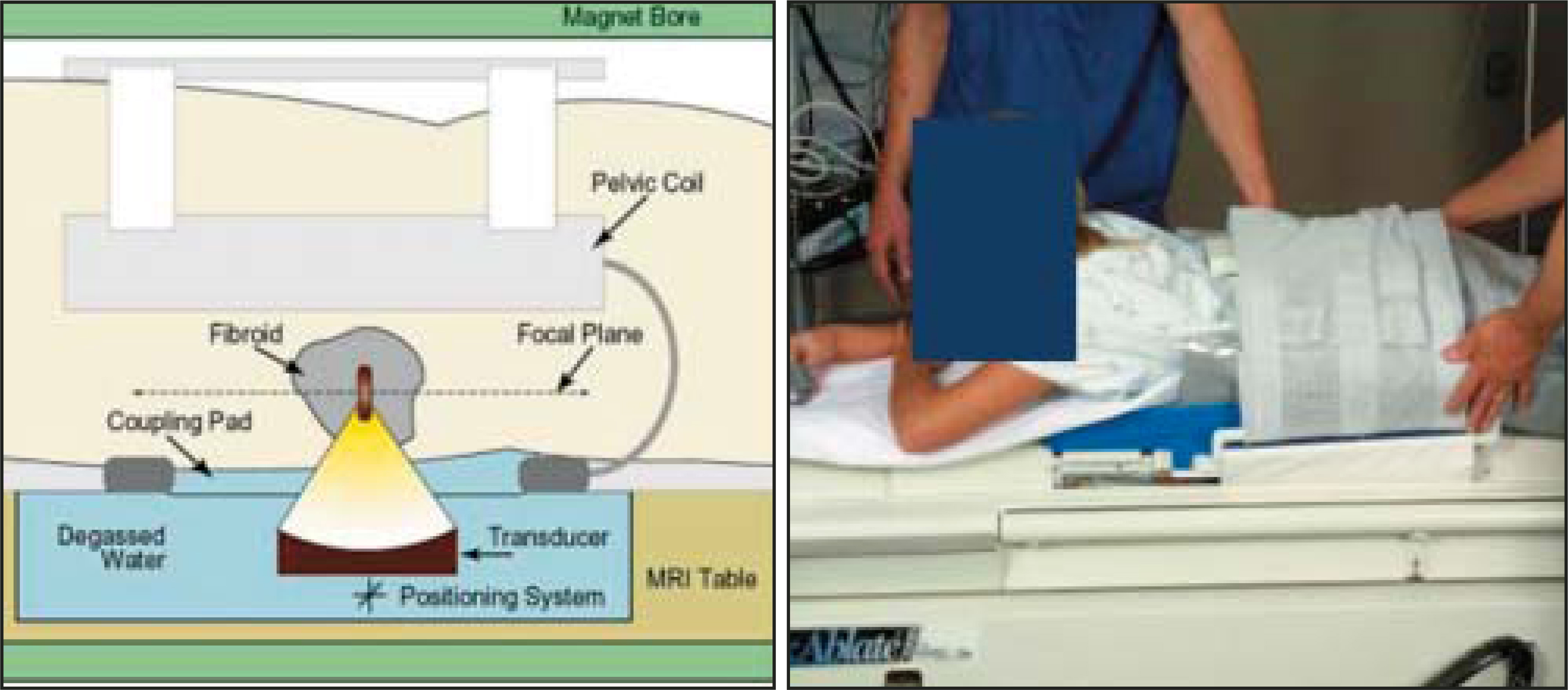
Minimally invasive therapies are also being vigorously pursued. A recently published feasibility study from Brigham and Women’s Hospital reported their experience on a small cohort of patients treated with MRI-guided focused ultrasound ablation.42 The focused ultrasound system used, ExAblate 2000 (InSightec-TxSonics, Dallas, TX) is designed for the patient to lie prone in the MR scanner with the uterus positioned over a water tank containing the focused piezoelectric transducer array (Figure 6). The transducer can then be adjusted to appropriately target the fibroid(s). MR imaging helps to define the size, location, and volume of each fibroid for targeted sonications. In addition, temperature-sensitive MR images were used to calculate peak temperatures and the thermal dose delivered, which helps to assure coagulation of the targeted tissues. The relatively painless, less invasive, and more localized nature of this technique is exciting. One potential disadvantage may be the large number of sonications that would be required to treat a patient with multiple fibroids. In this study, the mean number of sonications was 20.6 to treat a relatively small volume. If larger volumes are treated in a similar manner, procedure times may become excessively long. This new technique may have promise for the future, as it does not use X-ray and may be used in patients who hope for future pregnancy.
Finally, advancements in the field of genetics may play an important role in the future treatment of fibroids. Scientists are attempting to identify chromosomal aberrations found in fibroids with a view toward developing drugs that could counteract the gene defect.
Conclusion
Many women suffer from fibroid-related symptoms each day. Not long ago, there were limited therapeutic options. The medical field has come a long way in developing alternative primary treatments to meet different needs and preferences of women with fibroids. Uterine artery embolization presently appears to be a safe and effective treatment alternative to other surgical therapies. Focused ultrasound, gene-targeted drugs, and other pharmacologic choices will continue to expand treatment modalities for the future.
References
- Surrey E. Steroidal and nonsteroidal “add-back” therapy: Extending safety and efficacy of gonadotropin-releasing hormone agonists in the gynecologic patient. Fertil Steril. 1995;64:673-685.
- Palomba S, Morelli M, Di Carlo C. Bone metabolism in postmenopausal women who were treated with a gonadotropin-releasing hormone agonist and tibolone. Fertil Steril. 2002;78:63-68.
- Lepine L, Hillis S, Marchbanks P. Hysterectomy surveillance—United States, 1980-1993. MMWR CDC Surveill Summ. 1997;46(4):1-15.
- Wilcox L, Koonin L, Pokras R. Hysterectomy in the United States. 1988-1990. Obstet Gynecol. 1994;83:539.
- Presis I. CD-ROM. 1989.
- Makinen J, Johansson J, Tomas C. Morbidity of 10 110 hysterectomies by type of approach. Hum Reprod. 2001;16:1473-1478.
- Maresh M, Metcalfe M, McPherson K. The VALUE national hysterectomy study: Description of the patients and their surgery. BJOG. 2002;109:302-312.
- Kovac S. Hysterectomy outcomes in patients with similar indications. Obstet Gynecol. 2000;95(6 Pt 1):787-793.
- American College of Surgeons. About hysterectomy: Surgical removal of the uterus, or womb.
- Management of Uterine Fibroids. Evidence Report. 2001:1-173.
- Carlson K, Miller B, Fowler F. The Maine Women’s Health Study: I. Outcomes of hysterectomy. Obstet Gynecol. 1994;83:556-565.
- Kjerulff K, Langenberg P, Rhodes J. Effectiveness of hysterectomy. Obstet Gynecol. 2000;95:319-326.
- Weber A, Walters M, Schover L. Functional outcomes and satisfaction after abdominal hysterectomy. Am J Obstet Gynecol. 1999;181:530-535.
- Dicker R, Greenspan J, Strauss L. Complications of abdominal and vaginal hysterectomy among women of reproductive age in the United States. The Collaborative Review of Sterilization. Am J Obstet Gynecol. 1982;144:841-848.
- Harris W. Early complications of abdominal and vaginal hysterectomy. Obstet Gynecol Surv. 1995;50:795-805.
- Lumsden M. Embolization versus myomectomy versus hysterectomy: Which is best, when?. Hum Reprod. 2002;17:253-259.
- Miller C. Myomectomy. Comparison of open and laparoscopic techniques. Obstet Gynecol Clin North Am. 2000;27:407-420.
- Dubuisson J, Fauconnier A, Fourchotte V. Laparoscopic myomectomy: Predicting the risk of conversion to an open procedure. Hum Reprod. 2001;16:1726-1731.
- Nezhat C, Nezhat F, Silfen S. Laparoscopic myomectomy. Int J Fertil. 1991;36:275-280.
- Seinera P, Arisio R, Decko A. Laparoscopic myomectomy: Indications, surgical technique and complications. Hum Reprod. 1997;12:1927-1930.
- Birsan A, Deval B, Detchev R. Vaginal and laparoscopic myomectomy for large posterior myomas: Results of a pilot study. Eur J Obstet Gynecol Reprod Biol. 2003;110:89-93.
- Sawin S, Pilevsky N, Berlin J, Barnhart K. Comparability of perioperative morbidity between abdominal myomectomy and hysterectomy for women with uterine leiomyomas. Am J Obstet Gynecol. 2000;183:1448-1455.
- Hutchins F. Abdominal myomectomy as a treatment for symptomatic uterine fibroids. Obstet Gynecol Clin North Am. 1995;22:781-789.
- Mais V, Ajossa S, Guerriero S. Laparoscopic versus abdominal myomectomy: A prospective, randomized trial to evaluate benefits in early outcome. Am J Obstet Gynecol. 1996;174:654-658.
- Lethaby A, Vollenhoven B, Sowter M. Efficacy of pre-operative gonadotrophin hormone releasing analogues for women with uterine fibroids undergoing hysterectomy or myomectomy: A systematic review. BJOG. 2002;109:1097-1108.
- Fletcher H, Frederick J, Hardie M, Simeon D. A randomized comparison of vasopressin and tourniquet as hemostatic agents during myomectomy. Obstet Gynecol. 1996;87:1014-1018.
- LaMorte A, Lalwani S, Diamond M. Morbidity associated with abdominal myomectomy. Obstet Gynecol. 1993;82:897-900.
- Surgical alternatives to hysterectomy in the management of leiomyomas. Number 16, May 2000 (replaces educational bulletin number 192, May 1994). Int J Gynaecol Obstet. 2001;73:285-293.
- Uterine Leiomyomata. 1994:1-9.
- Nezhat F, Roemisch M, Nezhat C. Recurrence rate after laparoscopic myomectomy. J Am Assoc Gynecol Laparosc. 1998;5:237-240.
- Fedele L, Parazzini F, Luchini L. Recurrence of fibroids after myomectomy: A transvaginal ultrasonographic study. Hum Reprod. 1995;10:1795-1796.
- Buttram V, Reiter R. Uterine leiomyomata: Etiology, symptomatology, and management. Fertil Steril. 1981;36:433-445.
- Acien P, Quereda F. Abdominal myomectomy: Results of a simple operative technique. Fertil Steril. 1996;65:41-51.
- Lefebvre G, Allaire C, Jeffrey J. The management of uterine leiomyomas. J Obstet Gynaecol Can. 2003;25:396-418.
- Vilos G, Daly L, Tse B. Pregnancy outcome after laparoscopic electromyolysis. J Am Assoc Gynecol Laparosc. 1998;5:289-292.
- Nisolle M, Smets M, Malvaux V. Laparoscopic myolysis with the Nd:YAG laser. J Gynecol Surg. 1993;9:95-99.
- Rein M. Advances in uterine leiomyoma research: The progesterone hypothesis. Environ Health Perspect. 2000;108(5):791-793.
- Englund K. Sex steroid receptors in human myometrium and fibroids: Changes during the menstrual cycle and gonadotropin-releasing hormone treatment. J Clin Endocrinol Metab. 1998;83:4092-4096.
- Reinsch R, Murphy A, Morales A, Yen S. The effects of RU 486 and leuprolide acetate on uterine artery blood flow in the fibroid uterus: A prospective, randomized study. Am J Obstet Gynecol. 1994;170:623-627.
- Olive D. Role of progesterone antagonists and new selective progesterone receptor modulators in reproductive health. Obstet Gynecol Surv. 2002;57:S55-S63.
- Shozu M, Murakami K, Segawa T. Successful treatment of a symptomatic uterine leiomyoma in a perimenopausal woman with a nonsteroidal aromatase inhibitor. Fertil Steril. 2003;79:628-631.
- Tempany C, Stewart E, McDannold N. MR imaging-guided focused ultrasound surgery of uterine leiomyomas: A feasibility study. Radiology. 2003;226:897-905.
Citation
. Uterine artery embolization: Where does it stand in the management of uterine leiomyomas? Part 2. Applied Radiology. 2004;33(10):11-11. doi:10.37549/AR1289.