Variant of Left-Sided Scimitar Syndrome
Applied Radiology — Vol. 55 , Issue 2
Published: January 29, 2026
1 Creighton University School of Medicine, Phoenix, Arizona
* Corresponding author: Elise Dunning (elisedunning@creighton.edu)
Categories
Case Summary
A monochorionic diamniotic twin was born at 34 weeks, 1 day via C-section. On prenatal US, the patient was noted to have a ventricular septal defect (VSD), single umbilical artery, and oligohydramnios.
At birth, the patient’s weight was 1720 g and APGARs were 5 and 9 at 1 and 5 minutes. The patient had poor color, shallow breaths, and increased secretions, with initial oxygen saturations in the 50s and a heart rate in the 80s.
Chest x-ray (CXR) showed mild persistent gaseous distention of the upper esophagus with left apical atelectasis and relative hypoaeration of the left lung compared with the right.
Tracheoesophageal fistula (TEF) was identified via echocardiogram on the second day of life, along with the VSD, a large atrial septal defect (ASD), patent ductus arteriosus (PDA), and a hypoplastic left pulmonary artery. Routine CXRs showed persistent left lung hypoplasia.
An initial CT scan of the chest was completed at nearly 2 weeks of life given the abnormal pulmonary artery on echocardiogram.
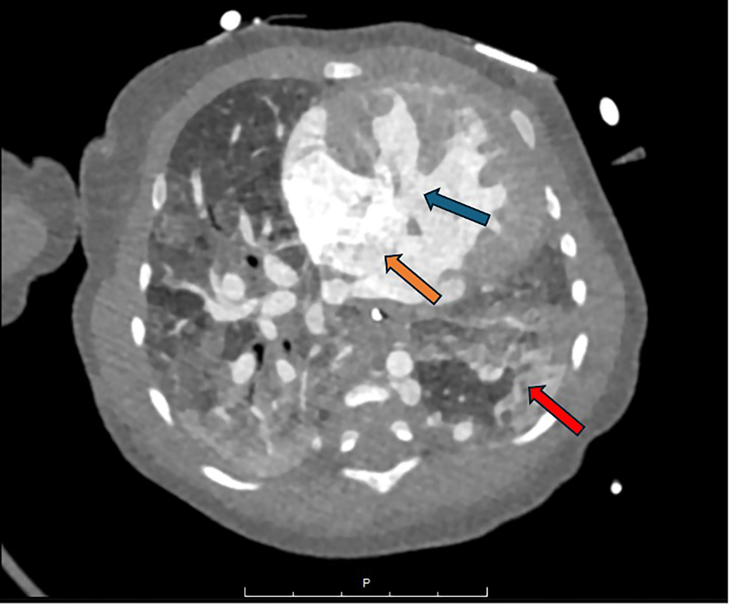
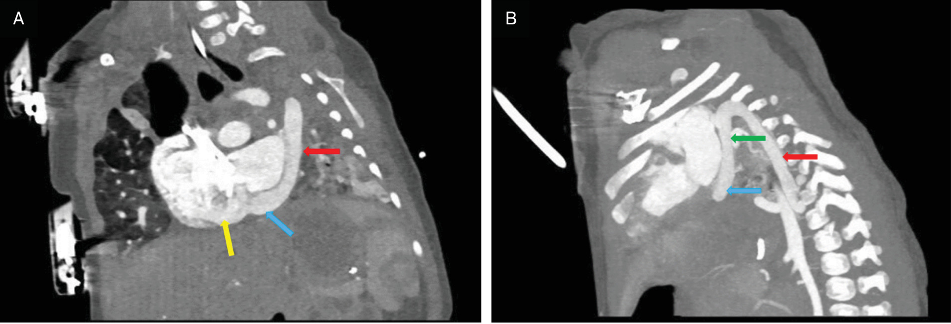
At 1.5 months of life, an echocardiogram was repeated, identifying an innominate malformation and pulmonary venous obstruction. A repeat CT scan (Figures 1, 2) was obtained at that time given the repeated echocardiogram findings.
During hospitalization, the patient had recurrent right pulmonary vein stenosis, as well as recurrent pulmonary infections requiring several rounds of antibiotics. After multiple surgeries and despite adequate mechanical ventilation, the patient developed severe pulmonary congestion and respiratory failure, succumbing at 7 months of age.
Imaging Findings
Initial chest CT scan with IV contrast at 2 weeks of life demonstrated left lung hypoplasia with diminutive left pulmonary veins, left pulmonary artery, and left mainstem bronchus, as well as tracheomalacia (images not included).
Chest CT scan with IV contrast at 1.5 months of life redemonstrated the left lung hypoplasia (Figures 1, 2), hypoplastic left mainstem bronchus and hypoplastic left pulmonary artery (Figure 3), as well as partial anomalous venous return of the left lung draining into the hemiazygos vein and back to the heart via a left superior vena cava (SVC) and left coronary sinus (Figures 2, 3).
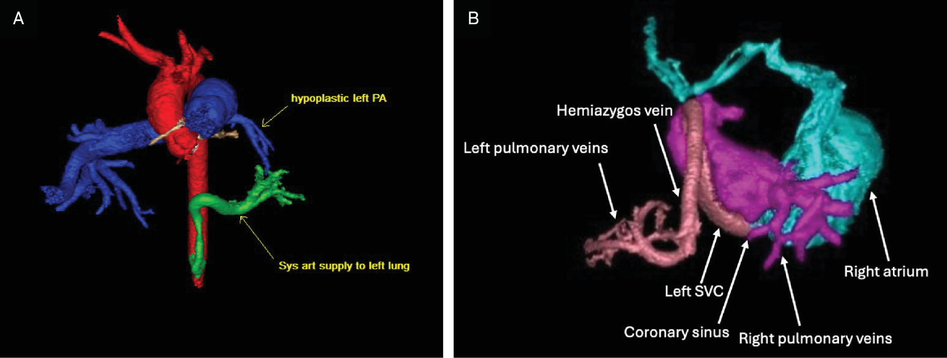
Systemic arterial supply to the left lower lobe of the lung was via an aortopulmonary collateral from the proximal abdominal aorta (Figures 2, 3). There was also a single right pulmonary vein that drained the superior and inferior right lung. There were associated congenital cardiac defects, including a VSD and ASD (Figure 1), as well as tracheomalacia resulting from compression by the brachiocephalic artery (not shown). Additional features, including a TEF and large PDA, were identified via echocardiogram (images not included).
Diagnosis
Left-sided scimitar syndrome variant.
The differential diagnosis includes scimitar syndrome and extralobar pulmonary sequestration resulting from other causes such as congenital pulmonary airway malformation. Other rare congenital anomalies include lobar atelectasis or unilateral absence of the pulmonary artery. Scimitar syndrome is less likely, given that the scimitar vein does not cross the diaphragm. There were associated cardiovascular defects and recurrent stenosis of the right pulmonary vein confluence.
Discussion
Scimitar syndrome, also known as congenital venolobar syndrome, Halasz syndrome, mirror-image lung syndrome, hypogenetic lung syndrome, or vena cava bronchovascular syndrome, is a rare congenital anomaly presenting in 1-3/100,000 live births. 1 It consists of partial anomalous venous return to the heart, resulting in a left-to-right shunt. 1 The name “scimitar” derives from the European term describing a broad variety of Middle Eastern curved swords, which the anomalous pulmonary vein can resemble on imaging. 1 The anomaly is classically described on the right side, and its distinct features are as follows 1 :
-
Partial or entire anomalous venous drainage of the right lung.
-
Variable degrees of the right lung and pulmonary artery hypoplasia.
-
Dextroposition of the heart.
-
Anomalous systemic blood supply to the ipsilateral lung. 1
-
Anomalies of the right bronchial tree, including sequestration. 2
Presentation ranges from asymptomatic to symptoms of cyanosis, respiratory distress, tachypnea, recurrent pneumonia, and heart failure. 1 Those with infantile onset may have demonstrated symptoms of respiratory failure secondary to pulmonary sequestration with recurrent lung infection, as well as features of right ventricular volume overload, pulmonary hypertension, subsequent heart failure, and sometimes pulmonary vein stenosis.
The prognosis for scimitar syndrome presenting in infancy is poor regardless of surgical intervention 1 ; a recent meta-analysis of scimitar syndrome prognosis and treatment found a mortality rate of 31.8%. 1
Left-sided scimitar syndrome is exceedingly rare, with only 6 cases reported in the literature. In these cases, there were aortopulmonary collaterals to the left lung; left lung and pulmonary artery hypoplasia; and at least one anomalous left pulmonary vein that can drain to various sites. 2 These cases also reported the left scimitar vein draining into the left hepatic vein immediately after crossing the diaphragm, 3 the pericardiophrenic vein, 4 the azygos vein, 5 the left hepatic vein close to the IVC, 6 and passing directly into the IVC. 7,8
In our patient’s case, the anomalous pulmonary venous system of the left lung drained into the hemiazygos vein, then into the left-sided superior vena cava, which then emptied into the coronary sinus and eventually passed into the right atrium.
Notably, all the previously reported cases had anomalous draining veins that crossed the diaphragm before returning to the heart; thus, this case does not meet the criteria for true left-sided scimitar syndrome. Although the draining vein in this case did not cross the diaphragm, however, it still resulted in the “scimitar” appearance typical of the condition on imaging. In addition, characteristic findings such as left lung hypoplasia, left pulmonary artery hypoplasia, and anomalous systemic arterial supply to the left lobe were all present in this case, as were congenital cardiac anomalies often associated with infantile scimitar syndrome. 1,3 Moreover, whereas right scimitar syndrome can be associated with left pulmonary vein stenosis, a rare but well-described phenomenon, our case was associated with recurrent right pulmonary vein stenosis. 9 Given the findings, our case arguably suggests an interesting variant of scimitar syndrome.
Conclusion
Left-sided scimitar syndrome is an exceptionally rare variant of a classically right-sided condition. The presentation in this case involved another anomaly, in which the draining vein did not cross the diaphragm, although the classic “scimitar” appearance was still present on imaging. Classic symptoms such as respiratory distress and recurrent pneumonia were present in this case, along with congenital cardiac anomalies and stenosis of the right pulmonary vein confluence. Surgical intervention is the only definitive treatment; however, survival rates remain poor for patients with infantile onset of the condition.
References
- Wang K, Xu X, Liu T, Gao W, Guo Y. Treatment and prognosis of Scimitar syndrome: a retrospective analysis in a single center of East China. Front Cardiovasc Med. 2022;9:973796. doi:10.3389/fcvm.2022.973796.
- Bo I, Carvalho J, Cheasty E, Rubens M, Rigby M. Variants of the scimitar syndrome. Cardiol Young. 2016;26(5):941-947. doi:10.1017/S1047951115001651.
- Pandey S, Bharath A, Mohata A. Left sided scimitar syndrome. Ann Pediatr Cardiol. 2023;16(4):303-305. doi:10.4103/apc.apc_73_23.
- Mardini M, Sakati N, Nyhan W. Anomalous left pulmonary venous drainage to the inferior vena cava and through the pericardiophrenic vein to the innominate vein: left-sided scimitar syndrome. Am Heart J. 1981;101(6):860-863. doi:10.1016/0002-8703(81)90630-x.
- Bratincsák A, Rao R, El-Said H. Unusual variant of a rare constellation: a left-sided scimitar syndrome with connection to the azygos vein. Congenit Heart Dis. 2010;5(2):174-177. doi:10.1111/j.1747-0803.2010.00401.x.
- Rutledge J, Hiatt P, Wesley Vick G, Grifka R. A sword for the left hand: an unusual case of left-sided scimitar syndrome. Pediatr Cardiol. 2001;22(4):350-352. doi:10.1007/s002460010245.
- Agayev A, Yekeler E. Left-sided scimitar syndrome. Pediatr Radiol. 2009;39(2):191. doi:10.1007/s00247-008-1031-6.
- Juraszek A, Cohn H, Van Praagh R, Van Praagh S. Isolated left-sided scimitar vein connecting all left pulmonary veins to the right inferior vena cava. Pediatr Cardiol. 2005;26(6):846-847. doi:10.1007/s00246-005-0920-9.
- Mendoza A, Herrera D, Caro A. Scimitar syndrome and left pulmonary vein stenosis: a serious and rare association. Ann Pediatr Cardiol. 2022;15(1):80-83. doi:10.4103/apc.apc_53_21.
Disclosures
The authors have no conflicts of interest to disclose. None of the authors received outside funding for the production of this original manuscript and no part of this article has been previously published elsewhere.
Citation
. Variant of Left-Sided Scimitar Syndrome. Applied Radiology. 2026;55(2). doi:10.37549/AR-D-25-0129.